Issue published September 22, 2026 Previous issue

- Volume 11, Issue 18
On the cover:
IFN-γ–producing self-reactive CD4+ T cells induce autoimmune adrenalitis in a mouse model of Addison’s disease
Andreyeva et al. generated a mouse model that uncovers how immune cells destroy the adrenal glands in Addison’s disease and identified IFN-γ as a promising target for future therapies. The cover image shows characteristic granuloma formation within the adrenal cortex. Immunofluorescence staining was performed for DAPI (blue) to visualize nuclei and CYP11A1 (red) to identify steroidogenic adrenocortical cells. Granulomatous lesions appear green because of the strong intrinsic autofluorescence of lipid-laden macrophages within the granulomas. Image credit: Arina Andreyeva and Ales Neuwirth.
Andreyeva et al. generated a mouse model that uncovers how immune cells destroy the adrenal glands in Addison’s disease and identified IFN-γ as a promising target for future therapies. The cover image shows characteristic granuloma formation within the adrenal cortex. Immunofluorescence staining was performed for DAPI (blue) to visualize nuclei and CYP11A1 (red) to identify steroidogenic adrenocortical cells. Granulomatous lesions appear green because of the strong intrinsic autofluorescence of lipid-laden macrophages within the granulomas. Image credit: Arina Andreyeva and Ales Neuwirth.
Go to section:
-
Research Letters
×
Abstract
Authors
Long Qian, Megan L. Baker, Laura Aponte Becerra, Cathleen Liang, Emma Koval, Kyra Shelton, Daris Javed, Gilbert Moeckel, Deepika Kumar, Avi Z. Rosenberg, Michael Kuperman, Tinyi Chu, Wassim Obeid, Xuefei Tian, Chirag R. Parikh, Leyuan Xu, Jonathan Barasch, Shuta Ishibe, Lloyd G. Cantley, Dennis G. Moledina
×Abstract
Authors
Ian A. Mellis, Madeline Wu, Kristin Daniel, Hsiang Hong, Yicheng Guo, David D. Ho


-
Research Articles
×
Abstract
ACE2 is a membrane-bound monocarboxypeptidase strongly expressed in the renal proximal tubule (PT) with high affinity to degrade the vasopressor angiotensin II (AngII). We employed a mouse model of PT-specific ACE2 deletion (PT ACE2–KO) to demonstrate that the renal PT is a critical site for ACE2 regulation of blood pressure (BP) via modulation of the intrarenal renin-angiotensin system (RAS). While deletion of ACE2 from the PT had a minimal effect on baseline physiology, PT ACE2–KO mice were more susceptible to AngII hypertension than control mice. At day 5 of AngII infusion, the enhanced BP response was associated with cardiac hypertrophy, increased renal AngII levels, failure to suppress epithelial sodium channel (ENaC) γ cleavage, and increased sodium pump activity in PT ACE2–KO mice. Control mice instead increased renal ACE2 expression to reduce renal AngII accumulation and suppress intrarenal RAS activation, which offered protection from hypertension and complications. Transcriptional analysis corroborated changes in intrarenal RAS components and revealed alterations in distinct physiological pathways during AngII hypertension in PT ACE2–KO mice. Our studies provide evidence for alterations in ENaC regulation to contribute to the development of AngII hypertension and support PT-derived ACE2 as an integral member of the intrarenal RAS.
Authors
Jacqueline M. Emathinger, Zhidan Xiang, Jonathan W. Nelson, Matthew W. Hagen, Nakyung Kim, David I. Ortiz-Melo, Natalie Mattocks, Jorge F. Giani, Dexter L. Lee, Hannah Hartman-Houstman, Donna L. Ralph, Alicia A. McDonough, Brianna Chen, Stan G. Louie, Thomas M. Coffman, Susan B. Gurley
×Abstract
BACKGROUND Primary laminopathies are a heterogeneous group of rare diseases caused by nuclear lamina dysfunction due to pathogenic LMNA variants. However, despite their ubiquitous expression, LMNA variants have rarely been linked to chronic kidney disease (CKD). Here, we systematically investigate clinical implications and functional underpinnings of a distinct LMNA missense variant [lamin A/C p.(Arg349Trp)] that has sporadically been found in patients with a complex phenotype including lipodystrophy, proteinuria, and focal segmental glomerulosclerosis (FSGS).METHODS In clinical and functional terms, we compare lamin A/C Arg349Trp with missense changes at Arg482, the most common hotspot residue for type 2 familial partial lipodystrophy (FPLD2). In particular, we assess renal endpoints in corresponding patient cohorts and investigate disease-associated alterations in vitro.RESULTS In contrast to patients with FPLD2, individuals with lamin A/C Arg349Trp experience high-grade proteinuria and a rapid decline of glomerular filtration rate with kidney failure at a median age of 43 years. Mechanistically, we demonstrate that Arg349Trp associates with an abrogation of the structural interaction between lamin A/C and nucleoporin 155, nuclear pore complex aggregation, and an alteration of TGF-β1–dependent signaling.CONCLUSION While patients with Lamin A/C Arg482 missense changes are at very low risk for progressive CKD, patients harboring Arg349Trp show nephrotic range proteinuria and kidney failure in midlife. Hence, high-grade proteinuric kidney disease is genotype specific, and patients with the Arg349Trp substitution require early renoprotective intervention to potentially halt progression and prevent kidney failure.FUNDING German Research Foundation, project IDs 502928386, 508310822, 539950728, 445703531, 451693578, 471294925, 496611648, and 539950728; Else Kröner-Fresenius Foundation (2019_A96).
Authors
Sebastian Sewerin, Charlotte Aurnhammer, Mohamed Hamed, Gwladys Revêchon, Ria Schönauer, Christin Findeisen, Konstanze Miehle, Šárka Tesařo vá, Theodoros Georgomanolis, Carsten Bergmann, Constantin Wolff, Marek Kollár, Baris Akinci, David Araújo-Vilar, Giovanni Ceccarini, Éva Csajbók, Alessandra Gambineri, Martin Heni, Thomas Scherer, Iztok Štotl, Ekaterina Sorkina, Marie-Christine Vantyghem, Elena Vorona, Martin Wabitsch, Julia von Schnurbein, Camille Vatier, Joëlle Roume, Yves Reznik, Maria Eriksson, Wolfram Antonin, Corinne Vigouroux, Jan Halbritter
×Abstract
Bone marrow–derived circulating monocytes continuously replenish intestinal macrophages, which become dysregulated in inflammatory bowel disease (IBD) and contribute to disease pathology. The origins of this dysregulation remain poorly understood. Here, we investigate the reprogramming of circulating monocytes in IBD prior to tissue recruitment using single-cell transcriptomic, epigenomic, and functional approaches. We characterize blood monocyte heterogeneity in newly diagnosed, treatment-naive IBD patients and healthy controls and show that monocytes in Crohn’s disease (CD) display a distinct transcriptional profile and altered distributions across inferred developmental trajectories; less pronounced changes were observed in ulcerative colitis (UC). We link CD-associated transcriptional changes to alterations in chromatin accessibility and identify NF-κB, EGR, KLF, and AP-1 family transcription factors as putative regulators of an inflammatory gene program in blood monocytes from patients with CD. We uncover a potential role for IFN-γ in priming blood monocytes for inflammatory function in CD by limiting their capacity to be regulated by IL-10. Finally, we show that the transcriptional and functional alterations in monocytes from patients with CD are maintained in monocyte-derived cells from the intestine. Together these data suggest that intestinal macrophage dysfunction in CD is, at least in part, pre-established by systemic signals prior to tissue recruitment.
Authors
Eve Hornsby, Radha Gadhok, Inva Hoti, Eva Wozniak, James R. Boot, Emma Connick, Paul Stevens, Holly Creed, Amy Lewis, Andrew Silver, James O. Lindsay, Andrew J. Stagg
×Abstract
CFTR in the lung epithelium contributes to the secretion of a surface liquid layer that is essential to lung homeostasis and defense. The understanding of how liquid is secreted in the lung is derived largely from studies of the airway epithelium. Comparatively little is known about liquid secretion mechanisms in the alveolar epithelium, including its cellular source. To define which cell type drives alveolar liquid secretion, we generated transgenic mice that expressed a Cftr-null allele in alveolar type 1 (AT1) cells, type 2 (AT2) cells, or both, then viewed liquid secretion in live alveoli using confocal microscopy of isolated, perfused lungs. Our findings show that liquid secretion was blocked in alveoli of all 3 transgenic mice, indicating that both AT1 and AT2 cells contribute to alveolar liquid secretion. Cftr-null expression in AT1 cells also blocked the secretion-mediated clearance of small particle and bacterial clusters from alveolar walls, indicating that AT1 cell CFTR contributes to alveolar defense. Together, these findings show alveolar liquid secretion depends on both AT1 and AT2 cell CFTR, and that CFTR in AT1 cells — a cell type not traditionally considered in liquid secretion mechanisms or CFTR-related lung diseases — contributes to lung liquid dynamics and host defense.
Authors
Sayahi Suthakaran, Sonya Homami, Deebly Chavez, Stephanie Tang, Sarah K.L. Moore, Chaya Sussman, Jimmy Zhang, Clemente J. Britto, Alice Prince, Alison J. May, Jaymin J. Kathiriya, Jaime L. Hook
×Abstract
Chronic kidney disease is a global health concern characterized by maladaptive repair processes that lead to kidney fibrosis. Following injury, early alterations in the extracellular matrix precede the development of kidney fibrosis and represent potential therapeutic targets to improve kidney repair. In this context, studies from our laboratory and others have shown that the matricellular protein SMOC2 can be targeted to decrease inflammation and tubulointerstitial fibrosis after kidney injury. Tubular epithelial cells (TECs), which are abundant and particularly susceptible to injury, play a central role in maladaptive repair; however, whether SMOC2 affects their function after kidney injury has not been explored. In this study, we showed that SMOC2 localized to the basement membrane of injured TECs across 3 murine models of kidney injury. Our in vitro studies demonstrate that SMOC2 induced a partial epithelial-to-mesenchymal (EMT) transition in TECs. We further demonstrated that its extracellular calcium-binding domain mediated binding to the decellularized extracellular matrix and accounted for most of its effects on TECs. Mechanistically, SMOC2 promoted partial EMT through an integrin-dependent pathway. Together, these findings provide mechanistic insight into how SMOC2 drives maladaptive repair by modulating TEC behavior and identify its calcium-binding domain as a key functional mediator.
Authors
Schrodinger Cenatus, Peng Gao, Nathalie Henley, Caroline Lamarche, Xue-Song Liu, Frédérick A. Mallette, Jonatan Barrera-Chimal, Casimiro Gerarduzzi
×Abstract
The mechanisms by which e-cigarette vaping (EV) affects lung health remain unclear. Clusters of EV-associated lung injury indicate that EV damages distal lung parenchyma and increases vulnerability to second-hit injury, including respiratory viral infections. Using human lung endothelial and epithelial cells and precision-cut lung slices, we investigated the mechanisms underlying distal lung cell injury and repair triggered by brief (24-hour) EV exposure. RNA-seq of lung tissue from golden Syrian hamsters evaluated the persistence of lung stress responses (10 days after 5 days of EV exposure) and the effect of EV on host defense against influenza A virus and SARS-CoV-2. EV disrupted the barrier function of human distal lung cells through JNK stress response signaling, triggered autophagy with impaired flux, suppressed mTOR signaling and cell proliferation, and culminated in apoptosis. Transcriptional responses in EV-exposed hamster lungs revealed persistent activation of JNK signaling, autophagy, barrier dysfunction, tissue remodeling, and impaired Th1 immunity. EV increased SARS-CoV-2 viral burden, downregulated antiviral genes (Ifit1, Isg15, Nfkbia), and amplified oxidative stress and IL-12 signaling. Short-term EV exposure triggered stress-induced distal lung cell injury with persistent changes in antiviral immunity and molecular pathways associated with tissue remodeling. That may increase susceptibility to viral infections and contribute to lung disease.
Authors
Tanner C. Rivera, Kelly S. Schweitzer, Christina Cornell, Jordan Nall, Nicholas Egersdorf, Courtney Moeder, Riley A. Cooney, Eszter K. Vladar, Steve D. Groshong, Gregory P. Downey, James P. Bridges, Richard Bowen, Hong Wei Chu, Irina Petrache
×Abstract
Severe SARS-CoV-2 infection is characterized by lung hyperinflammation, impaired IFN responses, and defective T cell activation, yet the molecular drivers of these immune dysregulations remain incompletely understood. Caspase-11 (CASP11), a key effector of the noncanonical inflammasome, has been shown to mediate an innate hyperinflammatory response and cytokine release in a mild SARS-CoV-2 infection model. However, the role played by CASP11 in severe SARS-CoV-2 disease and how it affects adaptive immunity has not been identified. Here, we found that CASP11 exacerbates severe SARS-CoV-2 pathogenesis by amplifying early innate immune responses while concurrently impairing antiviral CD8+ T cell immunity. Using global KO mice, hematopoietic BM chimeras, and Cx3cr1-expressing mononuclear phagocyte system cell–specific CASP11 deletion models, we show that targeting CASP11 reduces lung inflammation, promotes early NK cell–mediated IFN-γ production, and enhances robust virus-specific effector CD8+ T cell responses. This was associated with enhanced viral clearance and improved survival, even under lethal infection conditions. Importantly, CASP11-KO mice also exhibited faster resolution of postviral inflammation. These findings position CASP11 as a promising immunomodulatory target for acute and delayed manifestations of severe SARS-CoV-2 infection.
Authors
Mostafa M. Eltobgy, Mohamed M. Shamseldin, Owen D. Whitham, Heba M. Amer, Jeffrey R. Atkinson, Asmaa Badr, Jesse M. Hall, Gauruv Gupta, Yara Y. Hassan, Rabab El-Mergawy, Richard Perez, Sarah E. Faber, Maciej Pietrzak, Amy Webb, Xiaoli Zhang, Adam D. Kenney, Destiny Bissell, Jihad I. Omran, Shady Estfanous, Kylene P. Daily, Amir Yousif, Marisa R. Joldrichsen, Andrew McNamara, Mahesh KC, Mark E. Peeples, Emily A. Hemann, Hazem E. Ghoneim, Shahid M. Nimjee, Estelle Cormet-Boyaka, Jianrong Li, Prosper N. Boyaka, Jacob S. Yount, Benjamin M. Segal, Purnima Dubey, Amal O. Amer
×Abstract
The NF-κB signaling pathway coordinates inflammation, cell survival, and proliferation, while restraining excessive cell death to maintain immune homeostasis. Truncating mutations in RELA, encoding the NF-κB subunit p65, have been linked to autoinflammation and autoimmunity, but the underlying mechanisms remain incompletely defined. We investigated 6 patients from 5 unrelated families carrying heterozygous truncating RELA variants. Despite reduced p65 expression, patients exhibited a broad spectrum of inflammatory manifestations alongside elevated baseline and stimulus-induced proinflammatory cytokines. Functional analyses in patient-derived cells and mutant RELA-KI models showed that upstream NF-κB signaling was intact, but induction of inhibitory regulators such as IκBα and A20 was impaired. This defective feedback control shifted immune homeostasis toward amplified inflammatory responses that depended on the residual activity of the remaining functional RELA allele. Single-cell transcriptomics revealed distinct cell type–specific consequences: monocytes displayed constitutive type I IFN and NF-κB activation, B cells retained partial compensatory signaling, and T and NK cells exhibited transcriptional signatures of cell death pathways. Patient fibroblasts and mutant RELA-KI cells further confirmed enhanced TNF-induced inflammatory gene expression and hypersensitivity to apoptosis and necroptosis. These findings establish RELA haploinsufficiency as a cause of systemic immune dysregulation and link defective NF-κB feedback control to unchecked inflammation and inflammatory cell death.
Authors
Nadja Lucas, Sophia Weidler, Antonia A. Eicher, Baerbel Keller, Özlem Satirer, Adam Desrochers, Timothy J.S. Ramnarine, Mohammad Mokhtari, Sophie Elstner, Timmy Strauss, Simon W. Mages, Arek Kendirli, Oana Cristina Buzoianu, Tobias B. Haack, Lina Igel, Tim Niehues, Sandra von Hardenberg, Maria Fasshauer, Rami Abou Jamra, Hagen Ott, Ulrike Hüffmeier, Catharina Schuetz, Marisa Bijwaard, Susan Wagner, Paulina Switala, Sarah Koss, Jurek Schultz, Stefanie Kretschmer, Jasmin Kümmerle-Deschner, Christine Wolf, Johanna Klughammer, Min Ae Lee-Kirsch
×Abstract
BACKGROUND Dichloroacetate (DCA) is an orally administered structural analog of pyruvate, an endogenous pyruvate dehydrogenase kinase inhibitor.METHODS We conducted a phase III multicenter trial in 34 children with pyruvate dehydrogenase complex deficiency (PDCD). Participants were randomly allocated to 4 months of treatment with DCA or a placebo, followed by a 1-month washout period and crossover to the alternate arm, and could continue into an open-label extension period. DCA dosing was predetermined by pharmacogenomic analysis of GSTZ1, which modulates DCA metabolism. The primary endpoint was the observer-reported outcomes motor domain (ObsROmotor) score. Additional assessments evaluated motor function, plasma lactate levels, and survival.RESULTS Chronic DCA was well tolerated and safe. The primary endpoint, ObsROmotor, was not statistically significantly different between the treatment and placebo groups (P = 0.512). However, longer-term treatment, including the open-label extension, showed a statistically significant treatment effect (P = 0.002), especially in participants with higher baseline motor impairment (ObsROmotor ≥ 8; P = 0.001). DCA decreased plasma lactate –0.48 (0.82) mmol/L (–20%; P = 0.006). Survival of participants was significantly greater than that of a natural history cohort (log-rank P = 0.027).CONCLUSION Longer-term treatment with DCA, dosed based on GSTZ1 haplotype, is safe and was associated with a statistically significant improvement in patient motor function, plasma lactate, and survival.FUNDING NIH (R01FD005407; R42HD089804), University of Florida Department of Medicine, Saol Therapeutics.
Authors
Peter W. Stacpoole, Jose E. Abdenur, Jirair K. Bedoyan, Lorenzo Botto, Gregory M. Enns, Marni J. Falk, Rebecca Ganetzky, Cheryl Garganta, Kevin Glinton, Andrea Gropman, Sharon Hamm, Eugenia Henry, Nicola Longo, Richard Neiberger, Russell P. Saneto, Fernando Scaglia, Sub H. Subramony, Jerry Vockley, Richard E. Wagner
×Mitochonic acid 5 alleviates amyotrophic lateral sclerosis phenotypes via mitochondrial augmentation
Abstract
Amyotrophic lateral sclerosis (ALS) is a fatal neurodegenerative disease that urgently requires effective treatment. Mitochondrial dysfunction underlies ALS pathology and represents a potential therapeutic target. Here, we demonstrated the therapeutic potential of mitochonic acid 5 (MA-5), a novel mitochondria-targeted compound that ameliorated ALS phenotypes by enhancing mitochondrial function. In a Drosophila ALS model expressing a mutant human SOD1 (G85R), MA-5 significantly improved locomotor activity, with a trend toward restoration of mitochondrial integrity. In skin fibroblasts derived from ALS patients and motor neurons derived from induced pluripotent stem cells, MA-5 restored ATP production and increased mitochondrial motility. Multiomics analyses suggested that MA-5 modulated mitochondria-linked gene expression and downregulated the glycerophosphate shuttle, contributing to mitochondrial reactive oxygen species production. Transcriptomic analysis identified C7orf31 as a potential marker for monitoring the therapeutic effects of MA-5 and diagnosing ALS subtypes. These findings support MA-5 as a promising therapeutic candidate for ALS and propose C7orf31 as a potential biomarker for treatment monitoring and for disease subtyping.
Authors
Yoshitsugu Oikawa, Yuhan Luo, Naoki Suzuki, Tomoko Kasahara, Yoshiyasu Tongu, Yuki Yoshida, Tsukasa Tominari, Shogo Tanabe, Yoshiko Suto, Hitomi Kashiwagi, Saki Saito, Kensuke Ikeda, Chitose Suzuki, Arata Kuranaga, Tetsuya Akiyama, Satoru Morimoto, Yoshitsugu Aoki, Rieko Muramatsu, Tomoyoshi Soga, Masashi Aoki, Hideyuki Okano, Tetsuhiro Tanaka, Takaaki Abe, Erina Kuranaga, Takafumi Toyohara
×Abstract
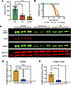
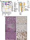
Autoimmune Addison’s disease (AD) is a rare but life-threatening disorder caused by immune-mediated destruction of the adrenal cortex, and progress in therapy has been limited by insufficient mechanistic insight. Here, we establish a model of experimental autoimmune adrenalitis that recapitulates key features of AD and reveals sex-dependent differences in disease manifestation within the model. Immunization with peptides derived from the adrenal self-antigen CYP11A1 induces corticosterone insufficiency. We show that autoimmune adrenalitis is driven by IFN-γ produced by self-reactive CD4+ T cells, promoting granulomatous inflammation in the adrenal cortex. Together, these findings identify IFN-γ as a central effector of autoimmune adrenalitis and suggest that targeting the IFN-γ pathway may represent a potential therapeutic strategy for AD.
Authors
Arina Andreyeva, Juraj Michalik, Veronika Niederlova, Veronika Cimermanova, Ales Drobek, Radislav Sedlacek, Jan Prochazka, Juraj Labaj, Olha Fedosieieva, Waldemar Kanczkowski, Peter Draber, André Sulen, Ondrej Stepanek, Aleš Neuwirth
×Abstract
Small molecules that modulate myofibril ATPase activity via the myosin regulatory light chain (RLC) display a broad spectrum of activity in their ability to enhance relaxation and slow contraction. EDG-7500 exhibits features consistent with a ‘diastolic-selective’ cardiac sarcomere modulator (d-CSM), and its ability to treat HCM was explored in engineered human tissue (EHT), human HCM cardiac strips, and an R403Q mutation swine model. In fibers, EDG-7500 preferentially inhibited myofibril ATPase activity and force at diastolic calcium levels, retained length-dependent force activation, accelerated relaxation, and exhibited a shallow, self-limiting inhibitory-exposure response to LV fractional shortening. Compared to CMIs, EDG-7500 moved myosin heads towards the thin filament and accelerated relaxation without decreasing force in mutated EHTs (R403Q). In human HCM cardiac strips, EDG-7500 did not alter myosin SRX state, but decreased Ca2+-sensitivity of force independent of mutation. In R403Q swine, chronic EDG-7500 normalized LV filling pressure and prevented pathological cardiac remodeling while preserving normal systolic function and cardiac reserve. EDG-7500 differentiates itself from CMIs by uniquely targeting both phases of the cardiac cycle, improving ventricular relaxation while preserving systolic function. This suggests optimal diastolic efficacy can be reached without balancing systolic impairment.
Authors
Craig A. Emter, Marcus Henze, Mike DuVall, Sarah Lehman, Lindsey Lee, Ben Barthel, Natalie A. Hawryluk, Molly Madden, Yangsong Wu, Amy Perry, Martin Beyer, Eric Wei, Cassady Rupert, Steve Roof, Angela Peter, Emily DiNatale, Sara Cantrell, Jessica Tolley, Stephen Schlachter, Jolanda van der Velden, Michelle Michels, Christine Seidman, Weikang Ma, Leslie Leinwand, Stuart Campbell, Julien Ochala, David Bluemke, Darla Tharp, Jonathan Seidman, Carlos L. del Rio, Marc Semigran, Marc Evanchik, Kevin Koch, Alan Russell
×Abstract
Surgical stress, such as liver ischemia/reperfusion (I/R) injury characterized by robust neutrophil infiltration and immune activation, induces sterile inflammation that reshapes tissue immunity and contributes to organ dysfunction, yet the intercellular circuits that spatially orchestrate these responses within the hepatic immune microenvironment remain incompletely defined. Here, we integrate single-cell RNA sequencing, spatial transcriptomics, high-dimensional spectral flow cytometry, and metabolomics to resolve the hepatic immune landscape following I/R at cellular and spatial resolution. While confirming extensive immune remodeling, we identify a dominant neutrophil–Kupffer cell communication axis mediated by neutrophil-derived thrombospondin-1 (TSP-1), which selectively engages CD36 on Kupffer cells. This interaction drives coordinated immune-metabolic reprogramming in Kupffer cells, characterized by suppression of oxidative phosphorylation, enhanced glycolysis, and remodeling of sphingolipid metabolism, including accumulation of hexosylceramides. Spatial analyses reveal preferential neutrophil–Kupffer cell colocalization within necrotic niches, and cross-species integration with human liver transplant datasets demonstrates conserved upregulation of the TSP-1/CD36 axis following reperfusion. Pharmacologic inhibition of TSP-1 attenuates liver injury and inflammatory responses in vivo. Together, these findings define a spatially organized, neutrophil-driven immune-metabolic circuit that governs functional reprogramming of Kupffer cells during surgical stress and identify the TSP-1/CD36 pathway as a conserved and targetable mediator of sterile liver injury.
Authors
Hongji Zhang, Xingping Huang, Preethi Jayakumar, Yunwei Zhang, Ti Yang, Xiaorong Guo, Grace Wu, Amrendra Kumar, Vijaya Bharti, Amblessed Onuma, Chengli Shen, Dequan Lou, Patricia S. Latham, James M. Crawford, Kong Chen, Anna E. Vilgelm, Lopa Mishra, Allan Tsung, Meihong Deng, Hai Huang
×Abstract
Mycoplasma pneumoniae pneumonia (MPP) can cause serious extrapulmonary complications, including life-threatening thrombosis. This study aimed to elucidate the roles of neutrophils and neutrophil extracellular traps (NETs) in vascular endothelial cell (EC) activation in pediatric MPP-associated thrombosis. We analyzed the relationship between neutrophils and thrombosis in children with MPP and used mouse models of neutrophilia (Csf3 plasmid injection), neutropenia (Csf3 deficient, Csf3–/–), and defective NETs formation (Pad4 deficient, Pad4–/–). The effects of neutrophils and NETs on EC activation were further examined in vivo, in vitro, and in human samples. Elevated neutrophil count was observed in patients with thrombosis and functioned as a potential diagnostic marker as well as a risk factor for MPP-associated thrombosis. EC activation was enhanced in MPP mice with neutrophilia but attenuated in neutropenic or Pad4–/– mice. NETs activated ECs through TLR2 and JAK/STAT3 signaling, and inhibition of NETs formation (Cl-amidine), TLR2 (C29), and JAK (upadacitinib) each attenuated this response. Strong correlations among neutrophils, NETs, EC activation, and thrombosis were observed in pediatric patients. These findings suggest that neutrophils promoted thrombosis in MPP via NETs-mediated EC activation involving TLR2 and JAK/STAT3 signaling. This study provides mechanistic insights into the inflammatory-thrombotic processes in MPP-associated thrombosis and offers a rationale for further investigation of neutrophils, NETs, TLR2, and JAK/STAT3 signaling in this context.
Authors
Xia Huang, Yifan Zhu, Yun Guo, Tian Lv, Haiyan Gu, Yingying Luo, Dan Li, Hang Sun, Deyu Zhao, Feng Liu
×Abstract
Right ventricular pressure overload (RVPO) is a critical pathophysiological feature of numerous pediatric cardiovascular diseases. Transverse tubules (T-tubules) form the foundation for efficient excitation-contraction coupling in mature cardiomyocytes. We hypothesized that RVPO impairs T-tubule maturation through the regulatory protein bridging integrator 1 (BIN1). In right ventricular samples from children with tetralogy of Fallot, characterized by RVPO, and in a neonatal rat RVPO model induced by pulmonary artery banding (PAB), T-tubule maturation was disrupted. RNA-seq revealed significant downregulation of T-tubule–associated genes, with Bin1 among the most suppressed. Bin1 overexpression restored T-tubule maturation in PAB rats. ATAC-seq showed reduced chromatin accessibility at Bin1 loci; motif analysis identified Mef2d (myocyte enhancer factor 2D) as the top enriched transcription factor. Mef2d knockdown rescued Bin1 expression and T-tubule maturation, and mutation of the Mef2d binding sites within the Bin1 promoter abolished the inhibitory effect of Mef2d on Bin1 promoter activity. This study delineates a phenomenon and a mechanism of cardiomyocyte maturation under pathological stress. The findings not only advance our understanding of this most pivotal event in postnatal cardiac development but also unveil a potential therapeutic direction for pediatric cardiovascular diseases associated with RVPO.
Authors
Yuqing Hu, Yiting Xue, Xudong Chen, Linghui Kong, Debao Li, Zheng Wang, Sixie Zheng, Siqi She, Hao Li, Sijuan Sun, Hao Chen, Lijun Chen, Peisen Ruan, Kai Wang, Lincai Ye
×Abstract
Systemic lupus erythematosus (SLE) is a progressive autoimmune disease that affects multiple organs and tissues, with lupus nephritis (LN) as one of its most severe complications. Although LN progression is associated with compromised permeability of human renal glomerular endothelial cells (HRGECs), the underlying mechanisms are not fully defined. Herein, we demonstrate that aberrant glycolysis drives this glomerular endothelial barrier defect by suppressing the transcription of tight junction (TJ) genes. Mechanistically, circulating self-DNA in SLE plasma acts as a ligand that activates the cGAS/STING pathway in HRGECs, driving aberrant glycolytic adaption. The resulting glycolytic product, lactate, serves as a substrate for protein lactylation, leading to extensive lactylation and subsequent ubiquitination of enhancer of zeste homolog 2 (EZH2). In consequence, EZH2 deficiency results in reduced H3K27me3 levels, thereby suppressing the transcription of TJ genes. In a self-DNA–induced SLE model, inhibition of cGAS/STING signaling or lactate production effectively restored the integrity of TJs of HRGECs and concurrently alleviated key LN symptoms. Together, lactate programs lactylation and ubiquitination of EZH2 to impair the glomerular endothelial barrier in human SLE.
Authors
Jiaxin Lei, Xingyu Zhai, Yixin Wang, Ying Li, Lei Li, Mengdi Liu, Jing Guo, Lingyi Li, Zhezhuyun Chen, Qinghua Cao, Zhichun Liu, Ting Liu, Lin Xu, Zhenke Wen
×Abstract
Prader-Willi syndrome (PWS) is a complex genetic disorder resulting from the deficiency of several maternally imprinted genes, including SNORD116, in the 15q11-q13 region. Loss of Snord116 in mice recapitulates some of the most salient clinical features of PWS, including growth hormone (GH) deficiency and hypogonadism. This study explored the impact of Snord116 deficiency on early postnatal pituitary development and growth in Snord116-KO mice. Snord116 was found to be expressed in both the anterior and posterior pituitary. Pituitary transcriptomes of Snord116-KO and WT mice at 2 developmental stages, P0 and 4 weeks of age, were interrogated and related to ex vivo analyses of GH secretion in the pituitaries of 5-week-old mice. Significant differences in pituitary transcriptomes were detected between Snord116-KO and WT mice at 4 weeks of age but not at P0. The differentially expressed genes and affected molecular pathways play important roles in regulating embryonic and postnatal pituitary development. Our results suggested that PWS GH deficiency was mainly due to pituitary hypoplasia and decreased GH production but not to reduced GH secretory function per se, implicating Snord116 in the specific molecular/cellular pathways that account for impaired postnatal pituitary development and GH deficiency in PWS.
Authors
Gabriel F. Batzli, Kaiying Guo, Fahrünisa Meryem Betül Erol, Charles A. LeDuc, Lisa C. Burnett, Rudolph L. Leibel, Yiying Zhang
×Abstract
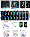
The glymphatic-meningeal pathway, important for brain homeostasis, depends on the drainage function of the cervical lymphatic system. Although new therapies aim to modulate this pathway, a lack of methods for quantifying lymphatic drainage function hinders our ability to understand how targeting the cervical lymph nodes may benefit brain health. To address this, we developed and applied a fluid transport model to dynamic contrast-enhanced MRI (DCE-MRI) data to visualize and quantify tracer-tagged lymph through the deep cervical lymph nodes (dcLNs). The model incorporated physical principles of solute transport to provide a biologically interpretable framework for analyzing microflows in real time. We applied this model to investigate the effects of chronic hypertension on dcLN drainage by comparing normotensive Wistar-Kyoto rats with spontaneously hypertensive stroke-prone (SHRSP) rats. In normal rats, the model revealed complex and tortuous lymph streams of a 200 kDa tracer transported through the sinus system of the dcLNs. In contrast, SHRSP rats exhibited significantly altered fluid dynamics, characterized by simpler stream patterns and reduced flow through the dcLNs. These findings demonstrated that untreated chronic hypertension adversely affects lymph node drainage function. This study provides potentially new insight into impaired lymphatic drainage as a mechanism linking systemic disease to brain health.
Authors
Kaiming Xu, Ankita Bhardwaj, Sunil Koundal, Qin Ren, Chenyu You, Xenophon Papademetris, Helene Benveniste, Tryphon T. Georgiou
×Abstract
Plasmodium falciparum sporozoite (PfSPZ) vaccines, composed of aseptic, purified, live parasites that arrest during or just after liver-stage development, show excellent safety and efficacy in humans. They can induce complete protection against Pf infection, mediated primarily by cellular immune responses against parasite antigens expressed in hepatocytes. Current PfSPZ vaccines rely on the West African PfNF54 parasite, which uniquely produces high numbers of PfSPZ in mosquitoes, facilitating manufacturing efficiency. However, PfNF54 has relatively low hepatocyte infectivity, limiting potency. We created hybrid pan-African Pf strains by genetically crossing PfNF54 with East African Pf strains. The hybrid, AV27, was selected for development based on balanced contribution of parental genomes, high PfSPZ production, and high liver-stage infectivity. As compared with NF54-based PfSPZ vaccines, we expect AV27-based vaccines will have greater and broader efficacy at lower doses due to higher liver-stage infectivity and inclusion of unique East African CD8+ T cell epitopes.
Authors
Lucia Pazzagli, Bethany Jenkins, Ankit Dwivedi, Asha Patil, Yonas Abebe, Tales V. Pascini, Urvashi Rai, Priya Gupta, Nastaran Rezakhani, Chakshu Gandhi, Yiwei Yang, Sudhir Kumar, Mohd Kamil, Gigliola Zanghí, Manuel Llinás, Stephen L. Hoffman, Joana C. Silva, Ashley M. Vaughan, B. Kim Lee Sim
×Abstract
Lineage plasticity, or transdifferentiation, is increasingly recognized as a resistance mechanism to androgen receptor (AR) inhibition in prostate cancer. Lineage plasticity is characterized by loss of AR signaling and epithelial differentiation, along with activation of stemness-associated pathways, epithelial-mesenchymal transition, or alternative differentiation programs such as neuroendocrine prostate cancer (NEPC). Loss of the tumor suppressors TP53 and RB1 is common in tumors exhibiting lineage plasticity; however, the mechanisms by which TP53/RB1 loss promotes this phenotype remain poorly understood, and effective treatments are limited. Using multiomic profiling of TP53/RB1-loss prostate cancer models, we identified alterations in chromatin accessibility, DNA methylation, and gene expression associated with lineage plasticity. Importantly, many pathways activated upon TP53/RB1 loss could be blocked through BET bromodomain inhibition. TP53/RB1-deficient cells also harbored widespread DNA methylation changes that silenced pathways linked with restraining lineage plasticity. Combined BET bromodomain and DNA methyltransferase (DNMT) inhibition was more effective than single-agent treatment in suppressing growth of TP53/RB1-loss models exhibiting a stem-like or NEPC program. This was partly explained by abrogation of discrete lineage plasticity pathways modulated by each agent. Altogether, our work suggests combined BET bromodomain and DNMT inhibition is a promising therapeutic approach for prostate tumors exhibiting lineage plasticity.
Authors
William K. Storck, Diana Flores, Anbarasu Kumaraswamy, Zhi Duan, Shrabastee Chakraborty, Chao Zhang, Eva Rodansky, Dhruv Khokhani, Olivia A. Swaim, Karan Bedi, Raymond G. Cavalcante, Canping Chen, Faming Zhao, Ya-Mei Hu, Zheng Xia, Ryan J. Rebernick, Marcin Cieslik, Rahul Mannan, Somnath Mahapatra, Arul M. Chinnaiyan, Aaron M. Udager, Joshua A. Kuleape, Catherine R. Alumkal, Hannah N. Beck, Peter S. Nelson, Colm Morrissey, Michael C. Haffner, Leigh Ellis, Yuzhuo Wang, Joel A. Yates, Joshi J. Alumkal
×Abstract
Autosomal dominant polycystic kidney disease (ADPKD) is a leading genetic cause of kidney failure, characterized by progressive cyst growth, inflammation, and interstitial fibrosis. Renal fibrosis, driven by myofibroblast activation and excessive extracellular matrix (ECM) deposition, is increasingly recognized as a key contributor to disease progression, yet targeted antifibrotic therapies remain limited. Here, we evaluated the therapeutic potential of pirfenidone to suppress fibrosis and disease progression in ADPKD. Single-nucleus RNA sequencing of human ADPKD kidneys identified fibroblasts as the predominant source of fibrous and adhesive ECM, with higher ECM-associated gene expression compared with that in normal kidney fibroblasts. In vitro, primary human ADPKD renal myofibroblasts displayed a similar profibrotic gene expression profile, and pirfenidone treatment suppressed ECM gene expression, cell proliferation, migration, and contractility. In the Pkd1RC/RC mouse model of ADPKD, pirfenidone reduced renal fibrosis, myofibroblast accumulation, ECM deposition, profibrotic gene expression, and associated signaling pathways and improved kidney function. Pirfenidone also reduced kidney enlargement but reduced cyst burden only in female mice. Collectively, these findings demonstrate that pirfenidone attenuates renal fibrosis and improves kidney function in ADPKD by suppressing myofibroblast activation and ECM production, supporting fibrosis as a therapeutic target and highlighting pirfenidone as a potential adjunct to cyst-directed therapies.
Authors
Viji Remadevi, Abeda Jamadar, Meekha M. Varghese, Haichun Yang, Sumedha Gunewardena, Darren P. Wallace, Reena Rao
×Abstract
Autosomal dominant leukodystrophy (ADLD) is a fatal adult-onset CNS demyelinating disorder for which no treatment exists. The majority of ADLD cases are caused by duplications of the lamin B1 (LMNB1) gene, resulting in increased LMNB1 expression. While reducing LMNB1 levels represents a logical therapeutic strategy, its efficacy has not been previously demonstrated in any in vivo model. Mouse models with oligodendrocyte-specific human LMNB1 (hLMNB1) overexpression recapitulate salient features of ADLD. Using a modified version of this model, where hLMNB1 can be inducibly downregulated, we demonstrated that hLMNB1 reduction can prevent or substantially ameliorate disease progression. Therapeutic effects were maximized when hLMNB1 reduction was induced before expected symptom onset, resulting in improvements in behavioral, biochemical, histopathological, and survival measures relative to those of untreated animals. Reducing hLMNB1 levels after symptom onset led to improved survival but mixed results for other disease phenotypes. In addition, we identified potential biomarkers that track disease progression. Furthermore, we demonstrated that near-complete knockdown of murine LMNB1 expression in adulthood did not result in any overt CNS phenotype. Together, these results provide a proof of concept supporting LMNB1 reduction as a therapeutic strategy and offer a rationale for treatments aimed at lowering levels of this protein in ADLD.
Authors
Nathan Herdman, Kaveh Moradi, Bruce Nmezi, Anushe Munir, Krizchelle A. Magtoto, Fang Liu, Mara Sullivan, Xuemei Zeng, Thomas K. Karikari, Quasar S. Padiath
×Abstract
Metastases in renal cell carcinoma (RCC) typically arise from large primary tumors. However, a subset of patients with small renal masses (SRMs; ≤4 cm) can develop metastatic disease. Identifying these tumors is clinically important, as many SRMs are managed with active surveillance, and their study may provide insight into the early acquisition of metastatic competence. It remains unclear whether these tumors acquire distinct metastatic programs or instead show premature activation of the same aggressive programs typically associated with larger tumors. Here, we performed integrated morphological and molecular profiling of a multiinstitutional cohort of metastatic SRMs, including whole-exome sequencing and RNA-Seq, using nonmetastatic primary tumors as controls. Among metastatic, non–clear cell SRMs, we identified NF2-altered tumors, ELOC-mutated RCC, and an mTOR-driven eosinophilic vacuolated tumor. Metastatic clear cell SRMs were enriched by multi-hit aggressive genotypes, including recurrent losses of chromosomes 8p, 9, and 14q, as well as co-occurring driver alterations (≥2 events), including BAP1 and mTOR pathway genes. Transcriptomic analyses revealed enrichment of the non-negative matrix factorization 3 (NMF3) subtype from the IMmotion151 trial-based taxonomy, along with metabolic rewiring and reduced cytotoxic immune effector function. Collectively, these findings identify molecular programs associated with metastatic competence in SRMs, highlight the importance of genomic studies of equivocal non-clear cell SRMs, and provide a biological framework for risk stratification in patients often considered for active surveillance.
Authors
Payal Kapur, Daria Beshnova, Hua Zhong, Ruby Sharma, Pooja Ghatalia, Angela Yoo, Daniel D. Le, Ratna Mukhopadhyay, Alana Christie, Jeffrey Miyata, Shuanzeng Wei, Rana R. McKay, Dinesh Rakheja, Satwik Rajaram, Robert G. Uzzo, A. Ari Hakimi, Zora Modrusan, James Brugarolas