
Research ArticleCell biologyInfectious diseasePulmonology
Open Access |  10.1172/jci.insight.194121
CFTR in alveolar type 1 cells contributes to lung liquid secretion and host defense
Sayahi Suthakaran,1,2 Sonya Homami,1,2 Deebly Chavez,1 Stephanie Tang,1,2 Sarah K.L. Moore,1 Chaya Sussman,1 Jimmy Zhang,1 Clemente J. Britto,3 Alice Prince,4 Alison J. May,5,6 Jaymin J. Kathiriya,1,5 and Jaime L. Hook1,5,7,8
1Division of Pulmonary, Critical Care, and Sleep Medicine, Department of Medicine, and
2Graduate School of Biomedical Sciences, Icahn School of Medicine at Mount Sinai, New York, New York, USA.
3Division of Pulmonary, Critical Care, and Sleep Medicine, Department of Medicine, Yale University School of Medicine, New Haven, Connecticut, USA.
4Department of Pediatrics, College of Physicians and Surgeons, Columbia University Medical Center, New York, New York, USA.
5Department of Stem Cell Biology and Regenerative Medicine,
6Department of Otolaryngology,
7Department of Microbiology, and
8Global Health and Emerging Pathogens Institute, Icahn School of Medicine at Mount Sinai, New York, New York, USA.
Address correspondence to: Jaime L. Hook, Departments of Medicine and Microbiology, One Gustave L. Levy Place, Box 1232, New York, New York 10029, USA. Phone: 212.241.4473; Email: jaime.hook@mssm.edu.
Authorship note: SS and SH are co-first authors and contributed equally to this work.
Find articles by Suthakaran, S. in: PubMed | Google Scholar
1Division of Pulmonary, Critical Care, and Sleep Medicine, Department of Medicine, and
2Graduate School of Biomedical Sciences, Icahn School of Medicine at Mount Sinai, New York, New York, USA.
3Division of Pulmonary, Critical Care, and Sleep Medicine, Department of Medicine, Yale University School of Medicine, New Haven, Connecticut, USA.
4Department of Pediatrics, College of Physicians and Surgeons, Columbia University Medical Center, New York, New York, USA.
5Department of Stem Cell Biology and Regenerative Medicine,
6Department of Otolaryngology,
7Department of Microbiology, and
8Global Health and Emerging Pathogens Institute, Icahn School of Medicine at Mount Sinai, New York, New York, USA.
Address correspondence to: Jaime L. Hook, Departments of Medicine and Microbiology, One Gustave L. Levy Place, Box 1232, New York, New York 10029, USA. Phone: 212.241.4473; Email: jaime.hook@mssm.edu.
Authorship note: SS and SH are co-first authors and contributed equally to this work.
Find articles by Homami, S. in: PubMed | Google Scholar
1Division of Pulmonary, Critical Care, and Sleep Medicine, Department of Medicine, and
2Graduate School of Biomedical Sciences, Icahn School of Medicine at Mount Sinai, New York, New York, USA.
3Division of Pulmonary, Critical Care, and Sleep Medicine, Department of Medicine, Yale University School of Medicine, New Haven, Connecticut, USA.
4Department of Pediatrics, College of Physicians and Surgeons, Columbia University Medical Center, New York, New York, USA.
5Department of Stem Cell Biology and Regenerative Medicine,
6Department of Otolaryngology,
7Department of Microbiology, and
8Global Health and Emerging Pathogens Institute, Icahn School of Medicine at Mount Sinai, New York, New York, USA.
Address correspondence to: Jaime L. Hook, Departments of Medicine and Microbiology, One Gustave L. Levy Place, Box 1232, New York, New York 10029, USA. Phone: 212.241.4473; Email: jaime.hook@mssm.edu.
Authorship note: SS and SH are co-first authors and contributed equally to this work.
Find articles by Chavez, D. in: PubMed | Google Scholar
1Division of Pulmonary, Critical Care, and Sleep Medicine, Department of Medicine, and
2Graduate School of Biomedical Sciences, Icahn School of Medicine at Mount Sinai, New York, New York, USA.
3Division of Pulmonary, Critical Care, and Sleep Medicine, Department of Medicine, Yale University School of Medicine, New Haven, Connecticut, USA.
4Department of Pediatrics, College of Physicians and Surgeons, Columbia University Medical Center, New York, New York, USA.
5Department of Stem Cell Biology and Regenerative Medicine,
6Department of Otolaryngology,
7Department of Microbiology, and
8Global Health and Emerging Pathogens Institute, Icahn School of Medicine at Mount Sinai, New York, New York, USA.
Address correspondence to: Jaime L. Hook, Departments of Medicine and Microbiology, One Gustave L. Levy Place, Box 1232, New York, New York 10029, USA. Phone: 212.241.4473; Email: jaime.hook@mssm.edu.
Authorship note: SS and SH are co-first authors and contributed equally to this work.
Find articles by Tang, S. in: PubMed | Google Scholar
1Division of Pulmonary, Critical Care, and Sleep Medicine, Department of Medicine, and
2Graduate School of Biomedical Sciences, Icahn School of Medicine at Mount Sinai, New York, New York, USA.
3Division of Pulmonary, Critical Care, and Sleep Medicine, Department of Medicine, Yale University School of Medicine, New Haven, Connecticut, USA.
4Department of Pediatrics, College of Physicians and Surgeons, Columbia University Medical Center, New York, New York, USA.
5Department of Stem Cell Biology and Regenerative Medicine,
6Department of Otolaryngology,
7Department of Microbiology, and
8Global Health and Emerging Pathogens Institute, Icahn School of Medicine at Mount Sinai, New York, New York, USA.
Address correspondence to: Jaime L. Hook, Departments of Medicine and Microbiology, One Gustave L. Levy Place, Box 1232, New York, New York 10029, USA. Phone: 212.241.4473; Email: jaime.hook@mssm.edu.
Authorship note: SS and SH are co-first authors and contributed equally to this work.
Find articles by Moore, S. in: PubMed | Google Scholar
1Division of Pulmonary, Critical Care, and Sleep Medicine, Department of Medicine, and
2Graduate School of Biomedical Sciences, Icahn School of Medicine at Mount Sinai, New York, New York, USA.
3Division of Pulmonary, Critical Care, and Sleep Medicine, Department of Medicine, Yale University School of Medicine, New Haven, Connecticut, USA.
4Department of Pediatrics, College of Physicians and Surgeons, Columbia University Medical Center, New York, New York, USA.
5Department of Stem Cell Biology and Regenerative Medicine,
6Department of Otolaryngology,
7Department of Microbiology, and
8Global Health and Emerging Pathogens Institute, Icahn School of Medicine at Mount Sinai, New York, New York, USA.
Address correspondence to: Jaime L. Hook, Departments of Medicine and Microbiology, One Gustave L. Levy Place, Box 1232, New York, New York 10029, USA. Phone: 212.241.4473; Email: jaime.hook@mssm.edu.
Authorship note: SS and SH are co-first authors and contributed equally to this work.
Find articles by Sussman, C. in: PubMed | Google Scholar
1Division of Pulmonary, Critical Care, and Sleep Medicine, Department of Medicine, and
2Graduate School of Biomedical Sciences, Icahn School of Medicine at Mount Sinai, New York, New York, USA.
3Division of Pulmonary, Critical Care, and Sleep Medicine, Department of Medicine, Yale University School of Medicine, New Haven, Connecticut, USA.
4Department of Pediatrics, College of Physicians and Surgeons, Columbia University Medical Center, New York, New York, USA.
5Department of Stem Cell Biology and Regenerative Medicine,
6Department of Otolaryngology,
7Department of Microbiology, and
8Global Health and Emerging Pathogens Institute, Icahn School of Medicine at Mount Sinai, New York, New York, USA.
Address correspondence to: Jaime L. Hook, Departments of Medicine and Microbiology, One Gustave L. Levy Place, Box 1232, New York, New York 10029, USA. Phone: 212.241.4473; Email: jaime.hook@mssm.edu.
Authorship note: SS and SH are co-first authors and contributed equally to this work.
Find articles by Zhang, J. in: PubMed | Google Scholar
1Division of Pulmonary, Critical Care, and Sleep Medicine, Department of Medicine, and
2Graduate School of Biomedical Sciences, Icahn School of Medicine at Mount Sinai, New York, New York, USA.
3Division of Pulmonary, Critical Care, and Sleep Medicine, Department of Medicine, Yale University School of Medicine, New Haven, Connecticut, USA.
4Department of Pediatrics, College of Physicians and Surgeons, Columbia University Medical Center, New York, New York, USA.
5Department of Stem Cell Biology and Regenerative Medicine,
6Department of Otolaryngology,
7Department of Microbiology, and
8Global Health and Emerging Pathogens Institute, Icahn School of Medicine at Mount Sinai, New York, New York, USA.
Address correspondence to: Jaime L. Hook, Departments of Medicine and Microbiology, One Gustave L. Levy Place, Box 1232, New York, New York 10029, USA. Phone: 212.241.4473; Email: jaime.hook@mssm.edu.
Authorship note: SS and SH are co-first authors and contributed equally to this work.
Find articles by
Britto, C.
in:
PubMed
|
Google Scholar
|

1Division of Pulmonary, Critical Care, and Sleep Medicine, Department of Medicine, and
2Graduate School of Biomedical Sciences, Icahn School of Medicine at Mount Sinai, New York, New York, USA.
3Division of Pulmonary, Critical Care, and Sleep Medicine, Department of Medicine, Yale University School of Medicine, New Haven, Connecticut, USA.
4Department of Pediatrics, College of Physicians and Surgeons, Columbia University Medical Center, New York, New York, USA.
5Department of Stem Cell Biology and Regenerative Medicine,
6Department of Otolaryngology,
7Department of Microbiology, and
8Global Health and Emerging Pathogens Institute, Icahn School of Medicine at Mount Sinai, New York, New York, USA.
Address correspondence to: Jaime L. Hook, Departments of Medicine and Microbiology, One Gustave L. Levy Place, Box 1232, New York, New York 10029, USA. Phone: 212.241.4473; Email: jaime.hook@mssm.edu.
Authorship note: SS and SH are co-first authors and contributed equally to this work.
Find articles by Prince, A. in: PubMed | Google Scholar
1Division of Pulmonary, Critical Care, and Sleep Medicine, Department of Medicine, and
2Graduate School of Biomedical Sciences, Icahn School of Medicine at Mount Sinai, New York, New York, USA.
3Division of Pulmonary, Critical Care, and Sleep Medicine, Department of Medicine, Yale University School of Medicine, New Haven, Connecticut, USA.
4Department of Pediatrics, College of Physicians and Surgeons, Columbia University Medical Center, New York, New York, USA.
5Department of Stem Cell Biology and Regenerative Medicine,
6Department of Otolaryngology,
7Department of Microbiology, and
8Global Health and Emerging Pathogens Institute, Icahn School of Medicine at Mount Sinai, New York, New York, USA.
Address correspondence to: Jaime L. Hook, Departments of Medicine and Microbiology, One Gustave L. Levy Place, Box 1232, New York, New York 10029, USA. Phone: 212.241.4473; Email: jaime.hook@mssm.edu.
Authorship note: SS and SH are co-first authors and contributed equally to this work.
Find articles by May, A. in: PubMed | Google Scholar
1Division of Pulmonary, Critical Care, and Sleep Medicine, Department of Medicine, and
2Graduate School of Biomedical Sciences, Icahn School of Medicine at Mount Sinai, New York, New York, USA.
3Division of Pulmonary, Critical Care, and Sleep Medicine, Department of Medicine, Yale University School of Medicine, New Haven, Connecticut, USA.
4Department of Pediatrics, College of Physicians and Surgeons, Columbia University Medical Center, New York, New York, USA.
5Department of Stem Cell Biology and Regenerative Medicine,
6Department of Otolaryngology,
7Department of Microbiology, and
8Global Health and Emerging Pathogens Institute, Icahn School of Medicine at Mount Sinai, New York, New York, USA.
Address correspondence to: Jaime L. Hook, Departments of Medicine and Microbiology, One Gustave L. Levy Place, Box 1232, New York, New York 10029, USA. Phone: 212.241.4473; Email: jaime.hook@mssm.edu.
Authorship note: SS and SH are co-first authors and contributed equally to this work.
Find articles by
Kathiriya, J.
in:
PubMed
|
Google Scholar
|
1Division of Pulmonary, Critical Care, and Sleep Medicine, Department of Medicine, and
2Graduate School of Biomedical Sciences, Icahn School of Medicine at Mount Sinai, New York, New York, USA.
3Division of Pulmonary, Critical Care, and Sleep Medicine, Department of Medicine, Yale University School of Medicine, New Haven, Connecticut, USA.
4Department of Pediatrics, College of Physicians and Surgeons, Columbia University Medical Center, New York, New York, USA.
5Department of Stem Cell Biology and Regenerative Medicine,
6Department of Otolaryngology,
7Department of Microbiology, and
8Global Health and Emerging Pathogens Institute, Icahn School of Medicine at Mount Sinai, New York, New York, USA.
Address correspondence to: Jaime L. Hook, Departments of Medicine and Microbiology, One Gustave L. Levy Place, Box 1232, New York, New York 10029, USA. Phone: 212.241.4473; Email: jaime.hook@mssm.edu.
Authorship note: SS and SH are co-first authors and contributed equally to this work.
Find articles by
Hook, J.
in:
PubMed
|
Google Scholar
|
Published August 11, 2026 - More info
JCI Insight. 2026;11(18):e194121. https://doi.org/10.1172/jci.insight.194121.
© 2026 Suthakaran et al. This work is licensed under the Creative Commons Attribution 4.0 International License. To view a copy of this license, visit http://creativecommons.org/licenses/by/4.0/.
Received: April 1, 2025; Accepted: August 5, 2026

-
Results
AT1 and AT2 cell–specific CFTRnull expression in transgenic mice. We bred widely used transgenic mouse strains to generate mice in which a Cftr-null allele and GFP expression were tamoxifen induced in adulthood in an AT1 or AT2 cell–specific manner. Thus, AT2-CFTRnull-GFP mice carried Sftpc-CreERT2 (26), Cftrfl10/fl10 (27), and ROSAmT/mG, a reporter allele that confers membrane-targeted TdTomato expression prior to Cre-mediated excision, and GFP expression afterward (28). Likewise, AT1-CFTRnull-GFP mice carried Ager-CreERT2 (29), Cftrfl10/fl10, and ROSAmT/mG; AT2-AT1-CFTRnull-GFP mice carried Sftpc-CreERT2, Ager-CreERT2, Cftrfl10/fl10, and ROSAmT/mG; and LMC (LMC-TdTomato) mice carried Cftrfl10/fl10 and ROSAmT/mG.
We used our established methods (6, 30) to view live alveoli of intact, perfused lungs of AT2-CFTRnull-GFP mice by real-time confocal microscopy (Figure 1A). GFP fluorescence was evident in plasma membranes of rounded cells that were located primarily at septal junctions (Figure 1B), consistent with AT2 cell morphology and location (31, 32). To test AT2 cell function, we used alveolar micropuncture to microinstill alveolar airspaces with LysoTracker dye, which is taken up by AT2 cells and localizes to AT2 cell lamellar bodies. Hyperinflation-induced LysoTracker fluorescence loss signals surfactant secretion (33). Our findings show lung hyperinflation caused time-dependent loss of LysoTracker fluorescence in GFP+ cells (Supplemental Figure 1, A and B; supplemental material available online with this article; https://doi.org/10.1172/jci.insight.194121DS1), affirming that GFP+ cells in AT2-CFTRnull-GFP mice function as AT2 cells. To quantify Cftr deletion efficiency, we isolated AT2 cells (Supplemental Figure 2, A–C) and performed qPCR of Cftr RNA using our established methods (34, 35). AT2 cells of an AT2-CFTRnull-GFP mouse had 100-fold reduction of Cftr RNA compared with AT2 cells of an LMC-TdTomato mouse (Supplemental Tables 1 and 2). Taking these findings together, we conclude that AT2 cells of AT2-CFTRnull-GFP mice are Cftr deficient and marked by GFP fluorescence.
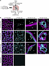 Figure 1
Alveolar epithelial cell type-specificity of GFP expression. (A) The illustration shows the experimental design of confocal imaging studies of live, intact, perfused lungs of transgenic mice. (B) Confocal images show plasma membrane GFP (cyan) and TdTomato (magenta) fluorescence in live lungs of mice of the indicated genotype. Dashed squares show locations of the high power views shown in the third and fourth columns. Closed arrowheads point out AT1 cell fluorescence, which is GFP+ (cyan) and distinct from neighboring TdTomato+ (magenta) endothelial cells only in AT1-CFTRnull-GFP and AT2-AT1-CFTRnull-GFP mice. Open arrowheads show plasma membrane fluorescence of AT2 cells, which is GFP+ (cyan) only in AT2-CFTRnull-GFP and AT2-AT1-CFTRnull-GFP mice. In the fourth column, cytosolic fluorescence of lysotracker green (LTG) delineates lamellar bodies (LB) and confirms AT2 cell identity. In the third row, arrows show example AT2 cells in an AT2-AT1-CFTRnull-GFP mouse. Air, airspace; ves, microvessel lumen. Scale bars: 100 (first and second columns), 3 (third columns), and 5 (fourth columns) μm. Images were replicated in lungs of 3 mice per genotype. Note, in the fourth column, both LTG and GFP are pseudocolored cyan.
By contrast, GFP fluorescence in live lungs of AT1-CFTRnull-GFP mice was evident in thin cells that line alveolar walls (Figure 1B), consistent with AT1 cell morphology and location. AT2 cells of AT1-CFTRnull-GFP mice had abundant Cftr RNA (Supplemental Figure 2D and Supplemental Table 1). Taking these data together, we interpret that CFTRnull expression in AT1-CFTRnull-GFP mice is AT1 cell specific.
In AT2-AT1-CFTRnull-GFP mice, GFP fluorescence was evident in plasma membranes of both AT2 and AT1 cells (Figure 1B), and AT2 cell qPCR showed more than 100-fold reduction of Cftr RNA (Supplemental Figure 2E and Supplemental Table 1). There was no GFP fluorescence in alveoli of LMC-TdTomato mice (Figure 1B). We interpret that AT2-AT1-CFTRnull-GFP mice are Cftr deficient in both AT2 and AT1 cells, while LMC-TdTomato mice are Cftr deficient in neither cell type.
CFTRnull expression is heterogeneous across alveoli of Ager-CreERT2–expressing mice. Tamoxifen-treated Ager-CreERT2–expressing mice show Cre recombination efficiency that varies across lung alveoli (29), leading to a heterogeneous distribution of Cre expression in which AT1 cells of some alveoli express Cre, while AT1 cells of other alveoli do not. In line with the published data, our images show that AT1 cell GFP fluorescence was heterogeneous across imaging fields of alveoli in lungs of AT1-CFTRnull-GFP mice (Figure 2, A and B), while TdTomato fluorescence was evident in all alveoli (Figure 2A). AT2-AT1-CFTRnull-GFP showed similar heterogeneity of GFP expression (data not shown). We categorized the proportion of GFP-expressing AT1 cells in imaging fields of AT1-CFTRnull-GFP mice as Low, Medium, or High if GFP expression was evident in less than 40%, 40%–80%, or more than 80%, respectively, of alveoli in the imaging field (Figure 2, A and B). We interpreted the GFP fluorescence categories to indicate sparse, moderate, and extensive CFTRnull expression, respectively, across imaging fields of alveoli.
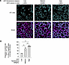 Figure 2
Cre recombination is spatially heterogeneous across alveoli of AT1-CFTRnull-GFP mice. (A and B) Confocal images (A) and group data (B) show GFP fluorescence (cyan) across imaging fields of at least 30 alveoli in live, perfused lungs of AT1-CFTRnull-GFP mice. In the images in A, cyan fluorescence marks Cre-expressing AT1 cells. Note, Cre expression is heterogeneous across alveoli, since some AT1 cells expressed TdTomato, but not GFP, as evident by TdTomato (magenta) fluorescence in GFP-free alveoli (examples: arrowheads). The group data in B quantify the heterogeneity by indicating the proportion of alveoli with GFP+ cells as a percentage of the total number of alveoli per imaging field. Circles in B indicate n and each represent 1 imaging field of at least 30 alveoli. Imaging fields were generated in lungs of 3 mice but presented according to their GFP fluorescence category. Data are shown as mean ± SEM; *P < 0.05 by ANOVA with post hoc Tukey testing. Scale bars: 100 μm.
CFTR in AT1 cells and AT2 cells contributes to alveolar liquid secretion. We next aimed to define the effect of AT1 and AT2 cell CFTRnull expression, and its heterogeneity, on alveolar function. To do this, we applied a technique that has been used previously to identify liquid secretion and absorption in alveoli of live, perfused lungs using upright confocal microscopy (5, 6). Briefly, the experiments are carried out by microinstilling alveolar airspaces with fluorophore-conjugated dextran in aqueous solution. The dextran-containing solution is visible in alveoli by confocal microscopy as a juxta-epithelial layer that pools at alveolar niches, where septa converge (5, 6). Since the fluorescence intensity of the dextran-containing solutions varies with dextran concentration (6), the solutions lose fluorescence in airspaces as they become diluted by nonfluorescent alveolar wall liquid (AWL) secreted by the alveolar epithelium (5, 6). Conversely, the solutions gain fluorescence when the epithelium absorbs liquid from airspaces (5, 6). Since secreted AWL flows toward airways and does not accumulate in alveoli (5), the widths of the dextran-containing solution pools are unchanged over time when AWL is secreted (5, 6). However, they diminish over time when airspace liquid is absorbed (5, 6).
Using this approach, we and others have reported alveoli of intact, perfused mouse lungs secrete AWL in a CFTR-dependent manner under baseline conditions (5, 6). A recent study from our group affirms CFTR dependence and adds that the direction of alveolar liquid transport depends on lung perfusion, since alveoli of lungs perfused at or near physiological levels secrete AWL, while alveoli of lungs in which perfusion is majorly reduced or halted absorb airspace liquid (36). Microvessel-to-airspace osmotic gradients do not seem to determine the direction of alveolar liquid transport (36). Because fluid leak from microvessels into airspaces in perfused lungs could dilute the dextran-containing solutions, we ruled out alveolar leak by showing that airspace dextran fluorescence decreased, then increased in the same alveoli, under conditions of continuous perfusion, in response to changes of pulmonary artery pressure (36). Since airspace dextran fluorescence could not increase in alveoli with barrier leak, these data align with reports indicating alveolar barrier function is intact in isolated, perfused lungs (37, 38). Finally, we have used airspace instillation of fluorophore-tagged antibodies to show alveolar macrophages are present in airspaces of isolated, perfused lungs (36), indicating that expected nonepithelial cell types are present in our preparation. Neutrophils are absent in airspaces under baseline conditions but recruited after intranasal instillation of S. aureus (6).
We now applied these methods to test the hypothesis that AT1 and AT2 cell CFTRnull expression inhibits AWL secretion. We microinstilled alveolar airspaces of LMC-TdTomato, AT2-AT1-CFTRnull-GFP, AT2-CFTRnull-GFP, and AT1-CFTRnull-GFP mice with Alexa Fluor 647–conjugated dextran (AF-dextran; 10 kDa; 10 mg/mL) (Figure 3A). We used AF-dextran because its fluorescence intensity varies with concentration in aqueous solution (Supplemental Figure 3, A and B), and its excitation and emission spectra are distinct from those of GFP and TdTomato. The contents of the airspace instillate and lung perfusate solutions for our experiments are shown in Supplemental Table 3. In alveoli of AT1-CFTRnull-GFP and AT2-AT1-CFTRnull-GFP mice, we restricted our analysis to imaging fields that had High-GFP AT1 cell fluorescence, in order to avoid confounding the experiments with alveoli that lacked CFTRnull expression.
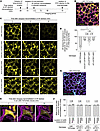 Figure 3
Real-time confocal imaging of liquid secretion in live alveoli. (A–G) Illustration in A shows the design of confocal imaging studies of liquid transport in live alveoli of intact, perfused lungs of transgenic mice of the genotypes indicated in B–G. As indicated in A, we microinstilled alveolar airspaces with Alexa Fluor 647–conjugated dextran (AF-dextran, yellow, 10 kDa, 10 mg/mL) to carry out the determinations of liquid transport shown in B–F. At the conclusion of each experiment, we added fluorescein isothiocyanate-conjugated dextran (FITC-dextran, cyan, 20 kDa, 5 mg/mL) to the lung perfusate solution to define alveolar barrier properties. The confocal image in B shows AF-dextran fluorescence in airspaces of alveoli (magenta) of a TdTomato-expressing LMC-TdTomato mouse at 2 minutes after AF-dextran microinstillation. Air, example airspace. Arrowheads point out example AF-dextran pools at alveolar niches. Images and group data in C and D show change of AF-dextran fluorescence over time. Fluorescence of alveolar walls is not shown. In D, circles indicate n and each represent one mouse in which mean fluorescence change was quantified in imaging fields of at least 30 alveoli; data are shown as mean ± SEM; *P < 0.05 by ANOVA with post hoc Tukey testing. High power views in E show widths of the example AF-dextran pool (white dashed lines and text) and airspace (magenta dashed lines and text) indicated by the dashed square in B. Group data in F show pool and airspace widths over time. In F, circles and bars are as in D; P is as indicated by 2-tailed t test. The confocal image in G shows fluorescence of FITC-dextran added to the lung perfusate solution. Images in B and G represent images generated in 3 mice each of LMC-TdTomato, AT2-AT1-CFTRnull-GFP, AT2-CFTRnull-GFP, and AT1-CFTRnull-GFP mouse groups. Scale bars: 100 (B and G), 50 (C), 20 (E) μm.
In alveoli of LMC-TdTomato mice, microinstilled AF-dextran lined alveolar walls and pooled at alveolar niches within minutes of microinstillation (Figure 3B), as expected. AF-dextran fluorescence subsequently decreased in a time-dependent manner (Figure 3, C and D), suggesting the alveolar epithelium secreted AWL. The widths of AF-dextran–containing liquid pools in alveoli were stable over time (Figure 3, E and F), also as expected, indicating that the volume of AF-dextran–containing liquid in airspaces did not increase during the imaging period. Stable airspace diameter during the same imaging period (Figure 3, E and F) indicates our measurements of AF-dextran pool width were not confounded by changes of alveolar morphology. To rule out the possibility that AF-dextran fluorescence loss was caused by leak of microvascular fluid into airspaces, we added fluorescein isothiocyanate–tagged (FITC-tagged) dextran (FITC-dextran, 20 kDa, 5 mg/mL) to the lung perfusate solution (Figure 3A), as we have done previously (6, 30). FITC-dextran fluorescence was visible in microvascular lumens but did not leak into alveolar airspaces (Figure 3G), indicating alveolar barrier function was intact during the imaging period. Together, these findings show alveoli secreted AWL in lungs of LMC-TdTomato mice.
By contrast, AF-dextran fluorescence loss in airspaces was blocked in alveoli of AT2-AT1-CFTRnull-GFP mice (Figure 3, C and D). Widths of AF-dextran–containing liquid pools and airspaces in alveoli did not change over time (Figure 3, E and F), and FITC-dextran in the lung perfusate solution did not leak into airspaces. These findings show CFTRnull expression in both AT1 and AT2 cells inhibits AWL secretion, and they confirm reports indicating that AWL secretion depends on CFTR in the alveolar epithelium (5, 6). Notably, AWL secretion was not completely lost in alveoli of AT2-AT1-CFTRnull-GFP mice (Figure 3, C and D). The residual secretion might reflect contributions to AWL secretion by non-CFTR Cl– channels in the alveolar epithelium.
AF-dextran fluorescence loss was also blocked in alveoli of AT2-CFTRnull-GFP mice and AT1-CFTRnull-GFP mice (Figure 3, C and D). Widths of AF-dextran–containing liquid pools and airspaces in alveoli of both AT2-CFTRnull-GFP and AT1-CFTRnull-GFP mice were steady over time (Figure 3, E and F), and FITC-dextran that was added to the lung perfusate did not leak into airspaces. Together, these findings show CFTRnull expression in each of AT2 and AT1 cells inhibited liquid secretion in alveoli. We conclude that CFTR in both AT2 and AT1 cells contributes to AWL secretion.
Heterogeneous recombination efficiency across alveoli of tamoxifen-treated Ager-CreERT2–expressing mice (29) provides an opportunity to relate the magnitude of AT1 cell CFTRnull expression in imaging fields of alveoli to AWL secretion rates in lungs of AT1-CFTRnull-GFP mice. To determine whether AWL secretion varies with the extent of AT1 cell CFTRnull expression, we reanalyzed the AF-dextran images that we generated in AT1-CFTRnull-GFP mice by GFP expression category, as defined in Figure 2, rather than by mouse, as in Figure 3. Figure 4, A–C, shows the relationship between AF-dextran fluorescence change and AT1 cell GFP expression. Our findings show that, in Low-GFP imaging fields, airspace AF-dextran fluorescence decreased rapidly (Figure 4, A–C), indicating that sparse AT1 cell CFTRnull expression across alveoli had no effect on AWL secretion. However, AF-dextran fluorescence loss was slower in Medium-GFP imaging fields, and it was slower still in High-GFP imaging fields (Figure 4, A–C). These findings show the rate of AWL secretion depends on AT1 cell CFTRnull expression, such that imaged regions of alveoli that had the most CFTRnull expression had the least AWL secretion, and vice versa. We conclude that the extent of CFTR expression in AT1 cells determines AWL secretion. This direct, “dose-dependent” relationship between AT1 cell CFTR expression and AWL secretion strengthens our conclusion that AT1 cell CFTR is a driver of liquid secretion in alveoli.
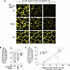 Figure 4
Dose-dependent relationship between AT1 cell Cftr deficiency and inhibition of AWL secretion in lungs of AT1-CFTRnull-GFP mice. (A–C) Confocal images (A) and group data (B and C) show AF-dextran fluorescence (yellow; 10 kDa, 10 mg/mL) after microinstillation in alveolar airspaces of lungs of AT1-CFTRnull-GFP mice. Experiments were carried out as indicated in Figure 3A. GFP fluorescence is not shown but was categorized in imaging fields of at least 30 alveoli as Low, Medium, and High as indicated in Figure 2. Images shown in the bottom row in A are reproduced from Figure 3C. In B and C, circles indicate n and each represent 1 trial of AF-dextran microinstillation in which mean fluorescence change was quantified in an imaging field of at least 30 alveoli. Trials were carried out in lungs of 3 mice but are presented according to the GFP fluorescence category (B) or the proportion of alveoli with GFP+ cells as a percentage of the total number of alveoli (C) in the imaging field. In C, the dashed line sets apart the 12 imaging fields in the High GFP fluorescence category, and the solid line was calculated by linear regression. Data are shown as mean ± SEM; *P < 0.05 by ANOVA with post hoc Tukey testing. Scale bars: 50 μm.
We followed up by testing whether microvessel-to-airspace osmotic gradients promote AWL secretion and could have affected our interpretations. Our data show the AF-dextran–containing airspace solution that we used to generate Figure 3 and Figure 4 was hyperosmolar to the lung perfusate solution (Supplemental Figure 4A and Supplemental Table 3), raising the possibility that the microvessel-to-airspace osmotic gradient present in our preparation, which is favorable to AWL secretion, drove the AF-dextran fluorescence loss we observed in our experiments (Figures 3 and 4). To test this possibility, we quantified AF-dextran fluorescence changes in alveoli of WT mice that we microinstilled with modified AF-dextran solution that abolished the microvessel-to-airspace osmotic gradient by reducing the osmolality of the microinstillate solution (Supplemental Figure 4, A–D, and Supplemental Table 3). Our data show the airspace fluorescence of the modified AF-dextran solution decreased, while the AF-dextran pool width and alveolar airspace width were steady (Supplemental Figure 4, C and D). These findings show AWL secretion occurred in the absence of a prosecretion osmotic gradient between microvessels and airspaces and, thus, affirm findings included in a separate report from our group (36). We conclude that the direction of liquid transport in alveoli occurs independently of microvessel-to-airspace osmotic gradients. These data strengthen our conclusion that AT1 and AT2 cells contribute to AWL secretion at homeostasis.
AT1 cell CFTR promotes particle clearance in alveoli. AWL secretion defends against particle stabilization in alveoli by generating pleura-to-airway liquid flow along alveolar walls that convectively transports particles toward the airways (5). We considered that AWL secretion specifically by AT1 cells might contribute to particle clearance mechanisms. To test this possibility, we used an established method (5, 30) in which we microinstilled fluorescent polystyrene beads (1 μm diameter) into airspaces of Medium-GFP regions of AT1-CFTRnull-GFP mice (Figure 5A). We analyzed the initial organization and movement of 80 bead groups by generating confocal images of the beads from 30 minutes to 3 hours after microinstillation, followed by measuring their displacement according to: (A) their initial location, defined as bead position against a flat alveolar septum or at an alveolar niche; and (B) their initial group size, defined as a small cluster (1-3 beads) or a microaggregate (4 or more beads).
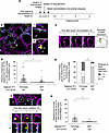 Figure 5
CFTR in AT1 cells promotes particle clearance in alveoli. (A–G) Illustration in A shows the design of the confocal imaging studies shown in B–G. As indicated in A, we microinstilled fluorescent beads (yellow) in airspaces of 3 imaging fields of at least 30 alveoli in live, perfused lungs of an AT1-CFTRnull-GFP mouse, then generated images over 2.5 hours to define bead location, group formation, and movement. GFP fluorescence of the imaging fields was in the Medium category. Images in B show the initial locations and size distributions of example bead groups that formed within 0.5 hours of beads microinstillation. Dashed squares show locations of the high-power views shown in i and ii. The arrowhead shows a small cluster of beads located adjacent to a GFP-expressing (cyan) flat alveolar septum, while the arrow points out a microaggregate located at a TdTomato-expressing (magenta) alveolar niche. mv, microvessel; air, airspace. Images and group data in C–G show spontaneous movement of all bead groups (C–E) or small clusters (F and G) in the 3 imaging fields over the first 3 hours after beads microinstillation. Bead groups were categorized according to whether the adjacent AT1 cell expressed WT CFTR (signaled by TdTomato fluorescence) or CFTRnull (signaled by GFP fluorescence), as indicated. Note in the images in C and F, beads located adjacent to WT CFTR-expressing AT1 cells moved (C and i in F), but beads adjacent to CFTRnull-expressing AT1 cells did not (ii in F). In D and G, circles indicate n and each represent 1 bead group. Data are shown as mean ± SEM; *P < 0.05 by 2-tailed t test. In E, stacked bars represent bead groups as proportions of the total number; *P < 0.05 by χ2 test. Scale bars: 30 (B), 3 (i and ii in B and C), and 15 (F) μm.
Our findings show the beads formed small clusters and microaggregates at flat septa and niches within minutes of microinstillation (Figure 5B and Supplemental Figure 5, A and B). Across imaged regions of alveoli, there was no difference in the number of bead groups that initially localized to alveolar walls lined by TdTomato- or GFP-expressing AT1 cells (Supplemental Figure 5A). The distribution of bead subgroups — that is, small clusters at flat septa, microaggregates at flat septa, small clusters at niches, and microaggregates at niches — was also equivalent (Supplemental Figure 5B). Although mean bead group size at TdTomato-expressing AT1 cells was greater than the mean size of bead groups at GFP-expressing AT1 cells (Supplemental Figure 5, C and D), the difference seemed to be driven by a handful of large microaggregates (Supplemental Figure 5E). We interpret that AT1 cell CFTRnull expression did not have a major effect on how microinstilled beads organized against alveolar walls.
We next analyzed the movement of beads that were initially located at TdTomato-expressing AT1 cells which, we interpret, expressed WT CFTR. Taking all bead groups together, we found the mean distance of bead displacement was 4 μm along alveolar walls during the imaging period (Figure 5, C–E). However, the distance of bead displacement was not uniform across bead subgroups. The subgroup that moved the least was microaggregates at niches (Supplemental Figure 5F). Bead subgroups that were made up of small clusters or that were located at flat septa moved short distances (Figure 5, F and G, and Supplemental Figure 5G), and the movement was either toward the airways or lateral. No bead groups moved toward the pleura. Together, these data largely support findings published by others that show the liquid flow generated by AWL secretion along alveolar walls transports beads in alveoli away from the pleura and toward the airways (5). In addition, our findings affirm our published data that show microaggregates at alveolar niches are stable in alveoli and resist alveolar clearance mechanisms (30).
We then considered the movement of beads that were located at GFP-expressing AT1 cells which, we interpret, expressed CFTRnull. As different from beads located at TdTomato-expressing AT1 cells, beads at GFP-expressing AT1 cells were largely immobile (Figure 5, D and E), indicating that AT1 cell CFTRnull expression blocked bead movement. Bead immobilization at CFTRnull-expressing AT1 cells was not caused by microenvironmental factors unrelated to CFTRnull expression, since in single alveoli lined on opposite walls by TdTomato- and GFP-expressing AT1 cells, only those beads located at TdTomato-expressing AT1 cells were displaced (Figure 5F). Like niche microaggregates at TdTomato-expressing AT1 cells, niche microaggregates at GFP-expressing AT1 cells moved little, but their relative immobilization was not different from other bead subgroups located at GFP-expressing AT1 cells (Supplemental Figure 5F). Notably, bead subgroups made up of small clusters or located at flat septa lined by GFP-expressing AT1 cells were almost totally immobile, and the distances of their displacements were markedly less than those of their counterparts at TdTomato-expressing AT1 cells (Figure 5, F and G, and Supplemental Figure 5G). These findings show AT1 cell CFTRnull expression blocked the displacement of small bead clusters in alveoli, particularly at flat septal locations. We suggest that AT1 cell CFTR contributes to alveolar defense against inhaled particles.
AT1 cell CFTR promotes S. aureus clearance in alveoli. Since AT1 cell CFTR contributed to alveolar defense against particles, we considered it might also contribute to alveolar defense against inhaled bacteria. To test this possibility, we intranasally instilled mice with GFP-expressing S. aureus (SAGFP) and then imaged the SAGFP in alveoli of live, perfused lungs from 1 to 4 hours after instillation (Figure 6A). We chose this timeframe because our published data show CD11b-expressing neutrophils and monocytes are absent from alveolar airspaces of SA-instilled lungs in this time period (6), reducing the possibility that innate immune responses would confound SAGFP clearance measures. To avoid fluorescence overlap, we instilled SAGFP in nonfluorescent transgenic mice (AT1-CFTRnull) that carried Ager-CreERT2 and Cftrfl10/fl10 but not ROSAmT/mG. Littermate control (LMC) mice carried Cftrfl10/fl10.
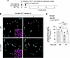 Figure 6
CFTR in AT1 cells promotes bacterial clearance in alveoli. (A–C) The illustration in A shows the experimental design of the confocal imaging studies of bacterial clearance shown in B and C. As indicated in A, mice were given intranasal instillations of SAGFP, then the lungs were excised for live imaging 10 minutes later. Confocal images (B) and group data (C) show microaggregates (arrows; MA) and small clusters (arrowheads; SC) of GFP-expressing S. aureus (SAGFP) in live alveoli of intact, perfused lungs of LMC or AT1-CFTRnull mice as indicated. Calcein red-orange (CR) dye was added to the perfusate solution to delineate alveolar walls (insets). Images included at least 30 alveoli and were generated in the same location at 1 and 4 hours after instillation. In the 4-hour images, magenta arrows and arrowheads point out the initial locations of microaggregates and small clusters that were displaced during the imaging period or were not found in the 4-hour images. White arrows and arrowheads in the 4 -hour images indicate microaggregates and small clusters that did not change. In C, circles indicate n and each represent the change of all small clusters or microaggregates present in one imaging field from 1 to 4 hours after i.n. SAGFP instillation. Imaging fields were generated in the lungs of 3 LMC and 3 AT1-CFTRnull mice. Since imaging fields had to be excluded from the analysis if no small clusters or microaggregates were present in the 1-hour image, the data are presented on a per-imaging-field basis rather than a per-mouse basis. Data are shown as mean ± SEM; *P < 0.05 by ANOVA with post hoc Tukey testing. Scale bar: 50 μm.
Confocal images of live alveoli of AT1-CFTRnull and LMC mice at 1 hour after SAGFP instillation show inhaled SAGFP organized against alveolar walls as small clusters of roughly 1–3 bacteria and microaggregates of 4 or more bacteria (Figure 6B). Microaggregates of SAGFP were resistant to alveolar clearance in both AT1-CFTRnull and LMC mice (Figure 6, B and C) and, thus, unaffected by AT1 cell CFTRnull expression. However, AT1 cell CFTRnull expression blocked the clearance of small clusters of SAGFP (Figure 6, B and C). These findings show AT1 cell CFTR contributes to the alveolar clearance of small bacterial clusters. We interpret that loss of AT1 cell CFTR caused retention of inhaled bacteria against alveolar walls, prolonging contact between small bacterial clusters and the alveolar epithelium.


