
Research ArticleCardiologyDevelopment
Open Access |  10.1172/jci.insight.204032
Right ventricular pressure overload disturbs T-tubule maturation via MEF2D’s transcriptional regulation of BIN1
Yuqing Hu,1 Yiting Xue,2 Xudong Chen,3 Linghui Kong,4 Debao Li,5 Zheng Wang,2 Sixie Zheng,2 Siqi She,2 Hao Li,2 Sijuan Sun,6 Hao Chen,2 Lijun Chen,1 Peisen Ruan,7 Kai Wang,1 and Lincai Ye2,8,9
1Department of Cardiology,
2Department of Thoracic and Cardiovascular Surgery, Shanghai Children’s Medical Center, Shanghai Jiao Tong University School of Medicine, Shanghai, China.
3Department of Thoracic and Cardiovascular Surgery, Shanghai Children’s Hospital, Shanghai Jiao Tong University School of Medicine, Shanghai, China.
4Department of Ultrasound, Women and Children’s Health Care Hospital of Linyi, Linyi, Shandong, China.
5Department of Pediatric Surgery, Children’s Hospital of Fudan University, National Children’s Medical Center, Shanghai, China.
6Department of Pediatric Intensive Care Unit, Shanghai Children’s Medical Center, School of Medicine, Shanghai Jiao Tong University, Shanghai, China.
7Department of Pediatric Intensive Care Unit, The Affiliated Women and Children’s Hospital of Ningbo University, Ningbo, Zhejiang, China.
8Institute of Pediatric Translational Medicine and
9Shanghai Pediatric Congenital Heart Disease Institute, Shanghai Children’s Medical Center, Shanghai Jiao Tong University School of Medicine, Shanghai, China.
Address correspondence to: Peisen Ruan, Department of Pediatric Intensive Care Unit, The Affiliated Women and Children’s Hospital of Ningbo University, Ningbo, Zhejiang, China. Phone: 86.21.58815377; Email: pts1223@sina.com. Or to: Kai Wang, Department of Cardiology, Shanghai Children’s Medical Center, Shanghai Jiao Tong University School of Medicine, 1678 Dongfang Road, Shanghai 200127, China. Phone: 86.21.38626649; Email: wangkai502@126.com. Or to: Lincai Ye, Shanghai Institute of Pediatric Congenital Heart Disease, Shanghai Children’s Medical Center, Shanghai Jiao Tong University School of Medicine, 1678 Dongfang Road, Shanghai 200127, China. Phone: 86.21.38626649; Email: ylc717@163.com.
Authorship note: YH, YX, XC, and LK contributed equally to this work and have been designated as co–first authors.
Find articles by
Hu, Y.
in:
PubMed
|
Google Scholar
|

1Department of Cardiology,
2Department of Thoracic and Cardiovascular Surgery, Shanghai Children’s Medical Center, Shanghai Jiao Tong University School of Medicine, Shanghai, China.
3Department of Thoracic and Cardiovascular Surgery, Shanghai Children’s Hospital, Shanghai Jiao Tong University School of Medicine, Shanghai, China.
4Department of Ultrasound, Women and Children’s Health Care Hospital of Linyi, Linyi, Shandong, China.
5Department of Pediatric Surgery, Children’s Hospital of Fudan University, National Children’s Medical Center, Shanghai, China.
6Department of Pediatric Intensive Care Unit, Shanghai Children’s Medical Center, School of Medicine, Shanghai Jiao Tong University, Shanghai, China.
7Department of Pediatric Intensive Care Unit, The Affiliated Women and Children’s Hospital of Ningbo University, Ningbo, Zhejiang, China.
8Institute of Pediatric Translational Medicine and
9Shanghai Pediatric Congenital Heart Disease Institute, Shanghai Children’s Medical Center, Shanghai Jiao Tong University School of Medicine, Shanghai, China.
Address correspondence to: Peisen Ruan, Department of Pediatric Intensive Care Unit, The Affiliated Women and Children’s Hospital of Ningbo University, Ningbo, Zhejiang, China. Phone: 86.21.58815377; Email: pts1223@sina.com. Or to: Kai Wang, Department of Cardiology, Shanghai Children’s Medical Center, Shanghai Jiao Tong University School of Medicine, 1678 Dongfang Road, Shanghai 200127, China. Phone: 86.21.38626649; Email: wangkai502@126.com. Or to: Lincai Ye, Shanghai Institute of Pediatric Congenital Heart Disease, Shanghai Children’s Medical Center, Shanghai Jiao Tong University School of Medicine, 1678 Dongfang Road, Shanghai 200127, China. Phone: 86.21.38626649; Email: ylc717@163.com.
Authorship note: YH, YX, XC, and LK contributed equally to this work and have been designated as co–first authors.
Find articles by Xue, Y. in: PubMed | Google Scholar
1Department of Cardiology,
2Department of Thoracic and Cardiovascular Surgery, Shanghai Children’s Medical Center, Shanghai Jiao Tong University School of Medicine, Shanghai, China.
3Department of Thoracic and Cardiovascular Surgery, Shanghai Children’s Hospital, Shanghai Jiao Tong University School of Medicine, Shanghai, China.
4Department of Ultrasound, Women and Children’s Health Care Hospital of Linyi, Linyi, Shandong, China.
5Department of Pediatric Surgery, Children’s Hospital of Fudan University, National Children’s Medical Center, Shanghai, China.
6Department of Pediatric Intensive Care Unit, Shanghai Children’s Medical Center, School of Medicine, Shanghai Jiao Tong University, Shanghai, China.
7Department of Pediatric Intensive Care Unit, The Affiliated Women and Children’s Hospital of Ningbo University, Ningbo, Zhejiang, China.
8Institute of Pediatric Translational Medicine and
9Shanghai Pediatric Congenital Heart Disease Institute, Shanghai Children’s Medical Center, Shanghai Jiao Tong University School of Medicine, Shanghai, China.
Address correspondence to: Peisen Ruan, Department of Pediatric Intensive Care Unit, The Affiliated Women and Children’s Hospital of Ningbo University, Ningbo, Zhejiang, China. Phone: 86.21.58815377; Email: pts1223@sina.com. Or to: Kai Wang, Department of Cardiology, Shanghai Children’s Medical Center, Shanghai Jiao Tong University School of Medicine, 1678 Dongfang Road, Shanghai 200127, China. Phone: 86.21.38626649; Email: wangkai502@126.com. Or to: Lincai Ye, Shanghai Institute of Pediatric Congenital Heart Disease, Shanghai Children’s Medical Center, Shanghai Jiao Tong University School of Medicine, 1678 Dongfang Road, Shanghai 200127, China. Phone: 86.21.38626649; Email: ylc717@163.com.
Authorship note: YH, YX, XC, and LK contributed equally to this work and have been designated as co–first authors.
Find articles by Chen, X. in: PubMed | Google Scholar
1Department of Cardiology,
2Department of Thoracic and Cardiovascular Surgery, Shanghai Children’s Medical Center, Shanghai Jiao Tong University School of Medicine, Shanghai, China.
3Department of Thoracic and Cardiovascular Surgery, Shanghai Children’s Hospital, Shanghai Jiao Tong University School of Medicine, Shanghai, China.
4Department of Ultrasound, Women and Children’s Health Care Hospital of Linyi, Linyi, Shandong, China.
5Department of Pediatric Surgery, Children’s Hospital of Fudan University, National Children’s Medical Center, Shanghai, China.
6Department of Pediatric Intensive Care Unit, Shanghai Children’s Medical Center, School of Medicine, Shanghai Jiao Tong University, Shanghai, China.
7Department of Pediatric Intensive Care Unit, The Affiliated Women and Children’s Hospital of Ningbo University, Ningbo, Zhejiang, China.
8Institute of Pediatric Translational Medicine and
9Shanghai Pediatric Congenital Heart Disease Institute, Shanghai Children’s Medical Center, Shanghai Jiao Tong University School of Medicine, Shanghai, China.
Address correspondence to: Peisen Ruan, Department of Pediatric Intensive Care Unit, The Affiliated Women and Children’s Hospital of Ningbo University, Ningbo, Zhejiang, China. Phone: 86.21.58815377; Email: pts1223@sina.com. Or to: Kai Wang, Department of Cardiology, Shanghai Children’s Medical Center, Shanghai Jiao Tong University School of Medicine, 1678 Dongfang Road, Shanghai 200127, China. Phone: 86.21.38626649; Email: wangkai502@126.com. Or to: Lincai Ye, Shanghai Institute of Pediatric Congenital Heart Disease, Shanghai Children’s Medical Center, Shanghai Jiao Tong University School of Medicine, 1678 Dongfang Road, Shanghai 200127, China. Phone: 86.21.38626649; Email: ylc717@163.com.
Authorship note: YH, YX, XC, and LK contributed equally to this work and have been designated as co–first authors.
Find articles by Kong, L. in: PubMed | Google Scholar
1Department of Cardiology,
2Department of Thoracic and Cardiovascular Surgery, Shanghai Children’s Medical Center, Shanghai Jiao Tong University School of Medicine, Shanghai, China.
3Department of Thoracic and Cardiovascular Surgery, Shanghai Children’s Hospital, Shanghai Jiao Tong University School of Medicine, Shanghai, China.
4Department of Ultrasound, Women and Children’s Health Care Hospital of Linyi, Linyi, Shandong, China.
5Department of Pediatric Surgery, Children’s Hospital of Fudan University, National Children’s Medical Center, Shanghai, China.
6Department of Pediatric Intensive Care Unit, Shanghai Children’s Medical Center, School of Medicine, Shanghai Jiao Tong University, Shanghai, China.
7Department of Pediatric Intensive Care Unit, The Affiliated Women and Children’s Hospital of Ningbo University, Ningbo, Zhejiang, China.
8Institute of Pediatric Translational Medicine and
9Shanghai Pediatric Congenital Heart Disease Institute, Shanghai Children’s Medical Center, Shanghai Jiao Tong University School of Medicine, Shanghai, China.
Address correspondence to: Peisen Ruan, Department of Pediatric Intensive Care Unit, The Affiliated Women and Children’s Hospital of Ningbo University, Ningbo, Zhejiang, China. Phone: 86.21.58815377; Email: pts1223@sina.com. Or to: Kai Wang, Department of Cardiology, Shanghai Children’s Medical Center, Shanghai Jiao Tong University School of Medicine, 1678 Dongfang Road, Shanghai 200127, China. Phone: 86.21.38626649; Email: wangkai502@126.com. Or to: Lincai Ye, Shanghai Institute of Pediatric Congenital Heart Disease, Shanghai Children’s Medical Center, Shanghai Jiao Tong University School of Medicine, 1678 Dongfang Road, Shanghai 200127, China. Phone: 86.21.38626649; Email: ylc717@163.com.
Authorship note: YH, YX, XC, and LK contributed equally to this work and have been designated as co–first authors.
Find articles by Li, D. in: PubMed | Google Scholar
1Department of Cardiology,
2Department of Thoracic and Cardiovascular Surgery, Shanghai Children’s Medical Center, Shanghai Jiao Tong University School of Medicine, Shanghai, China.
3Department of Thoracic and Cardiovascular Surgery, Shanghai Children’s Hospital, Shanghai Jiao Tong University School of Medicine, Shanghai, China.
4Department of Ultrasound, Women and Children’s Health Care Hospital of Linyi, Linyi, Shandong, China.
5Department of Pediatric Surgery, Children’s Hospital of Fudan University, National Children’s Medical Center, Shanghai, China.
6Department of Pediatric Intensive Care Unit, Shanghai Children’s Medical Center, School of Medicine, Shanghai Jiao Tong University, Shanghai, China.
7Department of Pediatric Intensive Care Unit, The Affiliated Women and Children’s Hospital of Ningbo University, Ningbo, Zhejiang, China.
8Institute of Pediatric Translational Medicine and
9Shanghai Pediatric Congenital Heart Disease Institute, Shanghai Children’s Medical Center, Shanghai Jiao Tong University School of Medicine, Shanghai, China.
Address correspondence to: Peisen Ruan, Department of Pediatric Intensive Care Unit, The Affiliated Women and Children’s Hospital of Ningbo University, Ningbo, Zhejiang, China. Phone: 86.21.58815377; Email: pts1223@sina.com. Or to: Kai Wang, Department of Cardiology, Shanghai Children’s Medical Center, Shanghai Jiao Tong University School of Medicine, 1678 Dongfang Road, Shanghai 200127, China. Phone: 86.21.38626649; Email: wangkai502@126.com. Or to: Lincai Ye, Shanghai Institute of Pediatric Congenital Heart Disease, Shanghai Children’s Medical Center, Shanghai Jiao Tong University School of Medicine, 1678 Dongfang Road, Shanghai 200127, China. Phone: 86.21.38626649; Email: ylc717@163.com.
Authorship note: YH, YX, XC, and LK contributed equally to this work and have been designated as co–first authors.
Find articles by Wang, Z. in: PubMed | Google Scholar
1Department of Cardiology,
2Department of Thoracic and Cardiovascular Surgery, Shanghai Children’s Medical Center, Shanghai Jiao Tong University School of Medicine, Shanghai, China.
3Department of Thoracic and Cardiovascular Surgery, Shanghai Children’s Hospital, Shanghai Jiao Tong University School of Medicine, Shanghai, China.
4Department of Ultrasound, Women and Children’s Health Care Hospital of Linyi, Linyi, Shandong, China.
5Department of Pediatric Surgery, Children’s Hospital of Fudan University, National Children’s Medical Center, Shanghai, China.
6Department of Pediatric Intensive Care Unit, Shanghai Children’s Medical Center, School of Medicine, Shanghai Jiao Tong University, Shanghai, China.
7Department of Pediatric Intensive Care Unit, The Affiliated Women and Children’s Hospital of Ningbo University, Ningbo, Zhejiang, China.
8Institute of Pediatric Translational Medicine and
9Shanghai Pediatric Congenital Heart Disease Institute, Shanghai Children’s Medical Center, Shanghai Jiao Tong University School of Medicine, Shanghai, China.
Address correspondence to: Peisen Ruan, Department of Pediatric Intensive Care Unit, The Affiliated Women and Children’s Hospital of Ningbo University, Ningbo, Zhejiang, China. Phone: 86.21.58815377; Email: pts1223@sina.com. Or to: Kai Wang, Department of Cardiology, Shanghai Children’s Medical Center, Shanghai Jiao Tong University School of Medicine, 1678 Dongfang Road, Shanghai 200127, China. Phone: 86.21.38626649; Email: wangkai502@126.com. Or to: Lincai Ye, Shanghai Institute of Pediatric Congenital Heart Disease, Shanghai Children’s Medical Center, Shanghai Jiao Tong University School of Medicine, 1678 Dongfang Road, Shanghai 200127, China. Phone: 86.21.38626649; Email: ylc717@163.com.
Authorship note: YH, YX, XC, and LK contributed equally to this work and have been designated as co–first authors.
Find articles by Zheng, S. in: PubMed | Google Scholar
1Department of Cardiology,
2Department of Thoracic and Cardiovascular Surgery, Shanghai Children’s Medical Center, Shanghai Jiao Tong University School of Medicine, Shanghai, China.
3Department of Thoracic and Cardiovascular Surgery, Shanghai Children’s Hospital, Shanghai Jiao Tong University School of Medicine, Shanghai, China.
4Department of Ultrasound, Women and Children’s Health Care Hospital of Linyi, Linyi, Shandong, China.
5Department of Pediatric Surgery, Children’s Hospital of Fudan University, National Children’s Medical Center, Shanghai, China.
6Department of Pediatric Intensive Care Unit, Shanghai Children’s Medical Center, School of Medicine, Shanghai Jiao Tong University, Shanghai, China.
7Department of Pediatric Intensive Care Unit, The Affiliated Women and Children’s Hospital of Ningbo University, Ningbo, Zhejiang, China.
8Institute of Pediatric Translational Medicine and
9Shanghai Pediatric Congenital Heart Disease Institute, Shanghai Children’s Medical Center, Shanghai Jiao Tong University School of Medicine, Shanghai, China.
Address correspondence to: Peisen Ruan, Department of Pediatric Intensive Care Unit, The Affiliated Women and Children’s Hospital of Ningbo University, Ningbo, Zhejiang, China. Phone: 86.21.58815377; Email: pts1223@sina.com. Or to: Kai Wang, Department of Cardiology, Shanghai Children’s Medical Center, Shanghai Jiao Tong University School of Medicine, 1678 Dongfang Road, Shanghai 200127, China. Phone: 86.21.38626649; Email: wangkai502@126.com. Or to: Lincai Ye, Shanghai Institute of Pediatric Congenital Heart Disease, Shanghai Children’s Medical Center, Shanghai Jiao Tong University School of Medicine, 1678 Dongfang Road, Shanghai 200127, China. Phone: 86.21.38626649; Email: ylc717@163.com.
Authorship note: YH, YX, XC, and LK contributed equally to this work and have been designated as co–first authors.
Find articles by She, S. in: PubMed | Google Scholar
1Department of Cardiology,
2Department of Thoracic and Cardiovascular Surgery, Shanghai Children’s Medical Center, Shanghai Jiao Tong University School of Medicine, Shanghai, China.
3Department of Thoracic and Cardiovascular Surgery, Shanghai Children’s Hospital, Shanghai Jiao Tong University School of Medicine, Shanghai, China.
4Department of Ultrasound, Women and Children’s Health Care Hospital of Linyi, Linyi, Shandong, China.
5Department of Pediatric Surgery, Children’s Hospital of Fudan University, National Children’s Medical Center, Shanghai, China.
6Department of Pediatric Intensive Care Unit, Shanghai Children’s Medical Center, School of Medicine, Shanghai Jiao Tong University, Shanghai, China.
7Department of Pediatric Intensive Care Unit, The Affiliated Women and Children’s Hospital of Ningbo University, Ningbo, Zhejiang, China.
8Institute of Pediatric Translational Medicine and
9Shanghai Pediatric Congenital Heart Disease Institute, Shanghai Children’s Medical Center, Shanghai Jiao Tong University School of Medicine, Shanghai, China.
Address correspondence to: Peisen Ruan, Department of Pediatric Intensive Care Unit, The Affiliated Women and Children’s Hospital of Ningbo University, Ningbo, Zhejiang, China. Phone: 86.21.58815377; Email: pts1223@sina.com. Or to: Kai Wang, Department of Cardiology, Shanghai Children’s Medical Center, Shanghai Jiao Tong University School of Medicine, 1678 Dongfang Road, Shanghai 200127, China. Phone: 86.21.38626649; Email: wangkai502@126.com. Or to: Lincai Ye, Shanghai Institute of Pediatric Congenital Heart Disease, Shanghai Children’s Medical Center, Shanghai Jiao Tong University School of Medicine, 1678 Dongfang Road, Shanghai 200127, China. Phone: 86.21.38626649; Email: ylc717@163.com.
Authorship note: YH, YX, XC, and LK contributed equally to this work and have been designated as co–first authors.
Find articles by Li, H. in: PubMed | Google Scholar
1Department of Cardiology,
2Department of Thoracic and Cardiovascular Surgery, Shanghai Children’s Medical Center, Shanghai Jiao Tong University School of Medicine, Shanghai, China.
3Department of Thoracic and Cardiovascular Surgery, Shanghai Children’s Hospital, Shanghai Jiao Tong University School of Medicine, Shanghai, China.
4Department of Ultrasound, Women and Children’s Health Care Hospital of Linyi, Linyi, Shandong, China.
5Department of Pediatric Surgery, Children’s Hospital of Fudan University, National Children’s Medical Center, Shanghai, China.
6Department of Pediatric Intensive Care Unit, Shanghai Children’s Medical Center, School of Medicine, Shanghai Jiao Tong University, Shanghai, China.
7Department of Pediatric Intensive Care Unit, The Affiliated Women and Children’s Hospital of Ningbo University, Ningbo, Zhejiang, China.
8Institute of Pediatric Translational Medicine and
9Shanghai Pediatric Congenital Heart Disease Institute, Shanghai Children’s Medical Center, Shanghai Jiao Tong University School of Medicine, Shanghai, China.
Address correspondence to: Peisen Ruan, Department of Pediatric Intensive Care Unit, The Affiliated Women and Children’s Hospital of Ningbo University, Ningbo, Zhejiang, China. Phone: 86.21.58815377; Email: pts1223@sina.com. Or to: Kai Wang, Department of Cardiology, Shanghai Children’s Medical Center, Shanghai Jiao Tong University School of Medicine, 1678 Dongfang Road, Shanghai 200127, China. Phone: 86.21.38626649; Email: wangkai502@126.com. Or to: Lincai Ye, Shanghai Institute of Pediatric Congenital Heart Disease, Shanghai Children’s Medical Center, Shanghai Jiao Tong University School of Medicine, 1678 Dongfang Road, Shanghai 200127, China. Phone: 86.21.38626649; Email: ylc717@163.com.
Authorship note: YH, YX, XC, and LK contributed equally to this work and have been designated as co–first authors.
Find articles by Sun, S. in: PubMed | Google Scholar
1Department of Cardiology,
2Department of Thoracic and Cardiovascular Surgery, Shanghai Children’s Medical Center, Shanghai Jiao Tong University School of Medicine, Shanghai, China.
3Department of Thoracic and Cardiovascular Surgery, Shanghai Children’s Hospital, Shanghai Jiao Tong University School of Medicine, Shanghai, China.
4Department of Ultrasound, Women and Children’s Health Care Hospital of Linyi, Linyi, Shandong, China.
5Department of Pediatric Surgery, Children’s Hospital of Fudan University, National Children’s Medical Center, Shanghai, China.
6Department of Pediatric Intensive Care Unit, Shanghai Children’s Medical Center, School of Medicine, Shanghai Jiao Tong University, Shanghai, China.
7Department of Pediatric Intensive Care Unit, The Affiliated Women and Children’s Hospital of Ningbo University, Ningbo, Zhejiang, China.
8Institute of Pediatric Translational Medicine and
9Shanghai Pediatric Congenital Heart Disease Institute, Shanghai Children’s Medical Center, Shanghai Jiao Tong University School of Medicine, Shanghai, China.
Address correspondence to: Peisen Ruan, Department of Pediatric Intensive Care Unit, The Affiliated Women and Children’s Hospital of Ningbo University, Ningbo, Zhejiang, China. Phone: 86.21.58815377; Email: pts1223@sina.com. Or to: Kai Wang, Department of Cardiology, Shanghai Children’s Medical Center, Shanghai Jiao Tong University School of Medicine, 1678 Dongfang Road, Shanghai 200127, China. Phone: 86.21.38626649; Email: wangkai502@126.com. Or to: Lincai Ye, Shanghai Institute of Pediatric Congenital Heart Disease, Shanghai Children’s Medical Center, Shanghai Jiao Tong University School of Medicine, 1678 Dongfang Road, Shanghai 200127, China. Phone: 86.21.38626649; Email: ylc717@163.com.
Authorship note: YH, YX, XC, and LK contributed equally to this work and have been designated as co–first authors.
Find articles by Chen, H. in: PubMed | Google Scholar
1Department of Cardiology,
2Department of Thoracic and Cardiovascular Surgery, Shanghai Children’s Medical Center, Shanghai Jiao Tong University School of Medicine, Shanghai, China.
3Department of Thoracic and Cardiovascular Surgery, Shanghai Children’s Hospital, Shanghai Jiao Tong University School of Medicine, Shanghai, China.
4Department of Ultrasound, Women and Children’s Health Care Hospital of Linyi, Linyi, Shandong, China.
5Department of Pediatric Surgery, Children’s Hospital of Fudan University, National Children’s Medical Center, Shanghai, China.
6Department of Pediatric Intensive Care Unit, Shanghai Children’s Medical Center, School of Medicine, Shanghai Jiao Tong University, Shanghai, China.
7Department of Pediatric Intensive Care Unit, The Affiliated Women and Children’s Hospital of Ningbo University, Ningbo, Zhejiang, China.
8Institute of Pediatric Translational Medicine and
9Shanghai Pediatric Congenital Heart Disease Institute, Shanghai Children’s Medical Center, Shanghai Jiao Tong University School of Medicine, Shanghai, China.
Address correspondence to: Peisen Ruan, Department of Pediatric Intensive Care Unit, The Affiliated Women and Children’s Hospital of Ningbo University, Ningbo, Zhejiang, China. Phone: 86.21.58815377; Email: pts1223@sina.com. Or to: Kai Wang, Department of Cardiology, Shanghai Children’s Medical Center, Shanghai Jiao Tong University School of Medicine, 1678 Dongfang Road, Shanghai 200127, China. Phone: 86.21.38626649; Email: wangkai502@126.com. Or to: Lincai Ye, Shanghai Institute of Pediatric Congenital Heart Disease, Shanghai Children’s Medical Center, Shanghai Jiao Tong University School of Medicine, 1678 Dongfang Road, Shanghai 200127, China. Phone: 86.21.38626649; Email: ylc717@163.com.
Authorship note: YH, YX, XC, and LK contributed equally to this work and have been designated as co–first authors.
Find articles by Chen, L. in: PubMed | Google Scholar
1Department of Cardiology,
2Department of Thoracic and Cardiovascular Surgery, Shanghai Children’s Medical Center, Shanghai Jiao Tong University School of Medicine, Shanghai, China.
3Department of Thoracic and Cardiovascular Surgery, Shanghai Children’s Hospital, Shanghai Jiao Tong University School of Medicine, Shanghai, China.
4Department of Ultrasound, Women and Children’s Health Care Hospital of Linyi, Linyi, Shandong, China.
5Department of Pediatric Surgery, Children’s Hospital of Fudan University, National Children’s Medical Center, Shanghai, China.
6Department of Pediatric Intensive Care Unit, Shanghai Children’s Medical Center, School of Medicine, Shanghai Jiao Tong University, Shanghai, China.
7Department of Pediatric Intensive Care Unit, The Affiliated Women and Children’s Hospital of Ningbo University, Ningbo, Zhejiang, China.
8Institute of Pediatric Translational Medicine and
9Shanghai Pediatric Congenital Heart Disease Institute, Shanghai Children’s Medical Center, Shanghai Jiao Tong University School of Medicine, Shanghai, China.
Address correspondence to: Peisen Ruan, Department of Pediatric Intensive Care Unit, The Affiliated Women and Children’s Hospital of Ningbo University, Ningbo, Zhejiang, China. Phone: 86.21.58815377; Email: pts1223@sina.com. Or to: Kai Wang, Department of Cardiology, Shanghai Children’s Medical Center, Shanghai Jiao Tong University School of Medicine, 1678 Dongfang Road, Shanghai 200127, China. Phone: 86.21.38626649; Email: wangkai502@126.com. Or to: Lincai Ye, Shanghai Institute of Pediatric Congenital Heart Disease, Shanghai Children’s Medical Center, Shanghai Jiao Tong University School of Medicine, 1678 Dongfang Road, Shanghai 200127, China. Phone: 86.21.38626649; Email: ylc717@163.com.
Authorship note: YH, YX, XC, and LK contributed equally to this work and have been designated as co–first authors.
Find articles by Ruan, P. in: PubMed | Google Scholar
1Department of Cardiology,
2Department of Thoracic and Cardiovascular Surgery, Shanghai Children’s Medical Center, Shanghai Jiao Tong University School of Medicine, Shanghai, China.
3Department of Thoracic and Cardiovascular Surgery, Shanghai Children’s Hospital, Shanghai Jiao Tong University School of Medicine, Shanghai, China.
4Department of Ultrasound, Women and Children’s Health Care Hospital of Linyi, Linyi, Shandong, China.
5Department of Pediatric Surgery, Children’s Hospital of Fudan University, National Children’s Medical Center, Shanghai, China.
6Department of Pediatric Intensive Care Unit, Shanghai Children’s Medical Center, School of Medicine, Shanghai Jiao Tong University, Shanghai, China.
7Department of Pediatric Intensive Care Unit, The Affiliated Women and Children’s Hospital of Ningbo University, Ningbo, Zhejiang, China.
8Institute of Pediatric Translational Medicine and
9Shanghai Pediatric Congenital Heart Disease Institute, Shanghai Children’s Medical Center, Shanghai Jiao Tong University School of Medicine, Shanghai, China.
Address correspondence to: Peisen Ruan, Department of Pediatric Intensive Care Unit, The Affiliated Women and Children’s Hospital of Ningbo University, Ningbo, Zhejiang, China. Phone: 86.21.58815377; Email: pts1223@sina.com. Or to: Kai Wang, Department of Cardiology, Shanghai Children’s Medical Center, Shanghai Jiao Tong University School of Medicine, 1678 Dongfang Road, Shanghai 200127, China. Phone: 86.21.38626649; Email: wangkai502@126.com. Or to: Lincai Ye, Shanghai Institute of Pediatric Congenital Heart Disease, Shanghai Children’s Medical Center, Shanghai Jiao Tong University School of Medicine, 1678 Dongfang Road, Shanghai 200127, China. Phone: 86.21.38626649; Email: ylc717@163.com.
Authorship note: YH, YX, XC, and LK contributed equally to this work and have been designated as co–first authors.
Find articles by Wang, K. in: PubMed | Google Scholar
1Department of Cardiology,
2Department of Thoracic and Cardiovascular Surgery, Shanghai Children’s Medical Center, Shanghai Jiao Tong University School of Medicine, Shanghai, China.
3Department of Thoracic and Cardiovascular Surgery, Shanghai Children’s Hospital, Shanghai Jiao Tong University School of Medicine, Shanghai, China.
4Department of Ultrasound, Women and Children’s Health Care Hospital of Linyi, Linyi, Shandong, China.
5Department of Pediatric Surgery, Children’s Hospital of Fudan University, National Children’s Medical Center, Shanghai, China.
6Department of Pediatric Intensive Care Unit, Shanghai Children’s Medical Center, School of Medicine, Shanghai Jiao Tong University, Shanghai, China.
7Department of Pediatric Intensive Care Unit, The Affiliated Women and Children’s Hospital of Ningbo University, Ningbo, Zhejiang, China.
8Institute of Pediatric Translational Medicine and
9Shanghai Pediatric Congenital Heart Disease Institute, Shanghai Children’s Medical Center, Shanghai Jiao Tong University School of Medicine, Shanghai, China.
Address correspondence to: Peisen Ruan, Department of Pediatric Intensive Care Unit, The Affiliated Women and Children’s Hospital of Ningbo University, Ningbo, Zhejiang, China. Phone: 86.21.58815377; Email: pts1223@sina.com. Or to: Kai Wang, Department of Cardiology, Shanghai Children’s Medical Center, Shanghai Jiao Tong University School of Medicine, 1678 Dongfang Road, Shanghai 200127, China. Phone: 86.21.38626649; Email: wangkai502@126.com. Or to: Lincai Ye, Shanghai Institute of Pediatric Congenital Heart Disease, Shanghai Children’s Medical Center, Shanghai Jiao Tong University School of Medicine, 1678 Dongfang Road, Shanghai 200127, China. Phone: 86.21.38626649; Email: ylc717@163.com.
Authorship note: YH, YX, XC, and LK contributed equally to this work and have been designated as co–first authors.
Find articles by
Ye, L.
in:
PubMed
|
Google Scholar
|
Published September 22, 2026 - More info
JCI Insight. 2026;11(18):e204032. https://doi.org/10.1172/jci.insight.204032.
© 2026 Hu et al. This work is licensed under the Creative Commons Attribution 4.0 International License. To view a copy of this license, visit http://creativecommons.org/licenses/by/4.0/.
Received: January 6, 2026; Accepted: July 30, 2026


-
Results
T-tubule maturation deficit in pediatric patients with RVPO. To determine whether RVPO affects T-tubule maturation in humans, we analyzed myocardial samples from pediatric patients with or without RVPO, as assessed by transvalvular pressure gradient (TPG) (Table 1 and Figure 1, A and B). All patients exhibited preserved RV function, evaluated by RV Fractional area change (FAC) % due to the general unavailability of cardiac MRI data. In the pressure-overload (PO) group, T-element density, T-tubule regularity, and T-tubule integrity index were significantly reduced (0.961 ± 0.283, 0.019 ± 0.009, and 0.068 ± 0.036, respectively) compared with the nonoverload group (Figure 1, C and D). These findings indicate that RVPO may impede T-tubule maturation in humans. It should be noted that hypoxia was observed in some patients (Table 1), and its potential influence on T-tubule maturation requires further investigation.
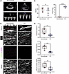 Figure 1
T-tubule maturation deficit in pediatric patients with RVPO. (A) Representative echocardiographic images from patients with ventricular septal defect (VSD, nonoverload group) and tetralogy of Fallot (TOF, pressure-overload group). (B) Quantification of Right Ventricular fractional area change (RV FAC) and transvalvular pressure gradient (TPG) measured by transthoracic echocardiography (n = 5 patients per group). (C) Representative images of T-tubules in the VSD and TOF groups. Scale bar: 25 μm. Zoomed-in view indicates red arrows that show T-elements, blue arrows indicate L-elements, and yellow lines denote the sarcolemma. (D) Quantification of T-element density, T-tubule regularity, and T-tubule integrity index (n = 6 rats per group, with 30 cardiomyocytes from 5 independent sections per rat). Data are presented as mean ± SD. P values were calculated by 2-tailed Student’s t test. **P < 0.01, ***P < 0.001, ****P < 0.0001.
Establishment of a neonatal rat model of RVPO. Given the central role of animal models in preclinical research (17, 18) and the prior lack of a neonatal RVPO model (19, 20), the effect of RVPO on postnatal RV development has remained unclear. We therefore established a neonatal RVPO model via pulmonary artery banding (PAB) on P1, as described previously (19, 21). At P7, pulmonary artery velocity-time integral (PA-VTI), peak velocity, and TPG were significantly elevated in PAB rats compared with sham controls (Supplemental Figure 1, A and B; supplemental material available online with this article; https://doi.org/10.1172/jci.insight.204032DS1). By P21, RV hypertrophy was evident, accompanied by ventricular septal shifting toward the left ventricle (LV) (Supplemental Figure 1, C and D). Although RV ejection fraction (RVEF) was generally preserved in PAB rats — consistent with observations in patients (3, 22) — a subset (~50%) exhibited reduced RVEF (Supplemental Figure 1, C and E). These results confirm the successful establishment of a neonatal RVPO model, providing a platform to investigate how RVPO remodels postnatal RV development.
RVPO disrupts T-tubule maturation in neonatal rats. We next assessed whether the neonatal RVPO model recapitulated the T-tubule defects observed in patients. As shown in Figure 2, A and B, T-element density, T-tubule regularity, and T-tubule integrity index were all significantly lower in the PAB group than in sham controls, indicating impaired T-tubule formation in neonatal rats and mirroring the human phenotype.
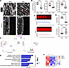 Figure 2
RVPO impairs T-tubule maturation in neonatal rats. (A) Representative T-tubule images from sham and PAB groups. Scale bar: 10 μm. Zoomed-in panels show green arrows that indicate T-elements, purple arrows that indicate L-elements, and yellow arrows that denote the sarcolemma. (B) Quantification of T-element density, T-tubule regularity, and T-tubule integrity index (n = 6 rats per group, with 30 cardiomyocytes from 5 independent sections per rat). (C) Upper panel: representative calcium transient traces from sham group cardiomyocytes. Lower panel: representative traces from PAB group cardiomyocytes. Note the elevated basal calcium signal intensity in PAB cardiomyocytes (~1.4) compared with sham (~1.0). (D) Quantification of calcium transient amplitude and time to peak (n = 15 cardiomyocytes per group; Mann-Whitney U test). (E) Principal component analysis of transcriptional profiles from sham and PAB samples. (F) Volcano plot of differentially expressed genes in PAB versus sham (adjusted P < 0.05, |log2FC| > 1). (G) Gene Ontology enrichment analysis of downregulated genes in PAB RVs. (H) Heatmap of downregulated genes in PAB-influenced RVs. Data are presented as mean ± SD. P values were calculated by 2-tailed Student’s t test. **P < 0.01, ***P < 0.001, ****P < 0.0001.
Since T-tubules are essential for efficient calcium handling in cardiomyocytes (14), we evaluated intracellular calcium dynamics. The amplitude of calcium transients was significantly reduced, and the time to peak was prolonged in PAB cardiomyocytes relative to sham (Figure 2, C and D). Moreover, PAB cells exhibited elevated basal cytosolic calcium levels (Figure 2C), suggesting impaired sarcoplasmic reticulum calcium reuptake and further supporting T-tubule dysfunction. Together, these functional data indicate that RVPO disrupts T-tubule maturation.
To explore the underlying mechanisms, we performed RNA-seq analysis of RV cardiomyocytes. Principal component analysis (PCA) revealed distinct transcriptomic profiles between PAB and sham groups (Figure 2E). A total of 686 genes were upregulated and 577 were downregulated in PAB samples (adjusted P < 0.05, |log2FC| > 1; Figure 2F). Gene ontology (GO) enrichment analysis indicated that downregulated genes were associated with processes including heart contraction, cardiac conduction, T-tubule organization, ion channel complex assembly, gated channel activity, and cation channel activity (Figure 2G). Notably, Bin1 and Jph2 — 2 key T-tubule regulators (12, 14) — were among the downregulated genes in T-tubule–related GO terms (Figure 2H).
Negative correlation between cardiomyocyte hypertrophy and T-tubule gene expression. Because T-tubule remodeling is frequently accompanied by cardiomyocyte hypertrophy (Supplemental Figure 1, C and D), we performed single-nucleus RNA-seq on RV tissues from sham and PAB rats at P1, P7, P14, and P21 (Supplemental Figure 2, A–D). After quality control and clustering, we constructed cardiomyocyte-specific metacells using the high dimensional weighted gene coexpression network analysis (hdWGCNA) framework to reduce technical noise. Pearson correlation analysis on metacell expression profiles revealed a significant negative correlation between hypertrophy marker genes (Nppa, Nppb) and T-tubule–related genes (Bin1, Jph2) across all time points (Supplemental Figure 2, E–H). These results suggest that the development of hypertrophy and the impairment of T-tubule maturation are inversely linked during postnatal RVPO, although causality remains to be determined.
RVPO alters chromatin accessibility of T-tubule–related genes. As chromatin accessibility governs transcriptional activity (23, 24), we performed ATAC-seq to assess its changes under RVPO. Approximately 57.57% of ATAC-seq peaks were shared between PAB and sham groups, while 29.28% (16,954 peaks) and 13.15% (6,002 peaks) were unique to PAB and sham groups, respectively (Figure 3, A and B). Genes with reduced accessibility in PAB (i.e., those corresponding to sham-only peaks) were enriched in biological processes such as heart contraction, mitochondrial depolarization, ion channel activity, and calcium signaling (Figure 3C) — findings consistent with RNA-seq findings and the impaired calcium handling observed in PAB cardiomyocytes (Figure 2, C–H).
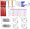 Figure 3
RVPO alters chromatin accessibility of T-tubule–associated genes. (A) Heatmap of shared and differentially accessible ATAC-seq peaks and associated genes. (B) Summary of differential gene expression and chromatin accessibility. “Up-regulated” and “Down-regulated” refer to RNA-seq results; “More accessible” and “Less accessible” refer to ATAC-seq results. (C) Enrichment analysis of genes with reduced chromatin accessibility in PAB RVs. (D) Chromatin accessibility profile around the transcription start site (TSS) of the Bin1 gene. (E) Representative BIN1 Western blot in sham and PAB groups. (F) Quantification of pBIN1 protein expression (n = 6 rats per group). (G) Quantification of cBIN1 protein expression (n = 6 rats per group). (H) Representative BIN1 Western blot in VSD and TOF patient samples. (I) Quantification of pBIN1 expression in human samples (n = 5 patients per group). (J) Quantification of cBIN1 expression in human samples (n = 5 patients per group). Data are presented as mean ± SD. P values were calculated by 2-tailed Student’s t test. **P < 0.01, ***P < 0.001.
Focusing on Bin1 and Jph2, we observed reduced chromatin accessibility at the Bin1 locus in PAB samples (Figure 3D), whereas JPH2 expression did not change in human RVPO samples but was downregulated in PAB rats (Supplemental Figure 3, A and B), and Jph2 accessibility was increased (Supplemental Figure 3C). Given this divergent regulation, Jph2 was excluded from subsequent mechanistic studies. Of note, this does not imply that Jph2 is less important than Bin1; it may be regulated through distinct pathways.
We next validated these findings at the protein level. Both pBIN1 and cBIN1 were downregulated in PAB rat RVs (Figure 3, E–G). Importantly, a similar reduction was observed in human RVPO samples (Figure 3, H–J), suggesting that both BIN1 isoforms contribute to RVPO-induced deficits in T-tubule maturation.
BIN1 overexpression rescues T-tubule maturation in neonatal RVPO rats. Given the downregulation of both pBIN1 and cBIN1, we asked whether restoring BIN1 expression could ameliorate T-tubule defects. We overexpressed full-length BIN1 mRNA in neonatal RVPO rats using AAV9 vectors under the cTnT promoter (Figure 4A). An infection rate of 20%–30% was maintained to avoid altering global cardiac function (Figure 4, B and C) (25, 26). As expected, AAV-mediated Bin1 delivery increased both pBIN1 and cBIN1 expression in cardiomyocytes (Figure 4, D–F), without affecting LV or RV function (Figure 4, G and H). This confirms that subsequent improvements in T-tubule integrity resulted directly from BIN1 upregulation rather than secondary hemodynamic changes.
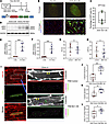 Figure 4
BIN1 overexpression rescues T-tubule maturation in neonatal RVPO rats. (A) Schematic of the BIN1 overexpression strategy. (B and C) AAV9 infection efficiency. (D) Representative BIN1 WES immunoblot from isolated RV cardiomyocytes. (E) Quantification of pBIN1 expression (n = 6 rats). (F) Quantification of cBIN1 expression (n = 6 rats). (G) Right ventricular fractional area change (RV FAC, %) of rats at P21. (H) Left ventricular ejection fraction (LVEF, %). (I) Representative T-tubule images from BIN1-overexpressing (BIN1-OE) and control cardiomyocytes in PAB RVs. Scale bar: 25 μm. Zoomed-in panels show green arrows that indicate T-elements, purple arrows that indicate L-elements, and yellow arrows that denote the sarcolemma. (J–L) Quantification of T-element density (J), T-tubule regularity (K), and T-tubule integrity index (L) (n = 6 rats per group, with 30 cardiomyocytes per rat). Data are presented as mean ± SD. P values were calculated by 2-tailed Student’s t test. *P < 0.05, **P < 0.01, ***P < 0.001, ****P < 0.0001.
To precisely evaluate BIN1’s role, we compared T-tubule architecture between BIN1-overexpressing (GFP+) cardiomyocytes and adjacent noninfected cells within the same RV microenvironment (Figure 4I). BIN1-overexpressing cells exhibited significantly higher T-element density, T-tubule regularity, and T-tubule integrity index (Figure 4, I–L), confirming that BIN1 restoration is sufficient to rescue T-tubule maturation in the setting of uncorrected RVPO. Furthermore, BIN1 overexpression also significantly reduced cardiomyocyte hypertrophy (Supplemental Figure 4)
MEF2D mediates BIN1 downregulation in neonatal RVPO rats. To investigate the cause of BIN1 downregulation, we analyzed transcription factor motif enrichment in the ATAC-seq data. The Mef2d motif was the most significantly enriched in PAB-specific accessible regions (Figure 5, A and B, and Table 2). Furthermore, chromatin accessibility at the Mef2d locus was higher in PAB than in sham rats (Figure 5, C and D), suggesting that Mef2d may contribute to Bin1 repression. Moreover, MEF2D expression was also increased in human RVPO samples (Supplemental Figure 5), indicating that this regulatory mechanism is conserved across species.
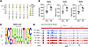 Figure 5
Mef2d motif is highly enriched in PAB rat RVs. (A) Dot plot showing enrichment of known transcription factor motifs. (B) Comparison of Mef2a, Mef2b, and Mef2d motif enrichment between sham and PAB RVs. (C) Sequence logo of the Mef2d motif. (D) Chromatin accessibility profile around the Mef2d TSS. Data are presented as mean ± SD. P values were calculated by 2-tailed Student’s t test. *P < 0.05.
We therefore knocked down MEF2D in neonatal RVPO rats by AAV (Figure 6A). With the AAV infection rate at 20%–30% (Figure 6, B and C), we observed that knockdown of MEF2D in cardiomyocytes (Figure 6, D and E) did not significantly compromise left or RV function (Figure 6, F and G), thus excluding potential secondary effects from heart failure (25, 26). Importantly, it markedly upregulated the expression of BIN1 isoforms, including both pBIN1 and cBIN1(Figure 6, H–J). As a result, T-tubule maturation was restored, as indicated by increased T-element density, regularity, and index of TT integrity of T-tubules (Figure 6, K–N). Consistent with BIN1 overexpression, MEF2D knockdown also attenuated cardiomyocyte hypertrophy (Supplemental Figure 4).
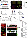 Figure 6
MEF2D knockdown rescues T-tubule maturation in neonatal RVPO rats. (A) Schematic of the MEF2D knockdown strategy. (B and C) AAV9 infection efficiency. (D) Representative MEF2D WES immunoblot from isolated RV cardiomyocytes. (E) Quantification of MEF2D expression (n = 6 rats). (F) Right ventricular fractional area change (RV FAC, %) of rats at P21. (G) Left ventricular ejection fraction (LVEF, %). (H) Representative BIN1 WES immunoblot from isolated cardiomyocytes. (I) Quantification of pBIN1 expression (n = 6 rats). (J) Quantification of cBIN1 expression (n = 6 rats). (K) Representative T-tubule images from MEF2D-knockdown (MEF2D-KD) and control cardiomyocytes in PAB RVs. Scale bar: 25 μm. Green arrows indicate T-elements, purple arrows indicate L-elements, and yellow arrows denote the sarcolemma. (L–N) Quantification of T-element density (L), T-tubule regularity (M), and T-tubule integrity index (N) (n = 6 rats per group, with 30 cardiomyocytes per rat). Data are presented as mean ± SD. P values were calculated by 2-tailed Student’s t test. *P < 0.05, **P < 0.01, ****P < 0.0001.
To determine whether MEF2D directly regulates Bin1 transcription, we mutated the predicted Mef2d-binding sites in the Bin1 promoter (Figure 7, A and B). Luciferase reporter assays showed that MEF2D significantly suppressed WT Bin1 promoter activity, whereas mutation of the Mef2d-binding sites abolished this repression (Figure 7B). These results establish a direct regulatory pathway: RVPO → MEF2D → BIN1 → T-tubule maturation (Figure 7C).
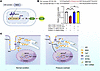 Figure 7
MEF2D inhibits Bin1 promoter activity. (A) Schematic of the dual-luciferase reporter assay. (B) Luciferase activity assays evaluating MEF2D regulation of the Bin1 promoter. Data were analyzed by Kruskal-Wallis test with post hoc Dunn’s test. **P < 0.01, ***P < 0.001. (C) Graphical summary of the RVPO/MEF2D/BIN1/T-tubule pathway identified in this study.


-
Discussion
This study elucidates a previously unrecognized molecular pathway through which RVPO disrupts a fundamental aspect of postnatal cardiac maturation: the development of the T-tubule network. We have identified and validated a RVPO/MEF2D/BIN1/T-tubule axis that operates in both pediatric patients with TOF and a corresponding neonatal rat model, providing a mechanistic explanation for the impaired cellular-level E-C coupling that may underpin long-term functional vulnerabilities in these hearts (3, 10).
The initial finding of disorganized and sparse T-tubules in the right ventricles of children with TOF directly links a common clinical condition (RVPO) to a core defect in cardiomyocyte ultrastructure. This observation is crucial because a mature T-tubule system is indispensable for synchronous calcium release and robust contraction (3, 10). The functional consequence of this structural deficit was confirmed in our neonatal RVPO model, which faithfully recapitulated the human phenotype. Cardiomyocytes from these rats exhibited compromised calcium handling, characterized by reduced amplitude and prolonged time-to-peak of calcium transients (Figure 2, C and D). These calcium kinetics are classic hallmarks of a deficient T-tubule network, where the loss of synchronous L-type calcium channel triggering leads to delayed and disorganized RyR activation (27, 28). The preservation of global RV function (as measured by FAC and LVEF) in the presence of such significant cellular-level dysfunction highlights a critical concept: the heart can compensate for impaired E-C coupling in its early stages, but this cellular deficit likely creates a substrate for arrhythmias and eventual contractile failure later in life, as seen in adult TOF survivors (1–2).
Our multiomics approach was pivotal in dissecting the mechanism upstream of this structural defect. The convergent evidence from RNA-seq and ATAC-seq was particularly powerful (1–2). RNA-seq revealed a broad downregulation of genes critical for E-C coupling and T-tubule integrity, while ATAC-seq demonstrated that this downregulation was associated with reduced chromatin accessibility in the regulatory regions of these same genes. This synergy suggests that RVPO instigates a transcriptional reprogramming that actively suppresses the cardiomyocyte maturation program. Among the downregulated genes, Bin1 emerged as a prime candidate. The role of Bin1 as a master regulator of membrane curvature and T-tubule formation is well established (3, 10), and its downregulation provides a direct molecular link to the observed structural deficits. Importantly, our study adds nuance by showing that both the tubule-forming engine pBIN1 and the microfold-sculpting cBIN1 are suppressed in RVPO, indicating a comprehensive disruption of the entire T-tubule maturation process. Consistently, we also observed reduced BIN1 expression in human TOF samples (Figure 3, H–J), confirming the clinical relevance of this finding.
The most significant mechanistic insight of this work is the identification of MEF2D as the transcriptional repressor responsible for BIN1 suppression. The enrichment of the Mef2d motif in the less-accessible chromatin regions of RVPO hearts, coupled with the increased openness of the Mef2d gene itself, painted a picture of MEF2D upregulation leading to the repression of its target genes. Our functional experiments confirmed this hypothesis. Knockdown of MEF2D in cardiomyocytes, while leaving global ventricular function intact, successfully restored BIN1 expression and, most importantly, rescued the T-tubule network. This finding is paradigm shifting. MEF2D, a transcription factor traditionally associated with promoting muscle differentiation and hypertrophy, is here revealed to act as a transcriptional repressor of a key maturation gene in the context of neonatal RVPO. The dual-luciferase assay provided the final piece of direct evidence, demonstrating that MEF2D binds to the Bin1 promoter to inhibit its activity. This establishes a clear, direct line from the pathological stimulus (RVPO) to a transcriptional regulator (MEF2D), to an effector protein (BIN1), and finally to a cellular phenotype (impaired T-tubule maturation).
To assess whether this regulatory axis is conserved across species, we examined MEF2D expression in human RVPO samples. We collected an additional 4 samples per group (Table 3) and found that MEF2D expression was also significantly elevated in patients with TOF compared with controls. These results, together with the BIN1 changes observed in human samples, strongly suggest that the RVPO/MEF2D/BIN1/T-tubule pathway is evolutionarily conserved and relevant to human disease.
Another important aspect of RVPO pathophysiology is cardiomyocyte hypertrophy. Using single-nucleus RNA-seq of rat right ventricles from P1 to P21, we observed a significant negative correlation between the expression of hypertrophy marker genes (Nppa, Nppb) and T-tubule–related genes (Bin1, Jph2). This finding suggests that hypertrophy and T-tubule remodeling are inversely related during postnatal development under pressure overload. Whether hypertrophy directly impairs T-tubule maturation or vice versa remains to be determined; nevertheless, our data highlight a potential reciprocal interaction that warrants further investigation.
The therapeutic implications of this axis are substantial. Our rescue of T-tubule structure through BIN1 overexpression, even in the persistent presence of RVPO, is a proof of concept that targeting this pathway can reverse cellular-level pathology. This suggests that strategies aimed at enhancing BIN1 expression or function — or inhibiting the repressive action of MEF2D — could be developed to promote myocardial maturation and resilience in children with RVPO, potentially improving their long-term outcomes independently of surgical correction of the overload itself.
Our preclinical experiments included both male and female neonatal rats with approximately equal sex distribution, and we observed no obvious sex-dependent differences in the RVPO-induced T-tubule defects or BIN1 downregulation in post hoc analyses. However, because all human tissue samples were derived from male patients, the generalizability of these findings to female pediatric patients, and the potential for sex-specific modulation of the MEF2D/BIN1 axis, warrant further investigation in larger, sex-balanced clinical cohorts.
Several important considerations arise from our work. First, the focus of this study was on the early, neonatal response to RVPO. It will be critical to investigate whether this MEF2D-mediated repression is sustained into adulthood and contributes to the progression of heart failure. Second, while we established the role of this axis in cardiomyocytes, the potential crosstalk with other cardiac cell types, such as fibroblasts and endothelial cells, remains an open question. Paracrine signals originating from these cells in the PO ventricle could further modulate the MEF2D/BIN1 axis in cardiomyocytes. Third, the upstream signal that triggers MEF2D upregulation in response to RVPO — whether mechanical stress, neurohormonal activation, or metabolic changes — is a vital area for future investigation. Fourth, we acknowledge that our identification of pBIN1 and cBIN1 isoforms was based on molecular weight differences reported in the literature (Supplemental Figure 6) (14) and the antibody manufacturer’s datasheet, as no commercially available antibodies specifically recognizing individual exons of BIN1 are currently available. Future studies using exon-specific antibodies or RNA probes will be valuable to further dissect the distinct functions of each isoform. Fifth, the absence of dedicated electrophysiological studies — such as electrocardiography (ECG) and programmed electrical stimulation — precludes a definitive assessment of arrhythmia susceptibility in this model. Therefore, while our calcium transient data suggest functional impairment, we refrain from making direct claims regarding arrhythmogenesis. Finally, although our in vivo data strongly support the role of MEF2D in T-tubule homeostasis, future in vitro studies using siRNA knockdown in adult cardiomyocytes or induced pluripotent stem cell–derived cardiomyocytes could provide complementary evidence and help elucidate the direct maintenance functions of MEF2D.
In conclusion, our study unveils a pathological mechanism wherein RVPO drives the upregulation of the transcription factor MEF2D, which in turn represses the expression of the key T-tubule sculpting protein BIN1, leading to a failure in T-tubule maturation and impaired calcium-mediated E-C coupling. The identification of this RVPO/MEF2D/BIN1/T-tubule axis not only deepens our understanding of the pathophysiology of pediatric heart diseases but also illuminates potential therapeutic avenues aimed at promoting proper myocardial maturation to safeguard the long-term health of these vulnerable young hearts (3, 10).


