
Immunology
Abstract
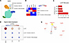
After encountering allogeneic nonself, monocytes differentiate to DCs, which in turn activate the adaptive immune system that drives transplant rejection. The downstream mechanisms of allorecognition are largely unknown. We analyzed scRNA-seq data sets to identify transcriptional changes in monocytes occurring after allostimulation. Hspa1a, which encodes HSP70, was upregulated in monocytes after allostimulation in contrast to syngeneic controls in mice lacking T, B, and NK cells. Similar findings were seen in scRNA-seq data derived from kidney biopsies of rejecting transplant patients. To validate the role of HSP70 in innate allorecognition and transplantation, we performed allogeneic bone marrow plug and kidney transplantation into WT, Hspa1a/Hspa1b–/–, and DSG-treated (HSP70 inhibitor) B6 mice and examined the graft immune infiltrate. Histology, T cell infiltration, and survival were assessed in kidney transplanted mice. In both models, DSG-treated or HSP70-KO recipients displayed significantly reduced infiltration by monocyte-derived DCs (mo-DC). Chronic rejection of Balb/c kidney grafts in WT B6 recipients was attenuated in DSG-treated and HSP70-KO recipients as indicated by a reduction in Banff score. Further experiments demonstrated that in vitro allostimulated immature HSP70-KO BMDC showed less maturation compared with WT BMDC. Targeting HSP70 in innate immune cells offers a novel approach to reduce chronic kidney graft rejection.
Authors
Zeping Gui, Neda Feizi, Berkay Demirors, Canxiang Lin, Hehua Dai, Mouhamad Al Moussawy, Andrew Friday, Steven M. Sanders, Latha Halesha, Amanda L. Williams, Faruk Sacirbegovic, Parmjeet S. Randhawa, Khodor I. Abou-Daya, Martin H. Oberbarnscheidt
Abstract
The role of CD8+ T cells in filarial infections remains poorly understood. Given the chronic nature of these infections, individuals living in endemic regions are frequently exposed to other pathogens including viruses. Because CD8 cells are essential for antiviral immunity, understanding how filarial infections shape the cytotoxic compartment is essential to elucidate their impact on bystander immunity. We evaluated the phenotype and function of CD8+ T cells from Loa loa-infected (Fil+) and -uninfected (Fil–) individuals at baseline and following cytomegalovirus (CMV) re-stimulation. Filarial infection was associated with increased activation and proliferation of CD8+ T cells, characterized by elevated expression of Ki-67, CD107a, and production of type 1 and 17 cytokines. Upon CMV re-stimulation, Fil+ individuals showed markedly reduced expansion of antigen-experienced (CD137+) and polyfunctional (CD137+IFN-γ+TNF-α+, CD137+IFN-γ+TNF-α+IL-2+) CD8+ T cells, along with decreased cytotoxic activity. Unbiased clustering analysis revealed markedly reduced frequency of the CD8+CD45RA+CD57+ subset in Fil+ individuals- a population further defined through transcriptomic profiling that showed enrichment for cytolytic gene signatures (GZMB, GNLY, GZMH, CD244, CX3CR1). These results demonstrated that filarial infection is associated with an altered CD8+ T cell profile that is associated with failure to mount effective viral-specific effector responses, including impaired cytokine production and cytotoxic activity.
Authors
Camila Queiroz-Glauss, Justin Lack, Daniel E. Sturdevant, Thomas B. Nutman
Abstract
Pneumocystis jirovecii pneumonia (PCP) remains a major cause of life-threatening respiratory failure in people with advanced HIV, yet the biological factors associated with mortality are incompletely understood. We performed integrated transcriptomic, proteomic, and metabolomic profiling of bronchoalveolar lavage fluid from 42 adults with HIV-associated PCP, relating molecular signatures to mortality. We found that fungal burden, measured by sputum immunofluorescence, Pneumocystis qPCR, and 1,3-β-D-glucan, did not differ between survivors and non-survivors or correlate with baseline gas exchange. Instead, non-survivors exhibited a bronchoalveolar immunometabolic failure signature characterised by marked depletion of immunoglobulins and complement, reduced pattern recognition receptors and fibroblast growth factor family proteins, and broad disruption of extracellular matrix organisation. Metabolomic profiling revealed enrichment of arginine-urea cycle and tricarboxylic acid pathways, with coordinated accumulation of arginine, citrulline, and TCA cycle intermediates consistent with nitrosative and mitochondrial stress. An integrated proteo-metabolomic score summarising this state was significantly higher in non-survivors and did not correlate with fungal burden or cytomegalovirus co-infection. In an immunodeficient precision-cut lung slice model, antigen-screened intravenous immunoglobulin restored macrophage-mediated Pneumocystis clearance, providing proof-of-concept for opsonic augmentation. Our findings indicate that mortality in HIV-PCP is defined by profound immunometabolic failure, highlighting humoral depletion and impaired alveolar repair as potential targets for host-directed adjunctive therapy.
Authors
Peter Rossi-Smith, Ayanda Trevor Mnguni, Dora Pungan, Robert J. Samuels, Vaishnavi R. Kumaran, Leena Syed, Trevor Ferris, Kamil Skirlo, Joseph N. Jarvis, Nelesh P. Govender, Graeme Meintjes, Sean Wasserman, Jay K. Kolls, Rachel P.J. Lai
Abstract
Persistent production of type I interferons (IFN-Is) is a hallmark of cutaneous lupus erythematosus (CLE). Ultraviolet (UV) light stimulates IFN-I response in the skin and exacerbates CLE. Here, we identify V-type immunoglobulin domain-containing suppressor of T cell activation (VISTA) as a negative regulator of both basal and UV-induced IFN-I response in the skin and show that VISTA limits skin photosensitivity in an IFN-I-dependent manner, in part through Stimulator of Interferon Genes (STING). Furthermore, we demonstrate a novel role for VISTA in keratinocytes both at steady state and in response to UV light. Conditional deletion of VISTA in epidermal keratinocytes results in a ~10-fold increase in basal skin IFN-I scores and a heightened UV-induced skin injury score, both of which are dependent on IFN-I signaling. VISTA-targeting monoclonal antibodies suppress the UV-induced IFN-I response in human keratinocytes and in mice expressing human VISTA in vivo, thereby reducing UV-induced skin injury scores. Together, these findings identify VISTA as a keratinocyte-intrinsic checkpoint that restrains STING-associated IFN-I response in the skin and suggest VISTA agonism as a therapeutic strategy to limit photosensitivity in CLE.
Authors
Zachary T. Peters, Lindsay K. Mendyka, J'Voughnn A. Blake, Himanshu B. Goswami, Angelique N. Cortez, Grace E. Crossland, Sicong Shan, Elizabeth C. Nowak, Myana Keusch, Mrinal K. Sarkar, Johann E. Gudjonsson, Christopher M. Burns, Dorothea T. Barton, Bruce R. Blazar, Tyler J. Curiel, Rodwell Mabaera, Victoria P. Werth, Andrea Kalus, Keith B. Elkon, Randolph J. Noelle, Sladjana Skopelja-Gardner
Abstract
Tuberculosis (TB) caused by Mycobacterium tuberculosis (Mtb) is the leading infectious cause of death globally. Despite wide use of antiretroviral therapy (ART) by people living with HIV, the risk of TB remains increased. To understand immune interactions within lung granulomas, we compared spatial transcriptomics of Mtb and simian immunodeficiency virus (SIV) in co-infected macaques with or without ART as a model for HIV/Mtb co-infection. Spatially differentiated transcriptional profiles were observed in Mtb-only granulomas, with myeloid cells enriched in metabolic/antimicrobial pathways within the inner ring and T cell co-stimulatory/activation pathways enriched in the outer ring. These spatially distinct patterns were lost in SIV/Mtb granulomas with higher enrichment in type I IFN pathways compared to Mtb-only granulomas. SIV/ART/Mtb granulomas had an intermediate transcriptional pattern without restoration to Mtb-only granulomas, despite a lack of viral replication. Cell-cell communication was reduced among SIV/Mtb co-infected groups. These data suggest that HIV disrupts spatially organized immune functions of granulomas, which are not fully restored by ART.
Authors
Jessica M. Medrano, Persis Sunny, Collin R. Diedrich, Pauline Maiello, Christopher Kline, Tara Rutledge, Edwin Klein, Joshua T. Mattila, Jishnu Das, Zandrea Ambrose, Philana Ling Lin
Abstract
BACKGROUND. This prospective, single-arm, multicenter phase 1/2 trial evaluated vorinostat added to standard graft-versus-host disease (GVHD) prophylaxis in pediatric, adolescent, and young adult (AYA) patients undergoing allogeneic hematopoietic cell transplantation (HCT) from HLA-matched related, HLA-matched unrelated, and haploidentical donors. METHODS. Patients aged 3–39 years received twice-daily vorinostat with tacrolimus/methotrexate after HLA-matched HCT from day −10 to +30, or with post-transplant cyclophosphamide/tacrolimus/mycophenolate mofetil after haploidentical HCT from day +5 to +30. The primary endpoint was cumulative incidence of grade II–IV acute GVHD by day +100. All outcomes were based on intention-to-treat analysis. RESULTS. Forty-three patients were enrolled; median age was 19 years, with 74% receiving HLA-matched and 26% haploidentical HCT. The recommended phase 2 dose was 60 mg/m² twice daily. No dose-limiting toxicities, unexpected vorinostat-related serious adverse events, or primary graft failures occurred. Median neutrophil and platelet recovery occurred at 14 and 18 days, respectively. Day +100 grade II–IV and III–IV acute GVHD were 14% (95% CI, 5.6, 26) and 4.7% (95% CI, 0.83, 14), respectively. One-year overall survival was 88.4% (95% CI, 79.3, 98.5), relapse 14% (95% CI: 5.6, 26), nonrelapse mortality 4.7% (95% CI: 0.8, 14), and GVHD-free/relapse-free survival 55.8% (95% CI: 42.8, 72.8). Correlative studies demonstrated on-target HDAC activity, with increased histone acetylation and lower proinflammatory cytokines. CONCLUSION. These findings support randomized evaluation of vorinostat-based GVHD prophylaxis in pediatric and AYA HCT. TRIAL REGISTRY. ClinicalTrials.gov, NCT03842696.
Authors
Nicolas Parnell, Xiao Cao, Jan H. Beumer, Thomas M. Braun, Yilei Cui, Gary J. Fisher, Guoqing Hou, Julianne Holleran, Tracey Churay, Michelle Rozwadowski, Luna Heider, Mark T. Vander Lugt, Carrie L. Kitko, April L. Rahrig, Ed Peres, Kirsten M. Williams, Julie-An Talano, Vanessa A. Fabrizio, Ghada Abusin, Gregory A. Yanik, John Magenau, Mary Riwes, Marcus J. Geer, Sarah Anand, Monalisa Ghosh, Attaphol Pawarode, Kristen Votruba, Pavan Reddy, Sung Won Choi
Abstract
Patients who survive sepsis are predisposed to new hospitalizations for respiratory failure, but the underlying mechanisms are unknown. Using a murine model in which prior sepsis predisposes to enhanced lung injury, we previously discovered that classical monocytes persist in the lungs after long-term recovery from sepsis and exhibit enhanced cytokine expression after secondary challenge with intra-nasal lipopolysaccharide. Here, we hypothesized that immune reprogramming of post-sepsis monocytes and altered ontogeny predispose to enhanced lung injury. Monocyte depletion and/or adoptive transfer was performed three weeks and three months after sepsis. Monocytes from post-sepsis mice were necessary and sufficient for enhanced LPS-induced lung injury and promoted neutrophil degranulation. Prior sepsis enhanced JAK-STAT signaling and AP-1 accessibility in monocytes and shifted monocytes toward the neutrophil-like monocyte lineage. Neutrophil-like monocytes demonstrated a pro-inflammatory phenotype with enhanced IL-1β expression and reduced phagocytic capacity. In human sepsis and/or pneumonia survivors, monocytes were predictive of 90-day mortality and exhibit transcriptional and proteomic neutrophil-like signatures. We conclude that sepsis reprograms monocytes into a pro-inflammatory phenotype and skews bone marrow progenitors and monocytes toward the neutrophil-like lineage, predisposing them to induce neutrophil degranulation and lung injury.
Authors
Scott J. Denstaedt, Breanna McBean, Alan P. Boyle, Brett C. Arenberg, Matthias Mack, Bethany B. Moore, Michael W. Newstead, Yamei Deng, Alexey I. Nesvizhskii, Benjamin H. Singer, Jennifer Cano, Hallie C. Prescott, Helen S. Goodridge, Rachel L. Zemans
Abstract
Early antiretroviral therapy (ART) limits viral reservoir establishment in infants but also constrains viral-specific immunity required to clear reactivated cells. In the early ART context, we evaluated whether combining adeno-associated virus serotype 9 (AAV9)–vectored eCD4-IgG1 with pharmacologic latency reversal using the second mitochondria-derived activator of caspases (SMAC) mimetic AZD5582 could promote reservoir reduction and control in simian immunodeficiency virus (SIV)–infected infant macaques. AAV9 delivery achieved sustained eCD4-IgG1 expression and AZD5582 induced on-ART viremia, but the combination did not reduce intact SIV proviral DNA relative to controls. Partial post-intervention control of viremia during Analytical Treatment Interruption (ATI) occurred in 2/6 infants receiving AAV9-eCD4-IgG1 + AZD5582 with restricted reservoir expansion during recrudescence at week 13. Intact reservoir size during ATI correlated inversely with on-ART viremia during AZD5582 treatment. Controllers did not show increased eCD4-IgG1 expression nor increased on-ART viremia during AZD5582 treatment, but harbored less pre-ART intact SIV DNA in PBMCs than non-controllers. Controllers also exhibited higher frequencies and greater polyfunctionality of virus-specific CD8+ T-cells prior to ATI. These findings implicate immune control of reservoir size and post-ART viral dynamics in early-treated infants and suggest that AAV9-eCD4-IgG1 + AZD5582 increases the likelihood of post-ART viral control.
Authors
Jairo A. Fonseca, Alexis C. King, Kaleaha S. Davis, Daniel O'Hagan, Adrian Khoei, Lucas Alves Britto Da Costa, Camryn Cockerham, Mackenzie Cottrell, Stephanie Ehnert, Jennifer S. Wood, Michael D. Alpert, Matthew R. Gardner, Jeffrey D. Lifson, Maud Mavigner, Mauricio A. Martins, Ann Chahroudi
Abstract
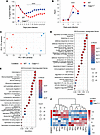
Ubiquitination is an important post-translational modification associated with essential cellular processes and implicated in regulation of immunity. Here we show that deletion of the E3 ubiquitin ligase Hectd3, germline or in hematopoietic compartment, including in CD11c+ cells, causes a more severe DSS-induced colitis and increased production of proinflammatory cytokines. Hectd3 mRNA levels were found reduced in colonic tissues of patients with ulcerative colitis (UC), which highlights a potential role for Hectd3 in regulating inflammatory responses in UC. We identified Myd88, a central adaptor in the TLR/IL1R signaling and inflammatory response, as a target for Hectd3 ubiquitination. We demonstrate that Hectd3 directly ubiquitinates Myd88 through K27-linked Poly-Ub chains in a nondegradative manner. Hectd3 KO GM-CSF-bone marrow derived cells treated with the TLR4 ligand lipopolysaccharide (LPS) produced more proinflammatory cytokines, show elevated phosphorylation of NF-κB and IRAK4, as well as elevated association of Myd88 with IRAK4, demonstrating that Hectd3 controls Myd88-IRAK4-NF-κB axis. Inhibition of Myd88 activity rescued colitis severity in the Hectd3 KO mice, including the elevated proinflammatory cytokine production. Thus, our results establish Myd88 as a new target for Hectd3 non-degradative polyubiquitination and restriction of immune colonic inflammation.
Authors
Shamima Islam, Shahnewaj Mannan, Ashley Zuniga, Upasana Parthasarathy, Valeriu B. Cismasiu, Leonardo Silvane, Sayan Chakraborty, Divya Priyanka Talada, Raghwendra Pratap Singh, Tomas Zelenka, Amreen Naveen, Sephra Chyanne Vickers, Michael G.M. Grant, Jonathan J. Cho, Theodore Drashansky, Alexander J. Kwiatkowski, Xintong Liu, Ross Tomaino, Olga A. Guryanova, Mariola J. Ferraro, Sang Yong Kim, Christian Jobin, Benjamin G. Keselowsky, Hongmin Li, Paulo C. Rodriguez, Martina Molgora, Amer A. Beg, Timothy I. Shaw, Zheng Ruan, Lixin Wan, Dorina Avram
Abstract
Severe SARS-CoV-2 infection is characterized by lung hyperinflammation, impaired IFN responses, and defective T cell activation, yet the molecular drivers of these immune dysregulations remain incompletely understood. Caspase-11 (CASP11), a key effector of the noncanonical inflammasome, has been shown to mediate an innate hyperinflammatory response and cytokine release in a mild SARS-CoV-2 infection model. However, the role played by CASP11 in severe SARS-CoV-2 disease and how it affects adaptive immunity has not been identified. Here, we found that CASP11 exacerbates severe SARS-CoV-2 pathogenesis by amplifying early innate immune responses while concurrently impairing antiviral CD8+ T cell immunity. Using global KO mice, hematopoietic BM chimeras, and Cx3cr1-expressing mononuclear phagocyte system cell–specific CASP11 deletion models, we show that targeting CASP11 reduces lung inflammation, promotes early NK cell–mediated IFN-γ production, and enhances robust virus-specific effector CD8+ T cell responses. This was associated with enhanced viral clearance and improved survival, even under lethal infection conditions. Importantly, CASP11-KO mice also exhibited faster resolution of postviral inflammation. These findings position CASP11 as a promising immunomodulatory target for acute and delayed manifestations of severe SARS-CoV-2 infection.
Authors
Mostafa M. Eltobgy, Mohamed M. Shamseldin, Owen D. Whitham, Heba M. Amer, Jeffrey R. Atkinson, Asmaa Badr, Jesse M. Hall, Gauruv Gupta, Yara Y. Hassan, Rabab El-Mergawy, Richard Perez, Sarah E. Faber, Maciej Pietrzak, Amy Webb, Xiaoli Zhang, Adam D. Kenney, Destiny Bissell, Jihad I. Omran, Shady Estfanous, Kylene P. Daily, Amir Yousif, Marisa R. Joldrichsen, Andrew McNamara, Mahesh KC, Mark E. Peeples, Emily A. Hemann, Hazem E. Ghoneim, Shahid M. Nimjee, Estelle Cormet-Boyaka, Jianrong Li, Prosper N. Boyaka, Jacob S. Yount, Benjamin M. Segal, Purnima Dubey, Amal O. Amer
No posts were found with this tag.


