
Clinical Research
Abstract
BACKGROUND. Body weight and insulin resistance in women increase in midlife. It is not known whether this is driven by the menopause transition or solely by chronological aging. We aimed to determine the role of the menopause transition and menopause-related BMI changes in the development of insulin resistance in midlife women. METHODS. Mixed effects, linear spline modeling of longitudinal data in 1315 women, 42- to 52-years-old, pre- or early perimenopausal in 1995 to 1996 with up to 16 follow up visits by 2018. Homeostatic-model-assessed-insulin-resistance (HOMA-IR) was measured up to 12 times. Date of final menstrual period (FMP) was prospectively determined. RESULTS. HOMA-IR increased faster over a 4-year interval extending from 1.5 years prior to 2.5 years after the FMP, than in the years before or after it; p value for both slope changes < 0.0001. In the referent woman (White, age 52 years at FMP, non-smoker, body mass index [BMI] 25.6 kg/m2 at baseline, and not on sex hormone therapy or statins), the average annualized rate of HOMA-IR increase, adjusted for covariates including changes in BMI, was 1.2%, 4.3%, and 0.9% respectively, before, during, and after the 4-year transition. CONCLUSION. Consistent with an independent effect of the menopause transition, insulin resistance increased significantly faster during the transition (from 1.5 years before the to 2.5 years after the FMP) than in the years preceding or following the transition. Future studies need to investigate mechanisms underpinning this observed association and determine the impact of lifestyle on dampening menopause-associated increases in insulin resistance. FUNDING. National Institutes of Health.
Authors
Arun S. Karlamangla, Wei Juan Han, Preethi Srikanthan, Albert Shieh, Duncan Thomas, Barbara Sternfeld, Monique M. Hedderson, Gail A. Greendale
Abstract
BACKGROUND. Hepatocellular carcinoma (HCC) exhibits molecular heterogeneity that challenges histopathologic classification and biomarker discovery. We assessed whether spatially resolved N-glycan imaging with machine learning could classify tumor regions and infer glutamine synthetase (GS) status. METHODS. In this retrospective study, MALDI mass spectrometry imaging of N-glycans was performed on formalin-fixed, paraffin-embedded sections from two independent cohorts (discovery, n = 88; validation, n = 60) with pathologist annotation. An XGBoost classifier was trained on 90 discriminative N-glycan features using patient-grouped cross-validation. Performance was assessed by AUC for pixel- and biopsy-level discrimination of tumor from adjacent non-tumor tissue, and for GS status classification. RESULTS. Pixel-level AUCs were 0.95 (cross-validation) and 0.89 (external validation); biopsy-level AUCs were 1.0 and 0.97, correctly identifying 97% of tumor-containing biopsies. Probability maps recapitulated pathologist-defined boundaries; UMAP embeddings captured inter- and intratumoral heterogeneity. Discriminative species (m/z 2393.846, 1905.634, 1743.579, 1809.639) reflected complex, fucosylated, branched remodeling. N-glycans bearing six GlcNAc residues were enriched in GS+ (n = 45) versus GS− (n = 17) tumors (P = 0.001) and discriminated GS status (AUC = 0.75), consistent with GLUL and MGAT5 upregulation in TCGA-LIHC. CONCLUSION. MALDI N-glycan imaging with machine learning enables spatially resolved, objective classification of HCC and links glycan phenotypes to tumor-associated metabolic programs. TRIAL REGISTRATION. Not applicable; retrospective analysis of archival, de-identified tissue. FUNDING. NIH/NCI R01CA285370, 1R01CA289381, R33CA267226, R01CA282022, R21CA263464, R21CA286287, R01CA253460, S10OD030212, R01CA251155, R01CA250227, U01CA271887, P50CA295495, P30CA138313, P20GM130457, P30DK123704, P30DK120531,R24DK139775; NIH/NIA R01AG078702; Smart State Endowment, State of South Carolina; LeDucq Foundation.
Authors
Muhammed F. Bayram, Jade K. Macdonald, Andrew DelaCourt, Peggi M. Angel, Richard R. Drake, Aatur Singhi, David Geller, Satdarshan P. Monga, Amit Singal, Anand Mehta
Abstract
BACKGROUND. Neurological Long COVID (n-LC) includes persistent cognitive and autonomic symptoms after SARS-CoV-2 infection. Prior studies of post-COVID conditions have described diverse humoral autoreactivity. It remains unclear whether n-LC is associated with a consistent CNS-directed humoral signature. METHODS. We performed a cross-cohort case-control analysis to detect autoantibodies in cerebrospinal fluid (CSF) and serum from n-LC participants. In the Yale COVID Mind Study cohort, CSF from n-LC participants and pre-pandemic and recovered controls was assessed using mouse brain immunofluorescence and proteome-wide phage immunoprecipitation sequencing (PhIP-Seq), followed by supervised modeling and orthogonal validation assays. In the Epidemiology, Immunology, and Clinical Characteristics of Emerging Infectious Diseases with Pandemic Potential (IDCRP EPICC) cohort, post-COVID sera collected prior to iPhone- or iPad-based cognitive screening were profiled by PhIP-Seq and compared between participants with and without cognitive impairment. RESULTS. CSF immunoreactivity on mouse brain tissue was observed in both n-LC and controls, with similar overall frequencies. PhIP-Seq identified sparse, patient-specific peptide reactivities to nuclear and neuronal proteins in CSF and serum. Supervised models provided limited discrimination between cases and controls. Candidate autoantigens had limited disease specificity on orthogonal testing. EPICC serum autoantibody profiling similarly failed to distinguish individuals with and without cognitive impairment. CONCLUSIONS. Across cohorts and compartments, n-LC was not associated with a shared CNS-directed autoantibody signature using the approaches employed. Observed heterogeneity may reflect biological diversity, although limited statistical power to detect a shared response cannot be excluded. FUNDING. Grants HU00012020067, HU00012120103, HU00011920111, R01NS125693, R01MH125737, and R01AI157488 from the Defense Health Program and NIH.
Authors
Debanjana Chakravarty, Ravi Dandekar, Vishal D. Lashkari, Iris Tilton, Lindsay McAlpine, Jennifer Chiarella, Allison Nelson, Thomas Ngo, PeiXi Chen, Chung-Yu Wang, Aditi Saxena, Bryan Castillo-Rojas, Kelsey Zorn, David R. Tribble, Timothy H. Burgess, Leah H. Rubin, Stephanie A. Richard, Brian K. Agan, Simon D. Pollett, Shelli Farhadian, Serena Spudich, Samuel J. Pleasure, Michael R. Wilson

Abstract
Authors
Long Qian, Megan L. Baker, Laura Aponte Becerra, Cathleen Liang, Emma Koval, Kyra Shelton, Daris Javed, Gilbert Moeckel, Deepika Kumar, Avi Z. Rosenberg, Michael Kuperman, Tinyi Chu, Wassim Obeid, Xuefei Tian, Chirag R. Parikh, Leyuan Xu, Jonathan Barasch, Shuta Ishibe, Lloyd G. Cantley, Dennis G. Moledina

Abstract
BACKGROUND Dichloroacetate (DCA) is an orally administered structural analog of pyruvate, an endogenous pyruvate dehydrogenase kinase inhibitor.METHODS We conducted a phase III multicenter trial in 34 children with pyruvate dehydrogenase complex deficiency (PDCD). Participants were randomly allocated to 4 months of treatment with DCA or a placebo, followed by a 1-month washout period and crossover to the alternate arm, and could continue into an open-label extension period. DCA dosing was predetermined by pharmacogenomic analysis of GSTZ1, which modulates DCA metabolism. The primary endpoint was the observer-reported outcomes motor domain (ObsROmotor) score. Additional assessments evaluated motor function, plasma lactate levels, and survival.RESULTS Chronic DCA was well tolerated and safe. The primary endpoint, ObsROmotor, was not statistically significantly different between the treatment and placebo groups (P = 0.512). However, longer-term treatment, including the open-label extension, showed a statistically significant treatment effect (P = 0.002), especially in participants with higher baseline motor impairment (ObsROmotor ≥ 8; P = 0.001). DCA decreased plasma lactate –0.48 (0.82) mmol/L (–20%; P = 0.006). Survival of participants was significantly greater than that of a natural history cohort (log-rank P = 0.027).CONCLUSION Longer-term treatment with DCA, dosed based on GSTZ1 haplotype, is safe and was associated with a statistically significant improvement in patient motor function, plasma lactate, and survival.FUNDING NIH (R01FD005407; R42HD089804), University of Florida Department of Medicine, Saol Therapeutics.
Authors
Peter W. Stacpoole, Jose E. Abdenur, Jirair K. Bedoyan, Lorenzo Botto, Gregory M. Enns, Marni J. Falk, Rebecca Ganetzky, Cheryl Garganta, Kevin Glinton, Andrea Gropman, Sharon Hamm, Eugenia Henry, Nicola Longo, Richard Neiberger, Russell P. Saneto, Fernando Scaglia, Sub H. Subramony, Jerry Vockley, Richard E. Wagner
Abstract
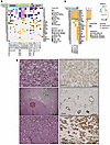
Metastases in renal cell carcinoma (RCC) typically arise from large primary tumors. However, a subset of patients with small renal masses (SRMs; ≤4 cm) can develop metastatic disease. Identifying these tumors is clinically important, as many SRMs are managed with active surveillance, and their study may provide insight into the early acquisition of metastatic competence. It remains unclear whether these tumors acquire distinct metastatic programs or instead show premature activation of the same aggressive programs typically associated with larger tumors. Here, we performed integrated morphological and molecular profiling of a multiinstitutional cohort of metastatic SRMs, including whole-exome sequencing and RNA-Seq, using nonmetastatic primary tumors as controls. Among metastatic, non–clear cell SRMs, we identified NF2-altered tumors, ELOC-mutated RCC, and an mTOR-driven eosinophilic vacuolated tumor. Metastatic clear cell SRMs were enriched by multi-hit aggressive genotypes, including recurrent losses of chromosomes 8p, 9, and 14q, as well as co-occurring driver alterations (≥2 events), including BAP1 and mTOR pathway genes. Transcriptomic analyses revealed enrichment of the non-negative matrix factorization 3 (NMF3) subtype from the IMmotion151 trial-based taxonomy, along with metabolic rewiring and reduced cytotoxic immune effector function. Collectively, these findings identify molecular programs associated with metastatic competence in SRMs, highlight the importance of genomic studies of equivocal non-clear cell SRMs, and provide a biological framework for risk stratification in patients often considered for active surveillance.
Authors
Payal Kapur, Daria Beshnova, Hua Zhong, Ruby Sharma, Pooja Ghatalia, Angela Yoo, Daniel D. Le, Ratna Mukhopadhyay, Alana Christie, Jeffrey Miyata, Shuanzeng Wei, Rana R. McKay, Dinesh Rakheja, Satwik Rajaram, Robert G. Uzzo, A. Ari Hakimi, Zora Modrusan, James Brugarolas
Abstract
Tregs play an essential role in immune tolerance, and Treg-promoting therapies are in development for the treatment of many inflammatory disorders. Interleukin-2 (IL-2)-based therapies increase Treg frequency, but little is known about impacts on Treg heterogeneity and function. We extended analyses of an IL-2 mutein (MK-6194) single–ascending-dose trial in healthy human participants by comprehensively defining Treg subsets and gene expression changes in vitro and in vivo. We found highly specific and dose-dependent activation and expansion of Tregs in clinical and pre-clinical studies. Following a single subcutaneous dose in humans, thymic-derived Tregs were selectively activated and expanded, while peripherally induced Tregs were unaffected. Expanded Tregs had increased expression of genes and proteins consistent with activation, suppressor function, and homing to non-lymphoid tissue, as well as increased transendocytosis activity, as measured by CTLA-4–dependent capture of CD80 and CD86 from non-Tregs. These results shed light onto underlying mechanisms by which Treg-targeted therapy may promote immune tolerance.
Authors
Laura A. Cooney, Mitch Fahning, Liliane Khoryati, Anna Kus, Sheila Scheiding, Lori Blanchfield, Matthew Lawrance, Basilin Benson, Kristina M. Harris, Gretchen A. Baltus, Shiuli Agarwal, Richard Wnek, Johannes F. Scheid, Kiki Cunningham-Bussel, Nancy D. Kim, S. Aubrey Stoch, Jyothsna Visweswaraiah, Nathan Higginson-Scott, Katalin Kis-Toth, Joanne L. Viney, Kevin L. Otipoby, Erik Sampson, Bridget Larkin, Daniel J. Campbell, S. Alice Long

Abstract
BACKGROUND. Loss of the Y chromosome (LOY) is a frequent event in male tumors and has been linked to cancer progression. However, the degree of mosaic LOY (mLOY) within normal tissues from men with or without cancer remains uncharacterized. METHODS. Here we used a FISH-based assay targeting X- and Y-chromosome centromeres to perform a pan-organ analysis of mLOY in 1,000 male tissue samples from 405 individuals representing 11 organs. Automated image processing generated a quantitative FISH-based mLOY score (YchrFISH) that we validated against a transcriptomic surrogate of Y-chromosome dosage from RNA-seq data. RESULTS. mLOY burden varied by tumor type, with highest degree in colorectal carcinoma. Across tissue groups, YchrFISH scores declined progressively from normal tissues of cancer-free men to histologically normal tissues adjacent to cancer and carcinoma (P < 0.0001). Paired analyses confirmed consistently greater mLOY in malignant compared with tumor-adjacent histologically normal tissue in different organs. Spatially resolved RNA-seq maps of bladders removed for cancer demonstrated a transcriptional gradient of Y-chromosome loss from normal urothelium through intraepithelial neoplasia to invasive carcinoma. CONCLUSION. mLOY gradients exist across histologically normal and malignant tissues, consistent with the concept of field cancerization. Our findings support epithelial mLOY as a biomarker of early malignant transformation and, to our knowledge, a previously unrecognized hallmark of male oncogenesis. FUNDING. NIH grants R35CA294022, P01CA163227, and P50CA97186 (the Pacific Northwest Prostate Cancer SPORE) and the Institute for Prostate Cancer Research.
Authors
Arkadiusz Gertych, Huihui Ye, Xingyu Chen, Eric Vail, V. Krishnan Ramanujan, Lauren Brady, Lawrence D. True, Peter S. Nelson, Peter R. Carroll, Dan Theodorescu
Abstract
Metastatic prostate cancer is a clinically and molecularly heterogeneous disease. Under the selective pressure of androgen receptor (AR)–directed therapies, resistant phenotypes frequently emerge, posing significant diagnostic and therapeutic challenges. Neuroendocrine prostate cancer (NEPC) is a clinically important phenotype characterized by lineage plasticity, neuroendocrine features, visceral metastases and poor prognosis. Accurately diagnosing NEPC remains difficult due to its histologic and molecular complexity but has high clinical relevance. In this study, we developed a deep learning model that leverages interpretable cellular features to improve feature extraction from H&E-stained tissue sections (NEURAL-PC). By incorporating a multiple instance learning (MIL) framework, NEURAL-PC enables robust NEPC classification solely from H&E tumor images, achieving an area under the receiver operating characteristic curve (AUROC) of 0.921 in independent external validation. In addition to its diagnostic utility, NEURAL-PC provides prognostic information that enables further subclassification of advanced prostate cancer across diverse datasets supporting its strong prognostic value and generalizability. Broadly, our work highlights a hybrid approach that integrates features across different domains, offering a promising strategy for developing reliable deep learning tools in pathology. Built on this framework, NEURAL-PC represents an extensively validated diagnostic and prognostic model for advanced prostate cancer.
Authors
Zhijun Chen, Erolcan Sayar, Daniela Guevara, Helen Richards, Haoyue Zhang, Radhika A. Patel, Agnes C. Gawne, Lucas J. Liu, Ilsa Coleman, Ruth Dumpit, Colm Morrissey, Michael T. Schweizer, Ruben Raychaudhuri, Laura S. Graham, Evan Y. Yu, Heather H. Cheng, Chien-Kuang C. Ding, Yuzhuo Wang, Peter Choyke, Baris Turkbey, Chantal Chanel-Vos, Christina Fedorov, John R. Otilano III, Troy Kane, Jyothi Manohar, Michael Sigouros, Jones T. Nauseef, Ana Molina, David Nanus, Scott T. Tagawa, Juan Miguel Mosquera, Himisha P. Beltran, Ruth Etzioni, Peter S. Nelson, Rama Soundararajan, Ana M. Aparicio, Cora N. Sternberg, Michael C. Haffner, Stephanie A. Harmon
Abstract
Autoimmune Addison’s disease (AD) is a rare but life-threatening disorder caused by immune-mediated destruction of the adrenal cortex, and progress in therapy has been limited by insufficient mechanistic insight. Here, we establish a model of Experimental Autoimmune Adrenalitis (EAA) that recapitulates key features of AD and reveals sex-dependent differences in disease manifestation within the model. Immunization with peptides derived from the adrenal self-antigen CYP11A1 induces corticosterone insufficiency. We show that autoimmune adrenalitis is driven by IFNG produced by self-reactive CD4+ T cells, promoting granulomatous inflammation in the adrenal cortex. Together, these findings identify IFNG as a central effector of autoimmune adrenalitis and suggest that targeting the IFNG pathway may represent a potential therapeutic strategy for AD.
Authors
Arina Andreyeva, Juraj Michalik, Veronika Niederlova, Veronika Cimermanova, Ales Drobek, Radislav Sedlacek, Jan Prochazka, Juraj Labaj, Olha Fedosieieva, Waldemar Kanczkowski, Peter Draber, André Sulen, Ondrej Stepanek, Aleš Neuwirth
No posts were found with this tag.


