
Research ArticleClinical ResearchImmunology
Open Access |  10.1172/jci.insight.193744
IL-2 mutein selectively expands thymic memory Tregs with increased transendocytosis in healthy participants
Laura A. Cooney,1 Mitch Fahning,2 Liliane Khoryati,2 Anna Kus,2 Sheila Scheiding,2 Lori Blanchfield,2 Matt Lawrance,2 Basilin Benson,2 Kristina M. Harris,1 Gretchen A. Baltus,3 Shiuli Agarwal,3 Richard Wnek,3 Johannes F. Scheid,3 Kiki Cunningham-Bussel,3 Nancy D. Kim,3 S. Aubrey Stoch,3 Jyothsna Visweswaraiah,4 Nathan Higginson-Scott,4 Katalin Kis-Toth,4 Joanne L. Viney,4 Kevin L. Otipoby,4 Erik Sampson,4 Bridget Larkin,4 Daniel J. Campbell,2,5 and S. Alice Long2
1Immune Tolerance Network, University of Michigan, Ann Arbor, Michigan, USA.
2Benaroya Research Institute at Virginia Mason, Seattle, Washington, USA.
3Merck & Co., Inc., Rahway, New Jersey, USA.
4Pandion Therapeutics, Inc., a subsidiary of Merck & Co., Inc., Rahway, New Jersey, USA.
5Department of Immunology, University of Washington School of Medicine, Seattle, Washington, USA.
Address correspondence to: Laura Cooney, NCRC 25-1622, 2800 Plymouth Road, Ann Arbor, Michigan, 48109, USA. Phone: 734.274.1149; Email: lcooney@immunetolerance.org. Or to: Daniel Campbell or S. Alice Long, 1201 9th Ave., Seattle, Washington, 98101, USA. Phone: 206.342.6500; Email: dcampbell@benaroyaresearch.org (DJC). Phone:.206.287.1034; Email: along@benaroyaresearch.org (SAL). LK’s present address is: Centre International de Recherche en Infectiologie, Lyon, France. KMH’s present address is: Pancreatic Cancer Action Network, Manhattan Beach, California, USA. JV, JLV, and KLO’s present address is: Seismic Therapeutic, Watertown, Massachusetts, USA. KKT’s present address is: NextPoint Therapeutics, Cambridge, Massachusetts, USA. ES’s present address is: PharmaEssentia Innovation Research Center, Bedford, Massachusetts, USA.
Find articles by Cooney, L. in: PubMed | Google Scholar
1Immune Tolerance Network, University of Michigan, Ann Arbor, Michigan, USA.
2Benaroya Research Institute at Virginia Mason, Seattle, Washington, USA.
3Merck & Co., Inc., Rahway, New Jersey, USA.
4Pandion Therapeutics, Inc., a subsidiary of Merck & Co., Inc., Rahway, New Jersey, USA.
5Department of Immunology, University of Washington School of Medicine, Seattle, Washington, USA.
Address correspondence to: Laura Cooney, NCRC 25-1622, 2800 Plymouth Road, Ann Arbor, Michigan, 48109, USA. Phone: 734.274.1149; Email: lcooney@immunetolerance.org. Or to: Daniel Campbell or S. Alice Long, 1201 9th Ave., Seattle, Washington, 98101, USA. Phone: 206.342.6500; Email: dcampbell@benaroyaresearch.org (DJC). Phone:.206.287.1034; Email: along@benaroyaresearch.org (SAL). LK’s present address is: Centre International de Recherche en Infectiologie, Lyon, France. KMH’s present address is: Pancreatic Cancer Action Network, Manhattan Beach, California, USA. JV, JLV, and KLO’s present address is: Seismic Therapeutic, Watertown, Massachusetts, USA. KKT’s present address is: NextPoint Therapeutics, Cambridge, Massachusetts, USA. ES’s present address is: PharmaEssentia Innovation Research Center, Bedford, Massachusetts, USA.
Find articles by Fahning, M. in: PubMed | Google Scholar
1Immune Tolerance Network, University of Michigan, Ann Arbor, Michigan, USA.
2Benaroya Research Institute at Virginia Mason, Seattle, Washington, USA.
3Merck & Co., Inc., Rahway, New Jersey, USA.
4Pandion Therapeutics, Inc., a subsidiary of Merck & Co., Inc., Rahway, New Jersey, USA.
5Department of Immunology, University of Washington School of Medicine, Seattle, Washington, USA.
Address correspondence to: Laura Cooney, NCRC 25-1622, 2800 Plymouth Road, Ann Arbor, Michigan, 48109, USA. Phone: 734.274.1149; Email: lcooney@immunetolerance.org. Or to: Daniel Campbell or S. Alice Long, 1201 9th Ave., Seattle, Washington, 98101, USA. Phone: 206.342.6500; Email: dcampbell@benaroyaresearch.org (DJC). Phone:.206.287.1034; Email: along@benaroyaresearch.org (SAL). LK’s present address is: Centre International de Recherche en Infectiologie, Lyon, France. KMH’s present address is: Pancreatic Cancer Action Network, Manhattan Beach, California, USA. JV, JLV, and KLO’s present address is: Seismic Therapeutic, Watertown, Massachusetts, USA. KKT’s present address is: NextPoint Therapeutics, Cambridge, Massachusetts, USA. ES’s present address is: PharmaEssentia Innovation Research Center, Bedford, Massachusetts, USA.
Find articles by Khoryati, L. in: PubMed | Google Scholar
1Immune Tolerance Network, University of Michigan, Ann Arbor, Michigan, USA.
2Benaroya Research Institute at Virginia Mason, Seattle, Washington, USA.
3Merck & Co., Inc., Rahway, New Jersey, USA.
4Pandion Therapeutics, Inc., a subsidiary of Merck & Co., Inc., Rahway, New Jersey, USA.
5Department of Immunology, University of Washington School of Medicine, Seattle, Washington, USA.
Address correspondence to: Laura Cooney, NCRC 25-1622, 2800 Plymouth Road, Ann Arbor, Michigan, 48109, USA. Phone: 734.274.1149; Email: lcooney@immunetolerance.org. Or to: Daniel Campbell or S. Alice Long, 1201 9th Ave., Seattle, Washington, 98101, USA. Phone: 206.342.6500; Email: dcampbell@benaroyaresearch.org (DJC). Phone:.206.287.1034; Email: along@benaroyaresearch.org (SAL). LK’s present address is: Centre International de Recherche en Infectiologie, Lyon, France. KMH’s present address is: Pancreatic Cancer Action Network, Manhattan Beach, California, USA. JV, JLV, and KLO’s present address is: Seismic Therapeutic, Watertown, Massachusetts, USA. KKT’s present address is: NextPoint Therapeutics, Cambridge, Massachusetts, USA. ES’s present address is: PharmaEssentia Innovation Research Center, Bedford, Massachusetts, USA.
Find articles by Kus, A. in: PubMed | Google Scholar
1Immune Tolerance Network, University of Michigan, Ann Arbor, Michigan, USA.
2Benaroya Research Institute at Virginia Mason, Seattle, Washington, USA.
3Merck & Co., Inc., Rahway, New Jersey, USA.
4Pandion Therapeutics, Inc., a subsidiary of Merck & Co., Inc., Rahway, New Jersey, USA.
5Department of Immunology, University of Washington School of Medicine, Seattle, Washington, USA.
Address correspondence to: Laura Cooney, NCRC 25-1622, 2800 Plymouth Road, Ann Arbor, Michigan, 48109, USA. Phone: 734.274.1149; Email: lcooney@immunetolerance.org. Or to: Daniel Campbell or S. Alice Long, 1201 9th Ave., Seattle, Washington, 98101, USA. Phone: 206.342.6500; Email: dcampbell@benaroyaresearch.org (DJC). Phone:.206.287.1034; Email: along@benaroyaresearch.org (SAL). LK’s present address is: Centre International de Recherche en Infectiologie, Lyon, France. KMH’s present address is: Pancreatic Cancer Action Network, Manhattan Beach, California, USA. JV, JLV, and KLO’s present address is: Seismic Therapeutic, Watertown, Massachusetts, USA. KKT’s present address is: NextPoint Therapeutics, Cambridge, Massachusetts, USA. ES’s present address is: PharmaEssentia Innovation Research Center, Bedford, Massachusetts, USA.
Find articles by
Scheiding, S.
in:
PubMed
|
Google Scholar
|

1Immune Tolerance Network, University of Michigan, Ann Arbor, Michigan, USA.
2Benaroya Research Institute at Virginia Mason, Seattle, Washington, USA.
3Merck & Co., Inc., Rahway, New Jersey, USA.
4Pandion Therapeutics, Inc., a subsidiary of Merck & Co., Inc., Rahway, New Jersey, USA.
5Department of Immunology, University of Washington School of Medicine, Seattle, Washington, USA.
Address correspondence to: Laura Cooney, NCRC 25-1622, 2800 Plymouth Road, Ann Arbor, Michigan, 48109, USA. Phone: 734.274.1149; Email: lcooney@immunetolerance.org. Or to: Daniel Campbell or S. Alice Long, 1201 9th Ave., Seattle, Washington, 98101, USA. Phone: 206.342.6500; Email: dcampbell@benaroyaresearch.org (DJC). Phone:.206.287.1034; Email: along@benaroyaresearch.org (SAL). LK’s present address is: Centre International de Recherche en Infectiologie, Lyon, France. KMH’s present address is: Pancreatic Cancer Action Network, Manhattan Beach, California, USA. JV, JLV, and KLO’s present address is: Seismic Therapeutic, Watertown, Massachusetts, USA. KKT’s present address is: NextPoint Therapeutics, Cambridge, Massachusetts, USA. ES’s present address is: PharmaEssentia Innovation Research Center, Bedford, Massachusetts, USA.
Find articles by Blanchfield, L. in: PubMed | Google Scholar
1Immune Tolerance Network, University of Michigan, Ann Arbor, Michigan, USA.
2Benaroya Research Institute at Virginia Mason, Seattle, Washington, USA.
3Merck & Co., Inc., Rahway, New Jersey, USA.
4Pandion Therapeutics, Inc., a subsidiary of Merck & Co., Inc., Rahway, New Jersey, USA.
5Department of Immunology, University of Washington School of Medicine, Seattle, Washington, USA.
Address correspondence to: Laura Cooney, NCRC 25-1622, 2800 Plymouth Road, Ann Arbor, Michigan, 48109, USA. Phone: 734.274.1149; Email: lcooney@immunetolerance.org. Or to: Daniel Campbell or S. Alice Long, 1201 9th Ave., Seattle, Washington, 98101, USA. Phone: 206.342.6500; Email: dcampbell@benaroyaresearch.org (DJC). Phone:.206.287.1034; Email: along@benaroyaresearch.org (SAL). LK’s present address is: Centre International de Recherche en Infectiologie, Lyon, France. KMH’s present address is: Pancreatic Cancer Action Network, Manhattan Beach, California, USA. JV, JLV, and KLO’s present address is: Seismic Therapeutic, Watertown, Massachusetts, USA. KKT’s present address is: NextPoint Therapeutics, Cambridge, Massachusetts, USA. ES’s present address is: PharmaEssentia Innovation Research Center, Bedford, Massachusetts, USA.
Find articles by Lawrance, M. in: PubMed | Google Scholar
1Immune Tolerance Network, University of Michigan, Ann Arbor, Michigan, USA.
2Benaroya Research Institute at Virginia Mason, Seattle, Washington, USA.
3Merck & Co., Inc., Rahway, New Jersey, USA.
4Pandion Therapeutics, Inc., a subsidiary of Merck & Co., Inc., Rahway, New Jersey, USA.
5Department of Immunology, University of Washington School of Medicine, Seattle, Washington, USA.
Address correspondence to: Laura Cooney, NCRC 25-1622, 2800 Plymouth Road, Ann Arbor, Michigan, 48109, USA. Phone: 734.274.1149; Email: lcooney@immunetolerance.org. Or to: Daniel Campbell or S. Alice Long, 1201 9th Ave., Seattle, Washington, 98101, USA. Phone: 206.342.6500; Email: dcampbell@benaroyaresearch.org (DJC). Phone:.206.287.1034; Email: along@benaroyaresearch.org (SAL). LK’s present address is: Centre International de Recherche en Infectiologie, Lyon, France. KMH’s present address is: Pancreatic Cancer Action Network, Manhattan Beach, California, USA. JV, JLV, and KLO’s present address is: Seismic Therapeutic, Watertown, Massachusetts, USA. KKT’s present address is: NextPoint Therapeutics, Cambridge, Massachusetts, USA. ES’s present address is: PharmaEssentia Innovation Research Center, Bedford, Massachusetts, USA.
Find articles by Benson, B. in: PubMed | Google Scholar
1Immune Tolerance Network, University of Michigan, Ann Arbor, Michigan, USA.
2Benaroya Research Institute at Virginia Mason, Seattle, Washington, USA.
3Merck & Co., Inc., Rahway, New Jersey, USA.
4Pandion Therapeutics, Inc., a subsidiary of Merck & Co., Inc., Rahway, New Jersey, USA.
5Department of Immunology, University of Washington School of Medicine, Seattle, Washington, USA.
Address correspondence to: Laura Cooney, NCRC 25-1622, 2800 Plymouth Road, Ann Arbor, Michigan, 48109, USA. Phone: 734.274.1149; Email: lcooney@immunetolerance.org. Or to: Daniel Campbell or S. Alice Long, 1201 9th Ave., Seattle, Washington, 98101, USA. Phone: 206.342.6500; Email: dcampbell@benaroyaresearch.org (DJC). Phone:.206.287.1034; Email: along@benaroyaresearch.org (SAL). LK’s present address is: Centre International de Recherche en Infectiologie, Lyon, France. KMH’s present address is: Pancreatic Cancer Action Network, Manhattan Beach, California, USA. JV, JLV, and KLO’s present address is: Seismic Therapeutic, Watertown, Massachusetts, USA. KKT’s present address is: NextPoint Therapeutics, Cambridge, Massachusetts, USA. ES’s present address is: PharmaEssentia Innovation Research Center, Bedford, Massachusetts, USA.
Find articles by
Harris, K.
in:
PubMed
|
Google Scholar
|
1Immune Tolerance Network, University of Michigan, Ann Arbor, Michigan, USA.
2Benaroya Research Institute at Virginia Mason, Seattle, Washington, USA.
3Merck & Co., Inc., Rahway, New Jersey, USA.
4Pandion Therapeutics, Inc., a subsidiary of Merck & Co., Inc., Rahway, New Jersey, USA.
5Department of Immunology, University of Washington School of Medicine, Seattle, Washington, USA.
Address correspondence to: Laura Cooney, NCRC 25-1622, 2800 Plymouth Road, Ann Arbor, Michigan, 48109, USA. Phone: 734.274.1149; Email: lcooney@immunetolerance.org. Or to: Daniel Campbell or S. Alice Long, 1201 9th Ave., Seattle, Washington, 98101, USA. Phone: 206.342.6500; Email: dcampbell@benaroyaresearch.org (DJC). Phone:.206.287.1034; Email: along@benaroyaresearch.org (SAL). LK’s present address is: Centre International de Recherche en Infectiologie, Lyon, France. KMH’s present address is: Pancreatic Cancer Action Network, Manhattan Beach, California, USA. JV, JLV, and KLO’s present address is: Seismic Therapeutic, Watertown, Massachusetts, USA. KKT’s present address is: NextPoint Therapeutics, Cambridge, Massachusetts, USA. ES’s present address is: PharmaEssentia Innovation Research Center, Bedford, Massachusetts, USA.
Find articles by Baltus, G. in: PubMed | Google Scholar
1Immune Tolerance Network, University of Michigan, Ann Arbor, Michigan, USA.
2Benaroya Research Institute at Virginia Mason, Seattle, Washington, USA.
3Merck & Co., Inc., Rahway, New Jersey, USA.
4Pandion Therapeutics, Inc., a subsidiary of Merck & Co., Inc., Rahway, New Jersey, USA.
5Department of Immunology, University of Washington School of Medicine, Seattle, Washington, USA.
Address correspondence to: Laura Cooney, NCRC 25-1622, 2800 Plymouth Road, Ann Arbor, Michigan, 48109, USA. Phone: 734.274.1149; Email: lcooney@immunetolerance.org. Or to: Daniel Campbell or S. Alice Long, 1201 9th Ave., Seattle, Washington, 98101, USA. Phone: 206.342.6500; Email: dcampbell@benaroyaresearch.org (DJC). Phone:.206.287.1034; Email: along@benaroyaresearch.org (SAL). LK’s present address is: Centre International de Recherche en Infectiologie, Lyon, France. KMH’s present address is: Pancreatic Cancer Action Network, Manhattan Beach, California, USA. JV, JLV, and KLO’s present address is: Seismic Therapeutic, Watertown, Massachusetts, USA. KKT’s present address is: NextPoint Therapeutics, Cambridge, Massachusetts, USA. ES’s present address is: PharmaEssentia Innovation Research Center, Bedford, Massachusetts, USA.
Find articles by Agarwal, S. in: PubMed | Google Scholar
1Immune Tolerance Network, University of Michigan, Ann Arbor, Michigan, USA.
2Benaroya Research Institute at Virginia Mason, Seattle, Washington, USA.
3Merck & Co., Inc., Rahway, New Jersey, USA.
4Pandion Therapeutics, Inc., a subsidiary of Merck & Co., Inc., Rahway, New Jersey, USA.
5Department of Immunology, University of Washington School of Medicine, Seattle, Washington, USA.
Address correspondence to: Laura Cooney, NCRC 25-1622, 2800 Plymouth Road, Ann Arbor, Michigan, 48109, USA. Phone: 734.274.1149; Email: lcooney@immunetolerance.org. Or to: Daniel Campbell or S. Alice Long, 1201 9th Ave., Seattle, Washington, 98101, USA. Phone: 206.342.6500; Email: dcampbell@benaroyaresearch.org (DJC). Phone:.206.287.1034; Email: along@benaroyaresearch.org (SAL). LK’s present address is: Centre International de Recherche en Infectiologie, Lyon, France. KMH’s present address is: Pancreatic Cancer Action Network, Manhattan Beach, California, USA. JV, JLV, and KLO’s present address is: Seismic Therapeutic, Watertown, Massachusetts, USA. KKT’s present address is: NextPoint Therapeutics, Cambridge, Massachusetts, USA. ES’s present address is: PharmaEssentia Innovation Research Center, Bedford, Massachusetts, USA.
Find articles by Wnek, R. in: PubMed | Google Scholar
1Immune Tolerance Network, University of Michigan, Ann Arbor, Michigan, USA.
2Benaroya Research Institute at Virginia Mason, Seattle, Washington, USA.
3Merck & Co., Inc., Rahway, New Jersey, USA.
4Pandion Therapeutics, Inc., a subsidiary of Merck & Co., Inc., Rahway, New Jersey, USA.
5Department of Immunology, University of Washington School of Medicine, Seattle, Washington, USA.
Address correspondence to: Laura Cooney, NCRC 25-1622, 2800 Plymouth Road, Ann Arbor, Michigan, 48109, USA. Phone: 734.274.1149; Email: lcooney@immunetolerance.org. Or to: Daniel Campbell or S. Alice Long, 1201 9th Ave., Seattle, Washington, 98101, USA. Phone: 206.342.6500; Email: dcampbell@benaroyaresearch.org (DJC). Phone:.206.287.1034; Email: along@benaroyaresearch.org (SAL). LK’s present address is: Centre International de Recherche en Infectiologie, Lyon, France. KMH’s present address is: Pancreatic Cancer Action Network, Manhattan Beach, California, USA. JV, JLV, and KLO’s present address is: Seismic Therapeutic, Watertown, Massachusetts, USA. KKT’s present address is: NextPoint Therapeutics, Cambridge, Massachusetts, USA. ES’s present address is: PharmaEssentia Innovation Research Center, Bedford, Massachusetts, USA.
Find articles by Scheid, J. in: PubMed | Google Scholar
1Immune Tolerance Network, University of Michigan, Ann Arbor, Michigan, USA.
2Benaroya Research Institute at Virginia Mason, Seattle, Washington, USA.
3Merck & Co., Inc., Rahway, New Jersey, USA.
4Pandion Therapeutics, Inc., a subsidiary of Merck & Co., Inc., Rahway, New Jersey, USA.
5Department of Immunology, University of Washington School of Medicine, Seattle, Washington, USA.
Address correspondence to: Laura Cooney, NCRC 25-1622, 2800 Plymouth Road, Ann Arbor, Michigan, 48109, USA. Phone: 734.274.1149; Email: lcooney@immunetolerance.org. Or to: Daniel Campbell or S. Alice Long, 1201 9th Ave., Seattle, Washington, 98101, USA. Phone: 206.342.6500; Email: dcampbell@benaroyaresearch.org (DJC). Phone:.206.287.1034; Email: along@benaroyaresearch.org (SAL). LK’s present address is: Centre International de Recherche en Infectiologie, Lyon, France. KMH’s present address is: Pancreatic Cancer Action Network, Manhattan Beach, California, USA. JV, JLV, and KLO’s present address is: Seismic Therapeutic, Watertown, Massachusetts, USA. KKT’s present address is: NextPoint Therapeutics, Cambridge, Massachusetts, USA. ES’s present address is: PharmaEssentia Innovation Research Center, Bedford, Massachusetts, USA.
Find articles by Cunningham-Bussel, K. in: PubMed | Google Scholar
1Immune Tolerance Network, University of Michigan, Ann Arbor, Michigan, USA.
2Benaroya Research Institute at Virginia Mason, Seattle, Washington, USA.
3Merck & Co., Inc., Rahway, New Jersey, USA.
4Pandion Therapeutics, Inc., a subsidiary of Merck & Co., Inc., Rahway, New Jersey, USA.
5Department of Immunology, University of Washington School of Medicine, Seattle, Washington, USA.
Address correspondence to: Laura Cooney, NCRC 25-1622, 2800 Plymouth Road, Ann Arbor, Michigan, 48109, USA. Phone: 734.274.1149; Email: lcooney@immunetolerance.org. Or to: Daniel Campbell or S. Alice Long, 1201 9th Ave., Seattle, Washington, 98101, USA. Phone: 206.342.6500; Email: dcampbell@benaroyaresearch.org (DJC). Phone:.206.287.1034; Email: along@benaroyaresearch.org (SAL). LK’s present address is: Centre International de Recherche en Infectiologie, Lyon, France. KMH’s present address is: Pancreatic Cancer Action Network, Manhattan Beach, California, USA. JV, JLV, and KLO’s present address is: Seismic Therapeutic, Watertown, Massachusetts, USA. KKT’s present address is: NextPoint Therapeutics, Cambridge, Massachusetts, USA. ES’s present address is: PharmaEssentia Innovation Research Center, Bedford, Massachusetts, USA.
Find articles by Kim, N. in: PubMed | Google Scholar
1Immune Tolerance Network, University of Michigan, Ann Arbor, Michigan, USA.
2Benaroya Research Institute at Virginia Mason, Seattle, Washington, USA.
3Merck & Co., Inc., Rahway, New Jersey, USA.
4Pandion Therapeutics, Inc., a subsidiary of Merck & Co., Inc., Rahway, New Jersey, USA.
5Department of Immunology, University of Washington School of Medicine, Seattle, Washington, USA.
Address correspondence to: Laura Cooney, NCRC 25-1622, 2800 Plymouth Road, Ann Arbor, Michigan, 48109, USA. Phone: 734.274.1149; Email: lcooney@immunetolerance.org. Or to: Daniel Campbell or S. Alice Long, 1201 9th Ave., Seattle, Washington, 98101, USA. Phone: 206.342.6500; Email: dcampbell@benaroyaresearch.org (DJC). Phone:.206.287.1034; Email: along@benaroyaresearch.org (SAL). LK’s present address is: Centre International de Recherche en Infectiologie, Lyon, France. KMH’s present address is: Pancreatic Cancer Action Network, Manhattan Beach, California, USA. JV, JLV, and KLO’s present address is: Seismic Therapeutic, Watertown, Massachusetts, USA. KKT’s present address is: NextPoint Therapeutics, Cambridge, Massachusetts, USA. ES’s present address is: PharmaEssentia Innovation Research Center, Bedford, Massachusetts, USA.
Find articles by Stoch, S. in: PubMed | Google Scholar
1Immune Tolerance Network, University of Michigan, Ann Arbor, Michigan, USA.
2Benaroya Research Institute at Virginia Mason, Seattle, Washington, USA.
3Merck & Co., Inc., Rahway, New Jersey, USA.
4Pandion Therapeutics, Inc., a subsidiary of Merck & Co., Inc., Rahway, New Jersey, USA.
5Department of Immunology, University of Washington School of Medicine, Seattle, Washington, USA.
Address correspondence to: Laura Cooney, NCRC 25-1622, 2800 Plymouth Road, Ann Arbor, Michigan, 48109, USA. Phone: 734.274.1149; Email: lcooney@immunetolerance.org. Or to: Daniel Campbell or S. Alice Long, 1201 9th Ave., Seattle, Washington, 98101, USA. Phone: 206.342.6500; Email: dcampbell@benaroyaresearch.org (DJC). Phone:.206.287.1034; Email: along@benaroyaresearch.org (SAL). LK’s present address is: Centre International de Recherche en Infectiologie, Lyon, France. KMH’s present address is: Pancreatic Cancer Action Network, Manhattan Beach, California, USA. JV, JLV, and KLO’s present address is: Seismic Therapeutic, Watertown, Massachusetts, USA. KKT’s present address is: NextPoint Therapeutics, Cambridge, Massachusetts, USA. ES’s present address is: PharmaEssentia Innovation Research Center, Bedford, Massachusetts, USA.
Find articles by Visweswaraiah, J. in: PubMed | Google Scholar
1Immune Tolerance Network, University of Michigan, Ann Arbor, Michigan, USA.
2Benaroya Research Institute at Virginia Mason, Seattle, Washington, USA.
3Merck & Co., Inc., Rahway, New Jersey, USA.
4Pandion Therapeutics, Inc., a subsidiary of Merck & Co., Inc., Rahway, New Jersey, USA.
5Department of Immunology, University of Washington School of Medicine, Seattle, Washington, USA.
Address correspondence to: Laura Cooney, NCRC 25-1622, 2800 Plymouth Road, Ann Arbor, Michigan, 48109, USA. Phone: 734.274.1149; Email: lcooney@immunetolerance.org. Or to: Daniel Campbell or S. Alice Long, 1201 9th Ave., Seattle, Washington, 98101, USA. Phone: 206.342.6500; Email: dcampbell@benaroyaresearch.org (DJC). Phone:.206.287.1034; Email: along@benaroyaresearch.org (SAL). LK’s present address is: Centre International de Recherche en Infectiologie, Lyon, France. KMH’s present address is: Pancreatic Cancer Action Network, Manhattan Beach, California, USA. JV, JLV, and KLO’s present address is: Seismic Therapeutic, Watertown, Massachusetts, USA. KKT’s present address is: NextPoint Therapeutics, Cambridge, Massachusetts, USA. ES’s present address is: PharmaEssentia Innovation Research Center, Bedford, Massachusetts, USA.
Find articles by Higginson-Scott, N. in: PubMed | Google Scholar
1Immune Tolerance Network, University of Michigan, Ann Arbor, Michigan, USA.
2Benaroya Research Institute at Virginia Mason, Seattle, Washington, USA.
3Merck & Co., Inc., Rahway, New Jersey, USA.
4Pandion Therapeutics, Inc., a subsidiary of Merck & Co., Inc., Rahway, New Jersey, USA.
5Department of Immunology, University of Washington School of Medicine, Seattle, Washington, USA.
Address correspondence to: Laura Cooney, NCRC 25-1622, 2800 Plymouth Road, Ann Arbor, Michigan, 48109, USA. Phone: 734.274.1149; Email: lcooney@immunetolerance.org. Or to: Daniel Campbell or S. Alice Long, 1201 9th Ave., Seattle, Washington, 98101, USA. Phone: 206.342.6500; Email: dcampbell@benaroyaresearch.org (DJC). Phone:.206.287.1034; Email: along@benaroyaresearch.org (SAL). LK’s present address is: Centre International de Recherche en Infectiologie, Lyon, France. KMH’s present address is: Pancreatic Cancer Action Network, Manhattan Beach, California, USA. JV, JLV, and KLO’s present address is: Seismic Therapeutic, Watertown, Massachusetts, USA. KKT’s present address is: NextPoint Therapeutics, Cambridge, Massachusetts, USA. ES’s present address is: PharmaEssentia Innovation Research Center, Bedford, Massachusetts, USA.
Find articles by Kis-Toth, K. in: PubMed | Google Scholar
1Immune Tolerance Network, University of Michigan, Ann Arbor, Michigan, USA.
2Benaroya Research Institute at Virginia Mason, Seattle, Washington, USA.
3Merck & Co., Inc., Rahway, New Jersey, USA.
4Pandion Therapeutics, Inc., a subsidiary of Merck & Co., Inc., Rahway, New Jersey, USA.
5Department of Immunology, University of Washington School of Medicine, Seattle, Washington, USA.
Address correspondence to: Laura Cooney, NCRC 25-1622, 2800 Plymouth Road, Ann Arbor, Michigan, 48109, USA. Phone: 734.274.1149; Email: lcooney@immunetolerance.org. Or to: Daniel Campbell or S. Alice Long, 1201 9th Ave., Seattle, Washington, 98101, USA. Phone: 206.342.6500; Email: dcampbell@benaroyaresearch.org (DJC). Phone:.206.287.1034; Email: along@benaroyaresearch.org (SAL). LK’s present address is: Centre International de Recherche en Infectiologie, Lyon, France. KMH’s present address is: Pancreatic Cancer Action Network, Manhattan Beach, California, USA. JV, JLV, and KLO’s present address is: Seismic Therapeutic, Watertown, Massachusetts, USA. KKT’s present address is: NextPoint Therapeutics, Cambridge, Massachusetts, USA. ES’s present address is: PharmaEssentia Innovation Research Center, Bedford, Massachusetts, USA.
Find articles by Viney, J. in: PubMed | Google Scholar
1Immune Tolerance Network, University of Michigan, Ann Arbor, Michigan, USA.
2Benaroya Research Institute at Virginia Mason, Seattle, Washington, USA.
3Merck & Co., Inc., Rahway, New Jersey, USA.
4Pandion Therapeutics, Inc., a subsidiary of Merck & Co., Inc., Rahway, New Jersey, USA.
5Department of Immunology, University of Washington School of Medicine, Seattle, Washington, USA.
Address correspondence to: Laura Cooney, NCRC 25-1622, 2800 Plymouth Road, Ann Arbor, Michigan, 48109, USA. Phone: 734.274.1149; Email: lcooney@immunetolerance.org. Or to: Daniel Campbell or S. Alice Long, 1201 9th Ave., Seattle, Washington, 98101, USA. Phone: 206.342.6500; Email: dcampbell@benaroyaresearch.org (DJC). Phone:.206.287.1034; Email: along@benaroyaresearch.org (SAL). LK’s present address is: Centre International de Recherche en Infectiologie, Lyon, France. KMH’s present address is: Pancreatic Cancer Action Network, Manhattan Beach, California, USA. JV, JLV, and KLO’s present address is: Seismic Therapeutic, Watertown, Massachusetts, USA. KKT’s present address is: NextPoint Therapeutics, Cambridge, Massachusetts, USA. ES’s present address is: PharmaEssentia Innovation Research Center, Bedford, Massachusetts, USA.
Find articles by Otipoby, K. in: PubMed | Google Scholar
1Immune Tolerance Network, University of Michigan, Ann Arbor, Michigan, USA.
2Benaroya Research Institute at Virginia Mason, Seattle, Washington, USA.
3Merck & Co., Inc., Rahway, New Jersey, USA.
4Pandion Therapeutics, Inc., a subsidiary of Merck & Co., Inc., Rahway, New Jersey, USA.
5Department of Immunology, University of Washington School of Medicine, Seattle, Washington, USA.
Address correspondence to: Laura Cooney, NCRC 25-1622, 2800 Plymouth Road, Ann Arbor, Michigan, 48109, USA. Phone: 734.274.1149; Email: lcooney@immunetolerance.org. Or to: Daniel Campbell or S. Alice Long, 1201 9th Ave., Seattle, Washington, 98101, USA. Phone: 206.342.6500; Email: dcampbell@benaroyaresearch.org (DJC). Phone:.206.287.1034; Email: along@benaroyaresearch.org (SAL). LK’s present address is: Centre International de Recherche en Infectiologie, Lyon, France. KMH’s present address is: Pancreatic Cancer Action Network, Manhattan Beach, California, USA. JV, JLV, and KLO’s present address is: Seismic Therapeutic, Watertown, Massachusetts, USA. KKT’s present address is: NextPoint Therapeutics, Cambridge, Massachusetts, USA. ES’s present address is: PharmaEssentia Innovation Research Center, Bedford, Massachusetts, USA.
Find articles by Sampson, E. in: PubMed | Google Scholar
1Immune Tolerance Network, University of Michigan, Ann Arbor, Michigan, USA.
2Benaroya Research Institute at Virginia Mason, Seattle, Washington, USA.
3Merck & Co., Inc., Rahway, New Jersey, USA.
4Pandion Therapeutics, Inc., a subsidiary of Merck & Co., Inc., Rahway, New Jersey, USA.
5Department of Immunology, University of Washington School of Medicine, Seattle, Washington, USA.
Address correspondence to: Laura Cooney, NCRC 25-1622, 2800 Plymouth Road, Ann Arbor, Michigan, 48109, USA. Phone: 734.274.1149; Email: lcooney@immunetolerance.org. Or to: Daniel Campbell or S. Alice Long, 1201 9th Ave., Seattle, Washington, 98101, USA. Phone: 206.342.6500; Email: dcampbell@benaroyaresearch.org (DJC). Phone:.206.287.1034; Email: along@benaroyaresearch.org (SAL). LK’s present address is: Centre International de Recherche en Infectiologie, Lyon, France. KMH’s present address is: Pancreatic Cancer Action Network, Manhattan Beach, California, USA. JV, JLV, and KLO’s present address is: Seismic Therapeutic, Watertown, Massachusetts, USA. KKT’s present address is: NextPoint Therapeutics, Cambridge, Massachusetts, USA. ES’s present address is: PharmaEssentia Innovation Research Center, Bedford, Massachusetts, USA.
Find articles by Larkin, B. in: PubMed | Google Scholar
1Immune Tolerance Network, University of Michigan, Ann Arbor, Michigan, USA.
2Benaroya Research Institute at Virginia Mason, Seattle, Washington, USA.
3Merck & Co., Inc., Rahway, New Jersey, USA.
4Pandion Therapeutics, Inc., a subsidiary of Merck & Co., Inc., Rahway, New Jersey, USA.
5Department of Immunology, University of Washington School of Medicine, Seattle, Washington, USA.
Address correspondence to: Laura Cooney, NCRC 25-1622, 2800 Plymouth Road, Ann Arbor, Michigan, 48109, USA. Phone: 734.274.1149; Email: lcooney@immunetolerance.org. Or to: Daniel Campbell or S. Alice Long, 1201 9th Ave., Seattle, Washington, 98101, USA. Phone: 206.342.6500; Email: dcampbell@benaroyaresearch.org (DJC). Phone:.206.287.1034; Email: along@benaroyaresearch.org (SAL). LK’s present address is: Centre International de Recherche en Infectiologie, Lyon, France. KMH’s present address is: Pancreatic Cancer Action Network, Manhattan Beach, California, USA. JV, JLV, and KLO’s present address is: Seismic Therapeutic, Watertown, Massachusetts, USA. KKT’s present address is: NextPoint Therapeutics, Cambridge, Massachusetts, USA. ES’s present address is: PharmaEssentia Innovation Research Center, Bedford, Massachusetts, USA.
Find articles by
Campbell, D.
in:
PubMed
|
Google Scholar
|
1Immune Tolerance Network, University of Michigan, Ann Arbor, Michigan, USA.
2Benaroya Research Institute at Virginia Mason, Seattle, Washington, USA.
3Merck & Co., Inc., Rahway, New Jersey, USA.
4Pandion Therapeutics, Inc., a subsidiary of Merck & Co., Inc., Rahway, New Jersey, USA.
5Department of Immunology, University of Washington School of Medicine, Seattle, Washington, USA.
Address correspondence to: Laura Cooney, NCRC 25-1622, 2800 Plymouth Road, Ann Arbor, Michigan, 48109, USA. Phone: 734.274.1149; Email: lcooney@immunetolerance.org. Or to: Daniel Campbell or S. Alice Long, 1201 9th Ave., Seattle, Washington, 98101, USA. Phone: 206.342.6500; Email: dcampbell@benaroyaresearch.org (DJC). Phone:.206.287.1034; Email: along@benaroyaresearch.org (SAL). LK’s present address is: Centre International de Recherche en Infectiologie, Lyon, France. KMH’s present address is: Pancreatic Cancer Action Network, Manhattan Beach, California, USA. JV, JLV, and KLO’s present address is: Seismic Therapeutic, Watertown, Massachusetts, USA. KKT’s present address is: NextPoint Therapeutics, Cambridge, Massachusetts, USA. ES’s present address is: PharmaEssentia Innovation Research Center, Bedford, Massachusetts, USA.
Find articles by
Long, S.
in:
PubMed
|
Google Scholar
|
Published September 17, 2026 - More info
JCI Insight. 2026;11(20):e193744. https://doi.org/10.1172/jci.insight.193744.
© 2026 Cooney et al. This work is licensed under the Creative Commons Attribution 4.0 International License. To view a copy of this license, visit http://creativecommons.org/licenses/by/4.0/.
Received: May 22, 2025; Accepted: September 10, 2026

-
Results
Treg activation and expansion induced by IL-2 mutein is highly selective in preclinical models. To better define Treg expansion and MK-6194 specificity broadly defined in Scheid et al. (13), we first characterized MK-6194 selectivity in vitro by stimulating PBMCs from healthy donors, followed by flow cytometry for induction of STAT5 phosphorylation (pSTAT5). After stimulation with MK-6194 or recombinant wild-type IL-2 (0.25 pM to 20 nM) for 30 minutes, MK-6194 dose-dependently induced pSTAT5 in CD25hi Tregs but was approximately 10-fold less potent than wild-type recombinant IL-2 (Figure 1A). In contrast, MK-6194 induced little to no response in CD25loCD4+ Tconv cells or NK cells, whereas recombinant wild-type IL-2 induced pSTAT5 in all 3 cell types at higher doses (Figure 1A).
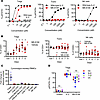 Figure 1
Selective and dose-dependent Treg expansion in vitro and in vivo following IL-2 mutein treatment. (A) Proportion of pSTAT5+ Tregs (CD3+CD4+FOXP3+CD25+CD127–), Tconv cells (CD3+CD4+FOXP3–), and NK cells (CD3–CD16+CD56+) following 30 minutes of stimulation with MK-6194 (red) or recombinant IL-2 (gray) (0.25 pM to 20 nM) using PBMCs isolated from healthy individuals. Data shown from 1 donor, representative of n = 5. (B) Frequency of Tregs (CD3+CD4+FOXP3+CD25+), Tconv (CD3+CD4+ FOXP3–), and NK cells (CD45+CD3–CD56+) from whole blood isolated on days 1, 3, 5, 6, 7, and 9 after treatment from NSG mice humanized with CD34+ hematopoietic stem cells. Mice were treated with 3 µg MK-6194 or vehicle (1-way ANOVA with Bonferroni’s multiple-comparison correction, *P < 0.05). Mean ± SD (n = 8). (C) Fold change in absolute number of PBMC subpopulations from cynomolgus monkeys 7 days after a single subcutaneous injection with 0.5 mg/kg MK-6194. Cell subsets were defined by flow cytometry as follows: Tregs (CD3+CD4+CD25+CD127–FOXP3+), naive CD4+ (CD3+CD4+CD28+CD95–), conventional CD4+ (CD3+CD4+CD127+CD25–), central memory (CD3+CD4+CD28+CD95+), effector memory (CD3+CD4+CD28–CD95+/–), CD8+ (CD3+CD8+), B cell (CD45+CD20+), and NK cell (CD45+CD159a+). Mean ± SD (n = 6). (D) Human PBMCs from healthy control donors (HC) or people with lupus (SLE), type 1 diabetes (T1D), or ulcerative colitis (UC) were stimulated in vitro with MK-6194 (90 nM, 7.6 ĩg/mL) or recombinant wild-type human IL-2 (5 ng/mL) for 30 minutes and pSTAT5 was assessed by flow cytometry. N = 5 per group. No significant differences between HC or autoimmune disease groups within a treatment condition by 2-way ANOVA. NS, not significant.
To explore the in vivo kinetics and selectivity of MK-6194, we used a NOD SCID gamma (NSG) mouse model reconstituted with human CD34+ hematopoietic stem cells (17). Reconstituted NSG mice received a single, subcutaneous dose of 3 μg MK-6194 (equivalent to approximately 0.15 mg/kg) or vehicle, and blood was collected on days 1, 3, 5, 6, 7, and 9 after treatment for immunophenotyping (Figure 1B). Consistent with human PBMCs, MK-6194 administration resulted in increased frequency of Tregs, peaking on day 7. In contrast, no significant changes in the frequencies of Tconv or NK cells were observed. A dose-response curve was also assessed in the humanized mouse model, comparing Treg expansion following a single dose of 0.04, 0.12, and 0.4 mg/kg of MK-6194 (Supplemental Figure 1; supplemental material available online with this article; https://doi.org/10.1172/jci.insight.193744DS1). Average Treg expansion increased linearly with increasing dose but was more heterogeneous at the highest dose. Furthermore, cynomolgus monkeys were administered a single subcutaneous dose of 0.5 mg/kg of MK-6194, and whole blood was collected on day 7 after treatment for immunophenotyping. Similarly to humanized mice, Tregs showed a significant expansion from baseline, whereas other potentially IL-2–responsive cell populations, including CD8+ T cells, NK cells, and central and effector memory CD4+ T cells, had little to no measurable change compared to baseline (Figure 1C).
To determine whether chronic autoimmune disease impacts the sensitivity and selectivity of the Treg response to MK-6194, PBMCs from healthy controls or donors with type 1 diabetes (T1D), systemic lupus erythematosus (SLE), or ulcerative colitis (UC) were stimulated with MK-6194 or wild-type IL-2 in vitro, and the response was measured by flow cytometry for pSTAT5. As shown in Figure 1D, MK-6194 induced similar levels of pSTAT5 in human Tregs, regardless of autoimmune status (no significant differences between participant groups by 2-way ANOVA). Similarly, autoimmune disease did not impact the Treg selectivity of MK-6194, as non-Treg populations, such as CD4+ memory, CD8+ memory, NK cells, and γδ T cells showed no difference in pSTAT5 induction following stimulation (Supplemental Figure 2). Thus, the presence of chronic inflammation did not alter proximal responses to MK-6194.
A single dose of IL-2 mutein induces selective, dose-dependent Treg expansion in healthy human participants. Humanized mouse models and nonhuman primates may not fully recapitulate responses in humans. Therefore, to assess the function and selectivity of MK-6194 in humans, healthy participants received a single subcutaneous injection in a phase Ia single ascending dose study, with fixed doses ranging from 1 to 10 mg (equivalent to approximately 0.01–0.15 mg/kg, 1 mg: n = 7, 3.5 mg: n = 6, 5 mg: n = 5, 7.5 mg: n = 5, 10 mg: n = 6, placebo: n = 7). Blood samples were obtained from participants 10 days before treatment (day –10), prior to treatment on day 1, and on days 8, 15, and 29 after treatment (Figure 2A). Samples from day –10 or prior to treatment on day 1 were used interchangeably to represent baseline based on sample availability. To assess the effects of treatment on the abundance of a wide range of cell subsets, PBMCs were immunolabeled with a 43-marker cytometry by time-of-flight (CyTOF) panel designed to identify immune cell populations, with a focus on IL-2–responsive cell types (Supplemental Table 5). Similar to previous findings (13), both the frequency and absolute number of total Tregs showed a dose-dependent expansion in the MK-6194–administered cohorts (Figure 2B). Absolute number of Tregs was highest on day 8 and remained significantly elevated above baseline through day 15 for all but the lowest dose of 1 mg (for day 15: 3.5 mg cohort, P < 0.001; 5 mg cohort, P = 0.022; 10 mg cohort, P < 0.001; see Supplemental Table 1). By day 29, both absolute Treg numbers and relative frequencies returned to baseline in all cohorts (Figure 2B, representative gating in Supplemental Figure 3A).
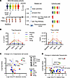 Figure 2
Selective and dose-dependent Treg expansion following IL-2 mutein treatment. (A) Study design of the human single ascending dose study, generated in BioRender. (B) Frequency among CD3+CD4+ T cells and absolute number of CD25+CD127loFOXP3+ Tregs before and after treatment (gating in Supplemental Figure 3A). Absolute number based on lymphocyte complete blood counts obtained from clinical laboratory tests. *Significant after adjusting for multiple testing (P values in Supplemental Table 1), using mixed model for repeated measures (n as depicted in A); dose cohort annotated by asterisk color. (C) Log2-transformed average fold change in absolute cell number from baseline to day 8, using hypothesis-based gating of IL-2–responsive populations (gating in Supplemental Figure 3B). Paired 2-tailed t test, significance of 0.01 to account for multiple testing (n as in A). Participant-level data in Supplemental Figure 4. Cell type represented by shape; dose represented by color. “CD4 Tconv” group includes total CD4+ Tconv, CD25+CD4+ Tconv, Ki67+CD4+ Tconv, and Th1 subsets. “CD8 Tconv” group includes total CD8+ Tconv, CD25+CD8+ Tconv, Ki67+CD8+ Tconv, and Tc1 subsets. “Innate” group includes γδ T cells and innate lymphoid cell 2–like cells. “NK” group includes CD56hi NK cells (NKhi) and CD56lo NK (NKdim) cells. (D) Log2-transformed average fold change from baseline to day 8 in relative frequency of DISCOV-R automated CyTOF metaclusters. The 18 metaclusters were grouped into categories of Tregs, CD4+ Tconv, CD8+ Tconv, Innate, NK, and B cells as in B, represented by shape. Dose represented by color, as in B. Paired 2-tailed t test, significance of 0.01 to account for multiple testing (n as in A). Metacluster definitions and participant-level data shown in Supplemental Figure 5.
To closely examine the effects of IL-2 mutein on non-Tregs, we assessed relative frequencies and absolute numbers of other predicted IL-2–responsive cell types at baseline versus day 8 after treatment with MK-6194. The cell types analyzed included naive and memory CD4+ and CD8+ Tconv cells, T helper type 1 (Th1) cells, cytotoxic CD8+ T (Tc1) cells, CD56hi and CD56dim NK cells, γδ T cells, and innate lymphoid cells (representative gating in Supplemental Figure 3B). We found a small increase in absolute numbers of CD56hi NK cells only in the highest-dose cohort and a small decrease in Tc1 only in the lowest-dose cohort (Figure 2C, participant-level data in Supplemental Figure 4). Changes in relative frequencies of lymphocyte populations were also examined using unbiased clustering of CyTOF data (clustering details in Supplemental Figure 5A). As with hypothesis-based gating, we found a significant, dose-dependent increase in the relative frequency of the Treg cluster (metacluster 8, CD25+FOXP3+CD127lo) from baseline to day 8 (Figure 2D). There were no significant effects on the relative frequency of any other cell cluster at any dose (Figure 2D). Thus, of all lymphocyte subsets, only Tregs showed consistent, dose-dependent changes in absolute numbers and relative frequencies following treatment.
Although we ruled out significant effects of MK-6194 on the frequency of non-Treg populations, we also wanted to confirm that there were no significant effects on the phenotype of other IL-2–responsive cells, such as increased activation or inflammatory signaling that might undermine therapeutic effects of Treg expansion. To assess the transcriptional profile of IL-2–responsive cell populations other than Tregs, lymphocytes expressing high levels of CD122 (IL-2Rβ) were sorted from the 10 mg group at baseline and day 8 for single-cell RNA sequencing (scRNA-seq; representative gating in Supplemental Figure 6). Analysis of RNA expression profiles via uniform manifold approximation and projection (UMAP) and Louvain clustering identified 10 distinct cell clusters (Figure 3A). Expression of lineage-defining genes such as CD3D, NCAM1, TCRGC2, and TRAC identified the majority of these cells as NK or T cells (Figure 3B). Most T cells analyzed were either CD8+ αβ T cells or γδ T cells. Tregs are generally CD122lo and thus were not captured in large enough numbers by our sorting strategy to permit analysis of Tregs in this dataset (Figure 3C, Supplemental Figure 6). Consistent with our CyTOF analyses, there were no significant changes in the relative abundance of any of the CD122hi cell clusters (Figure 3D). Similarly, differential gene expression analyses using pseudobulk data found no significant changes in gene expression in any of the 10 clusters after MK-6194 treatment (Figure 3E). Thus, these data confirm the Treg selectivity of MK-6194, with limited effects on other IL-2–responsive non-Treg populations, including NK cells, CD8+ T cells, and γδ T cells.
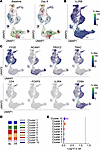 Figure 3
No changes in single-cell gene expression by CD122+ non-Tregs following IL-2 mutein treatment. (A) UMAP-based clustering of integrated scRNA-seq profiles of CD122+ cells sorted from either baseline or day 8 after MK-6194 treatment in the 10 mg dose cohort (n = 4). (B) Normalized expression of IL2RB (CD122) in each cell. (C) Normalized expression of the indicated lineage-specifying genes in each cell. (D) Comparison of cluster frequency between paired baseline (BL) and day 8 (D8) after MK-6194 treatment. No significant differences by the Wilcoxon test. (E) Differential gene expression from baseline versus day 8 after MK-6194 treatment in each pseudobulk cluster. Dashed horizontal line indicates threshold of P < 0.05, indicating no significant differences in gene expression.
IL-2 mutein treatment results in prolonged IL-2R signaling in Tregs. To examine proximal signaling of the IL-2R following MK-6194 treatment, we used flow cytometry to measure pSTAT5 in PBMCs isolated from study participants. PBMCs were immunolabeled immediately upon thawing with no additional stimulation in vitro, and thus pSTAT5 levels represent activation that occurred in vivo (Figure 4A). MK-6194 administration resulted in a dose-dependent increase in pSTAT5 levels in Tregs on day 8, which returned to baseline by day 15 (Figure 4B, representative gating in Supplemental Figure 7). Moreover, there was no detectable pSTAT5 increase in CD56hi NK cells, CD4+ memory, or CD8+ memory T cells on day 8 (Figure 4C), confirming the Treg specificity, as described above.
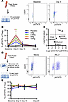 Figure 4
Prolonged, selective, dose-dependent pSTAT5 activation in Tregs following IL-2 mutein treatment in vivo. (A) Schematic and representative plots for ex vivo pSTAT5 staining, gated on CD4+CD25+CD127– Tregs, for 1 participant at baseline versus day 8 after IL-2 mutein treatment (representative gating in Supplemental Figure 7A). Schematic generated in BioRender. (B) Frequency of pSTAT5+ cells among Tregs for all individuals, all time points (n as depicted in Figure 2A) when stained directly ex vivo without restimulation. Significance: *P < 0.05 versus placebo by mixed effects model with Dunnett’s correction for multiple comparisons; dose group reflected by color. (C) Log2-transformed average fold change in frequency of pSTAT5+ cells among various cell subsets from baseline to day 8 for 10 mg cohort (representative gating in Supplemental Figure 7A) when stained directly ex vivo without restimulation. Differences between baseline and day 8 determined using a Wilcoxon matched pairs signed rank test (n = 5). Cell types represented by shapes. (D) Schematic and representative plots for pSTAT5 staining of Tregs following in vitro restimulation with recombinant human IL-2 (rhIL-2) versus media alone for 1 sample. Schematic generated in BioRender. (E) Frequency of pSTAT5+ cells among Tregs for all cohorts, all doses following restimulation in vitro with rhIL-2; dose represented by color as in A. No significant differences by mixed effects model with Dunnett’s correction for multiple comparisons (n as depicted in Figure 2A).
Prolonged signaling through the IL-2R is consistent with the extended half-life of MK-6194 and may have important downstream effects on Treg phenotype and function (13, 14). However, prolonged stimulation through some cytokine receptors results in receptor desensitization (18), and therefore we asked whether prior treatment with IL-2 mutein may impact Treg responsiveness to restimulation with IL-2 in vitro. PBMCs isolated from MK-6194–treated participants were restimulated with recombinant wild-type IL-2 in vitro, followed by staining for pSTAT5 by flow cytometry (Figure 4D, representative staining in Supplemental Figure 7B). At all doses and time points examined, MK-6194 administration had no significant impact on the ability of expanded Tregs to reactivate pSTAT5 in vitro, indicating that the IL-2R remains functionally responsive after IL-2 mutein stimulation in vivo (Figure 4E).
Thymus-derived memory Tregs are preferentially expanded in response to IL-2 mutein. To determine whether MK-6194 preferentially expands specific Treg subsets, we performed unbiased clustering of CyTOF data within the Treg population (gating in Supplemental Figure 3A). UMAP visualization shows 2 clusters of Helios+ memory tTregs, both lacking CD45RA and CCR7 and expressing high levels of CD25 and CCR4 (Figure 5A). One of these clusters expressed high levels of the skin-homing receptor CLA (henceforth referred to as CLA+ tTreg), and the other expressed high levels of the co-inhibitory receptor TIGIT (henceforth referred to as TIGIT+ tTreg). We also identified 1 Helios+CD45RA+ naive tTreg subset and 1 Helios– pTreg subset (Figure 5A, additional clustering detail in Supplemental Figure 8). Changes in the absolute numbers of these subsets on day 8 showed dose-dependent expansion of both CLA+ and TIGIT+ populations of memory tTregs, whereas there was no significant expansion of the naive tTreg or pTreg populations at any dose (Figure 5B).
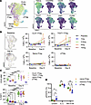 Figure 5
Preferential expansion of memory tTregs following IL-2 mutein treatment. (A) UMAP visualization of Treg (CD4+CD25hiFOXP3+) subclustering with heatmaps of selected subset-defining markers: green, high expression; blue, low expression. Includes integrated data from all cohorts at baseline and day 8. One cluster is defined as CD45RA+Helios+ naive tTregs (yellow), 2 clusters are defined as CD45RA–Helios+ memory tTregs (one with greater CLA expression, green; and the other with greater TIGIT expression, purple), and 1 cluster is defined as Helios– pTregs (blue). (B) Comparisons of Treg cluster frequencies between baseline and day 8. Contour plots, average ± SD of absolute counts for all cohorts across all individuals. CLA+ tTregs (time, P < 0.0001; dose, P = 0.0394) and TIGIT+ tTregs (time, P < 0.0001; dose, P = 0.0001) were significantly different by 1-way ANOVA. Mean ± SD of absolute cell counts for each Treg subset at baseline versus day 8. Dose indicated by color, Wilcoxon’s rank paired test. *P < 0.05. N as depicted in Figure 2A. (C) Comparison of CD25 mean metal intensity (MMI) and frequency of Ki67+ cells for all 3 tTreg clusters and 1 pTreg cluster. Data from the 3.5 mg and 10 mg cohorts combined. Repeated measures 1-way ANOVA with Bonferoni’s correction for multiple comparisons. **P < 0.01; ***P < 0.001; ****P < 0.0001. NS, no significant differences between any 2 groups (n = 11–12). (D) Healthy control human PBMCs were stimulated in vitro with MK-6194 (90 nM, 7.6 μg/mL) or recombinant wild-type human IL-2 (5 ng/mL) for 30 minutes, followed by flow cytometric analysis for induction of pSTAT5. Naive tTreg = CD45RA+Helios+, memory tTreg = CD45RA–Helios+, pTreg = CD45RA–Helios– (n = 5). *P < 0.05; **P < 0.01 by 2-way ANOVA with Tukey’s multiple-comparison test.
Although both CLA+ and TIGIT+ subsets of memory tTregs expanded in response to IL-2 mutein, there were differences between the two. Firstly, CLA+ tTregs were approximately twice as frequent as TIGIT+ tTregs at baseline (average 10.4 ± 4.6 SD versus 5.56 ± 3.1 SD cells × 106/L, respectively; P < 0.0001 by unpaired t test, n = 28, includes all cohorts). Secondly, TIGIT+ tTregs underwent approximately twice as much expansion as CLA+ tTregs (average 7.4 ± 6.8 SD versus 3.1 ± 1.3 SD fold increase, respectively; P = 0.0152 by unpaired t test, n = 17, includes 3.5 mg, 5 mg, and 10 mg cohorts). These observations raise the question of differential responsiveness to MK-6194 between Treg subsets. We predicted that response to treatment may be associated with higher surface levels of CD25 at baseline. However, the TIGIT+ tTreg subset expressed lower levels of CD25 and the cell proliferation marker Ki-67 at baseline compared with CLA+ tTregs (Figure 5C). Both memory tTreg subsets upregulated CD25 and Ki-67 expression on day 8 after treatment. Similarly to TIGIT+ tTregs, naive tTregs expressed low levels of CD25 and Ki67 at baseline and responded to IL-2 mutein treatment with a similar upregulation of CD25 and Ki67; however, unlike TIGIT+ tTregs, naive tTregs did not increase in absolute number (Figure 5B). Thus, MK-6194 activates both naive and memory tTregs but preferentially expands memory tTregs, with the TIGIT+ subset having greater proliferative potential associated with lower expression of activation markers at baseline. In contrast to memory tTregs, pTregs did not respond to MK-6194, showing no increase in abundance and no increase in CD25 or Ki67 expression, consistent with low CD25 expression at baseline (Figure 5C).
To determine whether the differential responsiveness between tTregs and pTregs begins at the level of IL-2R signaling, we examined the induction of pSTAT5 by naive tTregs (CD45RA+Helios+), memory tTregs (CD45RA–Helios+), and pTregs (CD45RA–Helios–) following in vitro stimulation with wild-type IL-2 or MK-6194. We found that although all Treg subsets showed STAT5 phosphorylation in response to MK-6194, responses in pTregs and naive tTregs were weaker than memory tTregs (Figure 5D). Thus, the selective expansion of memory tTregs after MK-6194 treatment may be due in part to enhanced responsiveness at the level of the IL-2R signaling.
IL-2 mutein administration results in transient transcriptional changes in circulating Tregs. To examine the transcriptional responses of Tregs to MK-6194 treatment, we performed bulk RNA-seq on CD4+CD25+CD127lo Tregs sorted from participants from the 1 mg (n = 5), 3.5 mg (n = 6), and 10 mg (n = 4) dosing cohorts at baseline and on days 8, 15, and 29 after treatment (representative sort gating in Supplemental Figure 9). Analysis identified 246 differentially expressed genes (DEGs) whose expression changed (log2[fold change] > 1, FDR < 0.05) following MK-6194 treatment (Figure 6A). The largest number of transcriptional changes was observed in the 10 mg dose cohort on day 8 (166 DEGs), with approximately half as many changes in the 3.5 mg dose cohort (82 DEGs) and few changes in the 1 mg cohort (13 DEGs) at the same time point. Consistent with the robust Treg expansion, DEGs included those regulating cell cycle progression such as CSK2, CDC25C, CDC45, CDKN2A, CDKN2B, and RBBP8, as well as genes controlling cell death, such as CASP4, FAS, and DAD1. DEGs also included genes that regulate cell signaling (PRKCB, AKT3, JUN, JUNB, JUND, FOSB, DGKE, MAPKAP3, and CD3D), migration (CCR7, SELL, CCR5, ITGAM, and CCR3), and cytokine responses (TGFBR2, IL1RL1, LTB, IL2RG, and IL32), as well as transcription factors that control Treg differentiation and function (TCF7, BHLHE40, MAF, MYB, and TBX21).
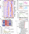 Figure 6
Transient activation-associated transcriptional changes in circulating Tregs following IL-2 mutein treatment. (A) Heatmap showing z-scored average expression of all DEGs in sorted Tregs (CD4+CD25+CD127lo) following MK-6194 treatment shown across all time points in the following dosing cohorts: 1 mg (n = 5), 3.5 mg (n = 6), and 10 mg (n = 4). Dendrogram shows 4 distinct patterns of gene expression change following treatment. B, baseline. (B) Graphs show log2-normalized mean gene expression of all genes in each gene expression category as indicated in A in the 10 mg dose cohort. Shaded region indicates 95% confidence interval. (C) Heatmap showing z-scored average expression of select STAT5-regulated genes at each time point in the 10 mg dose cohort. (D) Gene set enrichment analysis showing significant enrichment of genes in the Hallmark IL2 STAT5 signaling gene set in Tregs 8 days after MK-6194 treatment in the 10 mg dose cohort compared with baseline samples. (E) Graph showing log2 mean expression of Treg and Tconv signature genes in sorted Tregs at each time point in the 10 mg dose cohort. Significance was calculated using a linear mixed effect model. (F) Gene Ontology term enrichment analysis of DEGs between baseline and day 8 posttreatment samples from the 10 mg dose cohort. Dashed lines indicate FDR significance threshold.
Based on changes in gene expression observed in the 10 mg dose cohort, 4 subsets of DEGs were identified (Figure 6, A and B): (a) downregulated genes (45 genes); (b) genes transiently upregulated on day 8 that returned to baseline on day 15 (36 genes); (c) persistently upregulated genes with elevated expression on days 8 and 15 (128 genes); and (d) late genes whose expression increased on day 15 (36 genes). Nearly all changes in gene expression returned to baseline in all cohorts by day 29. As expected, IL-2 mutein treatment increased expression of canonical Treg and IL-2R/STAT5 target genes, including FOXP3, IL2RA (CD25), SOCS2, BCL2, and MYB, among others (Figure 6C), and gene set enrichment analysis showed significant enrichment of genes in the IL2 STAT5 signaling hallmark gene set on day 8 compared with baseline in the 10 mg dose cohort (Figure 6D). FOXP3 and CD25 are both transiently expressed following activation of human CD4+ Tconv cells, which can introduce challenges in distinguishing Tregs from activated Tconv cells. Therefore, to confirm that the CD4+CD25+CD127lo cells sorted following MK-6194 treatment consisted of bona fide stably committed Tregs, we examined expression of previously published “activation-independent” gene sets that reliably distinguish human Treg and Tconv populations (19). Indeed, we found that MK-6194 treatment significantly increased expression of the Treg gene signature (P < 0.001), whereas expression of the Tconv gene signature was low at baseline and did not significantly change after administration (Figure 6E), confirming at the transcriptional level that expanded CD25+ T cells are true Tregs and not activated CD25+ Tconv cells.
Among MK-6194–induced DEGs, Gene Ontology (GO) term enrichment identified pathways involved in cellular activation and proliferation, with upregulated pathways including “positive regulation of cell activation,” “regulation of cyclin-dependent protein kinase activity,” and “negative regulation of intrinsic apoptotic signaling pathway” (Figure 6F). Upregulated DEGs were also enriched in genes associated with the “oxidative phosphorylation,” “ATP metabolic processes,” and “proton membrane transport” pathways, indicating that MK-6194–expanded Tregs have undergone metabolic reprogramming to support their robust expansion and function. In contrast, DEGs downregulated by MK-6194 treatment were enriched in genes from the “negative regulation of mitotic cell cycle,” “T cell differentiation in the thymus,” and “regulation of RNA metabolic processes” pathways.
Treg expression of markers of suppressive function and tissue homing are enhanced by IL-2 mutein administration. In addition to controlling Treg abundance, IL-2R signaling regulates Treg activation state and immunosuppressive function (20). Indeed, we found elevated expression of genes associated with Treg activation and suppressor function such as CTLA4, ICOS, LRRC32 (GARP), TIGIT, HAVCR2 (TIM-3), IL1R2, TNFRSF4 (OX-40), MKI67, TNFRSF9 (41BB), and TNFRSF18 (GITR) on day 8 after 10 mg MK-6194 treatment (Figure 7A).
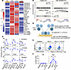 Figure 7
Increased expression of markers of Treg activation, function, and tissue-homing potential following IL-2 mutein treatment. (A) Heatmap showing z-scored average expression of select Treg activation genes at each time point in the 10 mg dose cohort (n = 4) by bulk RNA-seq of sorted CD4+CD25+CD127lo Tregs. (B) Gene set enrichment analysis showing enrichment of public consensus Treg gene sets in Tregs from baseline versus day 8 (D8) following 10 mg MK-6194 treatment. The public consensus Treg gene sets “healthy non-lymphoid tissue (NLT)” and “high in diseased NLT” are enriched on day 8 following MK-6194 treatment, and the gene set “low in diseased NLT” is enriched at baseline. No significant enrichment of the “Healthy LT/Blood” gene set at either time point. (C) Percentage of Tregs (CD4+CD25+CD127loFOXP3+) expressing the indicated markers at each time point in the 10 mg dose cohort as measured by CyTOF (mean ± SD, n = 6). Significant changes relative to baseline frequencies were calculated by 2-way ANOVA with Dunnett’s correction for multiple comparisons. *P < 0.05, **P < 0.01, ***P < 0.001, ****P < 0.0001. (D) Schematic of CTLA-4 transendocytosis assay. (E) Representative flow cytometric analysis of CD80-mCherry and CD86-GFP capture by Tregs (CD4+FOXP3+) from baseline and day 8 after 7.5 mg MK-6194 treatment as indicated (n = 4). Addition of blocking anti–CTLA-4 antibody demonstrates specificity of capture. (F) Graphs showing percentage of CD80–mCherry+CD86–GFP+ Tconv cells and Tregs in paired baseline and day 8 after 7.5 mg MK-6194 treatment samples (n = 4). Significance calculated by repeated-measures 1-way ANOVA with Holm-Šídák multiple-comparison test. **P < 0.01; *P < 0.05.
Tregs acquire different phenotypes and functions depending on where they reside (3), such as lymphoid tissue (LT) versus nonlymphoid tissue (NLT), and the disease state, such as healthy tissues versus solid tumors or sites of autoimmune inflammation. To assess the effects of MK-6194 on Treg adaptation to various human tissues and disease states, we performed gene set enrichment analysis of transcriptional changes induced by IL-2 mutein compared with 4 consensus human Treg gene sets (21): “High in diseased NLT” (based on 10 tumor gene sets and 2 synovial fluid gene sets), “Low in diseased NLT” (based on 7 gene sets comparing blood, healthy tissue, and diseased NLT), “Combined healthy NLT” (based on 9 gene sets including uterus, colon, skin, fat, and other tissues), and “Combined healthy LT” (based on 5 gene sets from spleen, lymph node, blood, and colon-associated LT). We found that the transcriptional signatures of Tregs from both healthy and diseased NLT were significantly enriched in MK-6194–expanded Tregs, whereas genes that were low in Tregs from diseased NLT were significantly enriched in the baseline Tregs prior to MK-6194 treatment (Figure 7B). Conversely, MK-6194 treatment had no effect on enrichment of the healthy LT Treg signature. Thus, MK-6194–expanded Tregs upregulate a gene signature consistent with residency and function in NLTs.
To confirm that these transcriptional changes in Tregs were also reflected at the protein level, we analyzed expression of markers of activation, suppressor function, and migration by CyTOF (Figure 7C). This analysis confirmed transiently increased protein expression of the activation and proliferation markers Ki-67, ICOS, and OX-40, as well as increased protein expression of CTLA-4, CD39, and TIGIT, suggesting elevated capacity for Treg suppressor function (22–25). Furthermore, the homing receptors α4β7 integrin and CXCR3 that direct Tregs to inflamed NLTs were upregulated following treatment, whereas the LT homing receptor CCR7 was downregulated, further suggesting preferential homing to NLT over LT.
In a previous study analyzing murine Tregs after treatment with a Treg-selective murine IL-2 mutein (26), we found that elevated expression of CTLA-4 was associated with enhanced ability to strip the co-stimulatory ligands CD80 and CD86 from the surface of antigen-presenting cells (APCs) via a process termed transendocytosis. This is an important Treg function that supports immune tolerance by reducing the ability of APCs to activate Tconv cells (25). Given the similar upregulation of CTLA-4 expression in MK-6194–expanded Tregs (Figure 7, A and C), we used an in vitro system to measure CTLA-4–dependent transendocytosis of CD80 and CD86 (Figure 7D). CD4+ T cells were isolated from 4 participants at baseline and day 8 after treatment with 7.5 mg MK-6194. Total CD4+ T cells were co-cultured for 6 hours with NIH3T3 cells transfected with human CD86-GFP and CD80-mCherry fusion proteins in the absence of further stimulation. Non-adherent cells were analyzed by flow cytometry to identify FOXP3+ Tregs that obtained GFP and mCherry fluorescence via transendocytosis (Figure 7D). We found that MK-6194 treatment significantly increased ligand capture by Tregs, as evidenced by increased frequency of GFP+mCherry+ cells, which was inhibited by addition of a CTLA-4–blocking antibody to the co-culture (Figure 7, E and F). By contrast, FOXP3– Tconv cells in the same cultures showed minimal ability to capture CD80 and CD86. Taken together, these results suggest that MK-6194–expanded Tregs are poised to migrate to NLTs and strip APCs of key costimulatory molecules.


