
Dermatology
Abstract
Persistent production of type I interferons (IFN-Is) is a hallmark of cutaneous lupus erythematosus (CLE). Ultraviolet (UV) light stimulates IFN-I response in the skin and exacerbates CLE. Here, we identify V-type immunoglobulin domain-containing suppressor of T cell activation (VISTA) as a negative regulator of both basal and UV-induced IFN-I response in the skin and show that VISTA limits skin photosensitivity in an IFN-I-dependent manner, in part through Stimulator of Interferon Genes (STING). Furthermore, we demonstrate a novel role for VISTA in keratinocytes both at steady state and in response to UV light. Conditional deletion of VISTA in epidermal keratinocytes results in a ~10-fold increase in basal skin IFN-I scores and a heightened UV-induced skin injury score, both of which are dependent on IFN-I signaling. VISTA-targeting monoclonal antibodies suppress the UV-induced IFN-I response in human keratinocytes and in mice expressing human VISTA in vivo, thereby reducing UV-induced skin injury scores. Together, these findings identify VISTA as a keratinocyte-intrinsic checkpoint that restrains STING-associated IFN-I response in the skin and suggest VISTA agonism as a therapeutic strategy to limit photosensitivity in CLE.
Authors
Zachary T. Peters, Lindsay K. Mendyka, J'Voughnn A. Blake, Himanshu B. Goswami, Angelique N. Cortez, Grace E. Crossland, Sicong Shan, Elizabeth C. Nowak, Myana Keusch, Mrinal K. Sarkar, Johann E. Gudjonsson, Christopher M. Burns, Dorothea T. Barton, Bruce R. Blazar, Tyler J. Curiel, Rodwell Mabaera, Victoria P. Werth, Andrea Kalus, Keith B. Elkon, Randolph J. Noelle, Sladjana Skopelja-Gardner
Abstract
Inflammatory cytokines reprogram keratinocyte metabolism, but the metabolic pathways that couple immune signals to pathological epidermal growth remain incompletely defined. Here, we identify GLS1-mediated glutaminolysis as a metabolic program preferentially induced in keratinocytes under type 3 inflammatory conditions. Integrated transcriptomic, metabolomic, genetic, and functional analyses showed that IL-17A induced GLS1 expression and glutaminolysis in keratinocytes. Keratinocyte-specific Gls1 deletion reduced the intracellular availability of arginine, proline, and methionine, impaired amino acid-dependent mTORC1 activation, disrupted redox homeostasis, and limited keratinocyte proliferation. Amino acid or antioxidant supplementation partially rescued these defects, whereas rapamycin blocked the amino acid-mediated proliferative rescue. Gls1 deletion did not impair steady-state skin development or homeostasis and did not alter MC903-induced type 2 dermatitis, but it delayed wound re-epithelialization and attenuated IMQ-induced psoriasiform inflammation. Loss of keratinocyte GLS1 also reduced epidermal chemokine expression and the accumulation of neutrophils and IL-17A-producing γδ T cells, revealing a role for glutaminolysis in amplifying epithelial-immune crosstalk. These findings define GLS1-mediated glutaminolysis as a context-specific metabolic checkpoint linking type 3 inflammation to keratinocyte proliferation and cutaneous immune amplification, and support locally or temporally controlled GLS1 inhibition as a potential therapeutic strategy for psoriasis.
Authors
Yongfei Hu, Hai Yu, Kaiyu Liang, Liyan Yuan, Wenjun Zhang, Zhe Zhuang, Suyun Ji, Xichun Xia
Abstract
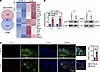
Allergic contact dermatitis (ACD), a recurrent inflammatory skin disorder, affects 21% of humans and is the second leading cause of occupational diseases in USA. ACD is initiated by the innate immune response to skin-contact sensitizers potentiated by the neuropeptide substance P (SP). Skin sensitizers stimulate SP-secreting sensory nerves and trigger proinflammatory functions of keratinocytes expressing the neurokinin 1 receptor (NK1R). Nevertheless, the neuroimmune regulation of hapten-initiated skin inflammation, remains incompletely elucidated. Using K14Cre/+NK1RKO mice skin-sensitized with 2,4-dinitrochlorobenzene (DNCB), we demonstrate that NK1R deletion exclusively in keratinocytes prevents hapten-initiated skin inflammation, impairs the mobilization of conventional dendritic cells (cDC) to draining lymph nodes (dLN) and blocks the elicitation of the contact hypersensitivity reaction (CHS) to the same extent observed in global Tac1KO (without SP) and NK1RKO mice. The DNCB effects were restored by skin co-administration of IL-1β and TNF-α. SP-NK1R signaling of mouse and human keratinocytes increased transcripts encoding proteins of the NLRP3 inflammasome. Although, DNCB and SP induced pro-IL-1β synthesis, only SP triggered intracellular Ca2+ increase, NFATc1 nuclear translocation and TNF-α synthesis, a cytokine mediating systemic inflammation in ACD. Our data identifying SP-NK1R-signaling of keratinocytes as a key mechanism for ACD provide relevant insight for therapies targeting skin neuroimmune interactions.
Authors
Sumeet Manandhar, Mohna Bandyopadhyay, Olga Tkacheva, William Shufesky, Greg Gibson, Simon C. Watkins, Adrian Morelli, Adriana T. Larregina
Abstract
Chronic, non-healing wounds are a severe diabetic complication. The underlying mechanisms are not fully understood, and the role of ATF7 in this context has not been well characterized. In our study, we utilized db/db diabetic mice and AAV-mediated keratinocyte-specific Atf7 overexpression in vivo. HaCaT keratinocyte/THP-1 macrophage cocultures under high glucose were used in vitro. Our results showed that ATF7 was upregulated in diabetic wounds. Keratinocyte-specific Atf7 overexpression accelerated diabetic wound closure, enhanced re-epithelialization, granulation tissue formation, and keratinocyte proliferation, while suppressing macrophage M1 polarization and inflammation. Multiomics screening identified NOTCH1 as a key ATF7 target. ATF7 transcriptionally repressed NOTCH1 by recruiting Suv39h1, increasing H3K9me3 at the NOTCH1 promoter. This reduced NOTCH1 protein and its active intracellular domain (N1ICD) within keratinocyte-derived exosomes. ATF7-overexpressing keratinocyte exosomes carried less N1ICD, leading to decreased N1ICD transfer to macrophages and subsequent inhibition of M1 polarization. Notably, local injection of exosomes from ATF7-overexpressing keratinocytes accelerated wound healing in db/db mice. In summary, ATF7 promotes diabetic wound healing by repressing NOTCH1 transcription via H3K9me3, thereby reducing exosomal N1ICD secretion from keratinocytes and inhibiting macrophage M1 polarization. This identifies the ATF7/NOTCH1/exosome axis as a therapeutic target.
Authors
Pengcheng Xu, Yuan Xue, Linlin Feng, Jingwen Kuang, Xiaochen Hu, Huiyi Tang, Biao Cheng, Limin Wei
Abstract
Authors
Aneri Shethji, Theresa Hutchins, Anwesha Sanyal, Tianhao Liu, Wei Chen, Kathryn S. Torok

Abstract
Authors
Peter Dimitrion, Jesse Veenstra, Deangelo Ferguson, Ping Wang, Jeffrey Cruz, Tasneem F. Mohammad, Ian Loveless, Aamir Siddiqui, Iltefat H. Hamzavi, Li Zhou, Indra Adrianto, Qing-Sheng Mi
Abstract
Significant loss of pigmentation can increase visual disability, skin cancer risk, and psychosocial stress. Tyrosinase (TYR) catalyzes the first and rate-limiting step of melanin synthesis. Inhibitors of TYR are well established and are currently used in clinical settings; however, there is a dearth of direct activators of TYR. Here, using a human TYR construct, we developed high-throughput screening methods, in cell confirmatory assays employing 13C-tyrosine tracing, and computational analysis techniques, and identified ampyrone (4-aminoantipyrine) as a TYR activator. Ampyrone increased the in vitro catalytic activity of the human recombinant intra-melanosomal domain of TYR (hTYR) and its hypomorphic variant, Pro406Leu (P406L), a cause of oculocutaneous albinism type 1B (OCA1B). Moreover, ampyrone induced melanin synthesis in both wild-type and OCA1B human melanocytes, mouse OCA2 melanocytes, as well as 3-dimensional (3D) human skin cultures. Computational studies provided additional insight into the effects of direct TYR agonists on enzyme activity. Our results identified ampyrone as a lead candidate for TYR activation, potentially supporting the development of therapies for patients with genetic and acquired diseases of hypopigmentation.
Authors
Monika B. Dolinska, Yuhong A. Wang, Nathan P. Coussens, Vijay K. Kalaskar, Zuhal Eraslan, Samuel J. Grondin, Joseph Bonica, Sarah Toay, Matthew D. Hall, Min Shen, Matthew Boxer, Qiuying Chen, Steven S. Gross, Nabeel Attarwala, Yingyos Jittayasothorn, Ramakrisha P. Alur, Dhyanam Shukla, Robin Kee, Charles DeYoung, Cuilee Sha, David R. Adams, Stacie K. Loftus, Tiziana Cogliati, Yuri V. Sergeev, Jonathan H. Zippin, Brian P. Brooks
Abstract
Authors
Natalie E. Andresen, Heehwa G. Son, Joongho J. Joh, Shadmehr Demehri
Abstract
Enhancing NAD+ levels with nicotinamide riboside (NR) confers anti-inflammatory effects in human disease, although immunoregulatory mechanisms remain poorly characterized. We previously showed that ex vivo NR supplementation of primary CD4+ T cells from psoriatic individuals dampened immune responsiveness. To validate this in vivo, we performed a randomized, placebo-controlled NR supplementation study in individuals with mild-to-moderate psoriasis. Participants received oral NR (500 mg twice daily) or matching placebo for 4 weeks, with blood samples collected at baseline and after supplementation. NR reduced Th17 immune responsiveness. Bulk CD4+ T cell RNA-seq identified induction of the SLIT-ROBO signaling pathway. NR supplementation increased circulating SLIT2 levels and enhanced SLIT2 production in dermal fibroblasts. Pharmacologic and genetic interrogation in CD4+ T cells and fibroblasts demonstrated that SLIT2, acting through the ROBO1 receptor, inhibited Rho GTPase signaling, thereby attenuating canonical Th17 polarization and fibroblast inflammatory activation. These findings indicate that NAD+ augmentation exerts anti-inflammatory effects in psoriasis through SLIT2-ROBO1-mediated crosstalk between dermal fibroblasts and circulating CD4+ T cells, leading to suppression of Th17-driven inflammation.
Authors
Kim Han, Rachael J. Klein, Thomas C. Recupero, Anna Chiara Russo, Rahul Sharma, Anand K. Gupta, Shahin Hassanzadeh, Rebecca D. Huffstutler, Pradeep K. Dagur, Bryan Fisk, Neelam R. Redekar, Michael N. Sack
Abstract
Impaired adhesion and differentiation of keratinocytes is a hallmark of several skin diseases, but only some of the factors that regulate these processes have been identified. Here, we studied the role of isoform-rich dermokine – a wound- and tumor-regulated protein – in keratinocytes using a combination of multi-omics and functional approaches. CRISPR/Cas9-induced knockout of dermokine isoforms in human keratinocytes inhibited differentiation of these cells in three-dimensional organotypic skin cultures, which was confirmed by quantitative proteomics. In two-dimensional monocultures, dermokine deficiency affected the proteome and phosphoproteome as revealed by mass spectrometry. We found reduced abundance of differentiation-specific proteins and increased phosphorylation of cell adhesion protein p120 (catenin-δ1). The adhesive strength of dermokine knockout keratinocytes was impaired, which was rescued by p120 knock-down or ROCK inhibition. Finally, we verified the correlation between decreased dermokine expression and increased p120 phosphorylation in human non-healing wounds. These results identify dermokine as regulator of keratinocyte adhesion and differentiation, involving at least in part its effect on p120 phosphorylation and ROCK. Our data point to a function of dermokine in the pathogenesis of chronic wounds.
Authors
Vahap Canbay, Till Wüstemann, Weihua Tian, Tobias A. Beyer, Camilla Reiter Elbæk, Michael Stumpe, Gaetana Restivo, Chatpakorn Christiansen, Anabel Migenda Herranz, Susanne Mailand, Jürg Hafner, Rune Busk Damgaard, Steffen Goletz, Jörn Dengjel, Ulrich auf dem Keller, Chiara Francavilla
No posts were found with this tag.


