
Research ArticleDermatologyInflammation
Open Access |  10.1172/jci.insight.201178
ATF7 drives diabetic wound healing via NOTCH1 repression and N1ICD-dependent macrophage polarization control
Pengcheng Xu,1 Yuan Xue,2 Linlin Feng,3 Jingwen Kuang,3 Xiaochen Hu,4 Huiyi Tang,5 Biao Cheng,6 and Limin Wei1
1Department of Breast Surgery, and
2Department of Thyroid and Head and Neck Oncology Surgery, The First Affiliated Hospital of Henan University of Science and Technology, Luoyang, China.
3The First Affiliated Hospital of Henan University of Science and Technology, Luoyang, China.
4Henan Key Laboratory of Cancer Epigenetics, Cancer Hospital, The First Affiliated Hospital of Henan University of Science and Technology, Luoyang, China.
5Department of Sports Medicine, Guangzhou Sport University, Guangzhou, China.
6Department of Burn and Plastic Surgery, General Hospital of Southern Theater Command, PLA, Guangzhou, China.
Authorship note: PX, YX, and LF contributed equally to this work.
Find articles by Xu, P. in: PubMed | Google Scholar
1Department of Breast Surgery, and
2Department of Thyroid and Head and Neck Oncology Surgery, The First Affiliated Hospital of Henan University of Science and Technology, Luoyang, China.
3The First Affiliated Hospital of Henan University of Science and Technology, Luoyang, China.
4Henan Key Laboratory of Cancer Epigenetics, Cancer Hospital, The First Affiliated Hospital of Henan University of Science and Technology, Luoyang, China.
5Department of Sports Medicine, Guangzhou Sport University, Guangzhou, China.
6Department of Burn and Plastic Surgery, General Hospital of Southern Theater Command, PLA, Guangzhou, China.
Authorship note: PX, YX, and LF contributed equally to this work.
Find articles by Xue, Y. in: PubMed | Google Scholar
1Department of Breast Surgery, and
2Department of Thyroid and Head and Neck Oncology Surgery, The First Affiliated Hospital of Henan University of Science and Technology, Luoyang, China.
3The First Affiliated Hospital of Henan University of Science and Technology, Luoyang, China.
4Henan Key Laboratory of Cancer Epigenetics, Cancer Hospital, The First Affiliated Hospital of Henan University of Science and Technology, Luoyang, China.
5Department of Sports Medicine, Guangzhou Sport University, Guangzhou, China.
6Department of Burn and Plastic Surgery, General Hospital of Southern Theater Command, PLA, Guangzhou, China.
Authorship note: PX, YX, and LF contributed equally to this work.
Find articles by Feng, L. in: PubMed | Google Scholar
1Department of Breast Surgery, and
2Department of Thyroid and Head and Neck Oncology Surgery, The First Affiliated Hospital of Henan University of Science and Technology, Luoyang, China.
3The First Affiliated Hospital of Henan University of Science and Technology, Luoyang, China.
4Henan Key Laboratory of Cancer Epigenetics, Cancer Hospital, The First Affiliated Hospital of Henan University of Science and Technology, Luoyang, China.
5Department of Sports Medicine, Guangzhou Sport University, Guangzhou, China.
6Department of Burn and Plastic Surgery, General Hospital of Southern Theater Command, PLA, Guangzhou, China.
Authorship note: PX, YX, and LF contributed equally to this work.
Find articles by Kuang, J. in: PubMed | Google Scholar
1Department of Breast Surgery, and
2Department of Thyroid and Head and Neck Oncology Surgery, The First Affiliated Hospital of Henan University of Science and Technology, Luoyang, China.
3The First Affiliated Hospital of Henan University of Science and Technology, Luoyang, China.
4Henan Key Laboratory of Cancer Epigenetics, Cancer Hospital, The First Affiliated Hospital of Henan University of Science and Technology, Luoyang, China.
5Department of Sports Medicine, Guangzhou Sport University, Guangzhou, China.
6Department of Burn and Plastic Surgery, General Hospital of Southern Theater Command, PLA, Guangzhou, China.
Authorship note: PX, YX, and LF contributed equally to this work.
Find articles by Hu, X. in: PubMed | Google Scholar
1Department of Breast Surgery, and
2Department of Thyroid and Head and Neck Oncology Surgery, The First Affiliated Hospital of Henan University of Science and Technology, Luoyang, China.
3The First Affiliated Hospital of Henan University of Science and Technology, Luoyang, China.
4Henan Key Laboratory of Cancer Epigenetics, Cancer Hospital, The First Affiliated Hospital of Henan University of Science and Technology, Luoyang, China.
5Department of Sports Medicine, Guangzhou Sport University, Guangzhou, China.
6Department of Burn and Plastic Surgery, General Hospital of Southern Theater Command, PLA, Guangzhou, China.
Authorship note: PX, YX, and LF contributed equally to this work.
Find articles by Tang, H. in: PubMed | Google Scholar
1Department of Breast Surgery, and
2Department of Thyroid and Head and Neck Oncology Surgery, The First Affiliated Hospital of Henan University of Science and Technology, Luoyang, China.
3The First Affiliated Hospital of Henan University of Science and Technology, Luoyang, China.
4Henan Key Laboratory of Cancer Epigenetics, Cancer Hospital, The First Affiliated Hospital of Henan University of Science and Technology, Luoyang, China.
5Department of Sports Medicine, Guangzhou Sport University, Guangzhou, China.
6Department of Burn and Plastic Surgery, General Hospital of Southern Theater Command, PLA, Guangzhou, China.
Authorship note: PX, YX, and LF contributed equally to this work.
Find articles by Cheng, B. in: PubMed | Google Scholar
1Department of Breast Surgery, and
2Department of Thyroid and Head and Neck Oncology Surgery, The First Affiliated Hospital of Henan University of Science and Technology, Luoyang, China.
3The First Affiliated Hospital of Henan University of Science and Technology, Luoyang, China.
4Henan Key Laboratory of Cancer Epigenetics, Cancer Hospital, The First Affiliated Hospital of Henan University of Science and Technology, Luoyang, China.
5Department of Sports Medicine, Guangzhou Sport University, Guangzhou, China.
6Department of Burn and Plastic Surgery, General Hospital of Southern Theater Command, PLA, Guangzhou, China.
Authorship note: PX, YX, and LF contributed equally to this work.
Find articles by Wei, L. in: PubMed | Google Scholar
Published August 24, 2026 - More info
JCI Insight. 2026;11(16):e201178. https://doi.org/10.1172/jci.insight.201178.
© 2026 Xu et al. This work is licensed under the Creative Commons Attribution 4.0 International License. To view a copy of this license, visit http://creativecommons.org/licenses/by/4.0/.
Received: October 10, 2025; Accepted: July 9, 2026

-
Results
Atf7 expression is upregulated in the wound tissues of diabetic mice. Differentially upregulated genes were screened from the GSE182906 (day 14) dataset. These genes were then intersected with the transcription factor (TF) dataset from the AnimalTFDB website, yielding 20 differentially upregulated TFs (Figure 1A). ATF7 was selected for further investigation based on its established roles in inflammation and proliferation. A full-thickness skin wound model was established using db/db and C57BL/6 mice. Quantitative real-time PCR (qRT-PCR) and Western blot analysis revealed that ATF7 expression was upregulated in the wound tissues of db/db mice compared with C57BL/6 control mice (Figure 1B). Subsequently, immunofluorescence (IF) costaining for ATF7 and the keratinocyte marker keratin 14 (K14) was performed on wound tissues. In C57BL/6 mice, ATF7 and K14 exhibited colocalization. Within the wound tissues of db/db mice, K14 expression was decreased while ATF7 expression was elevated (Figure 1C).
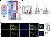 Figure 1
Atf7 expression is upregulated in wound tissues of diabetic mice. (A) Differentially upregulated genes (|logFC| ≥ 1.5, P < 0.05) were screened from the GSE182906 dataset (day 14) and intersected with the Mus musculus transcription factor (TF) dataset from AnimalTFDB. A cluster heatmap displays the expression profiles of the 20 identified TFs. (B) Full-thickness excisional skin wounds were created on the dorsum of 10-week-old male db/db and C57BL/6 mice. Atf7 mRNA and ATF7 protein expression in wound tissues was analyzed by qRT-PCR and Western blotting on days 7 and 14 after wounding. (C) Representative immunofluorescence images showing colocalization of ATF7 and the keratinocyte marker keratin 14 (K14) in wound tissues and quantitative analysis for double-positive cells. Nuclei were counterstained with DAPI. Scale bar: 50 μm (original magnification: ×400). Results are expressed as mean ± SD. ***P < 0.001 by 2-tailed, unpaired Student’s t test. Each experimental group consisted of 6 animals (n = 6).
Atf7 overexpression promotes diabetic wound healing. To investigate the functional role of ATF7 in vivo, we constructed an adeno-associated virus (AAV) carrying Atf7 under the control of the keratinocyte-specific K14 promoter. This vector was administered to mice to achieve keratinocyte-specific overexpression of ATF7 (Supplemental Figure 1B; supplemental material available online with this article; https://doi.org/10.1172/jci.insight.201178DS1). Subsequently, a full-thickness skin wound model was established in these mice. We found that AAV-mediated Atf7 overexpression accelerated wound closure rates in db/db mice compared with controls (Figure 2, A and B). Histological analysis of wound tissues on days 7 and 14 after wounding revealed that Atf7 overexpression resulted in a reduced distance between the wound edges in diabetic mice (Figure 2, C and D). This finding indicates enhanced re-epithelialization and wound contraction in the diabetic mice following Atf7 overexpression. Furthermore, Atf7-overexpressing wounds exhibited increased granulation tissue formation (Figure 2E). To assess cellular proliferation, we evaluated the expression of the proliferation marker Ki67. IF analysis revealed an increase in Ki67-positive cells within the wounds of Atf7-overexpressing db/db mice (Figure 2, F and G). Additionally, increased colocalization of ATF7 and K14 in wound tissues was observed following Atf7 overexpression (Figure 2H). Collectively, these results suggest that Atf7 overexpression promotes wound healing in diabetic mice.
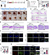 Figure 2
Atf7 overexpression promotes diabetic wound healing. (A) Schematic of the AAV2 vector construct. Keratinocyte-specific overexpression of Atf7 was achieved using an AAV2 serotype vector driven by the K14 promoter (AAV2K14-Atf7). Db/db mice (10-week-old males) received intradermal injections of AAV2K14-Atf7 or control vector at the wound margin site. Three weeks after injection, full-thickness excisional skin wounds were created on the dorsum. Representative wound photographs at indicated days after wounding (days 0, 3, 7, 11, and 14). (B) Quantitative analysis of wound closure rate. (C) Representative H&E-stained sections of wound tissues. Scale bar: 500 μm (original magnification: ×40). (D) Quantification of re-epithelialization ratio from H&E-stained sections. (E) Representative H&E-stained sections showing granulation tissue formation. Arrows highlight granulation tissue. Scale bar: 100 μm (original magnification: ×200). (F) Representative immunofluorescence images showing Ki67 expression (proliferation marker) in wound tissues. Nuclei counterstained with DAPI. Scale bar: 50 μm (original magnification: ×400). (G) The quantitative analysis of Ki67+ cells. (H) Representative immunofluorescence images showing colocalization of ATF7 and K14 in wound tissues and quantitative analysis for double-positive cells. Nuclei counterstained with DAPI. Scale bar: 50 μm (original magnification: ×400. Results are expressed as mean ± SD. *P < 0.05; **P < 0.01; ***P < 0.001 by 2-way ANOVA with Bonferroni’s post hoc test (B) or 2-tailed, unpaired Student’s t test (D, G, and H). Each experimental group consisted of 6 animals (n = 6).
Atf7 overexpression inhibits macrophage M1 polarization in diabetic wounds. To assess macrophage polarization states within the wound microenvironment, single-cell suspensions were prepared from skin wound tissues and analyzed by flow cytometry. The percentages of M1 macrophages, defined as MHCII+F4/80+CD115+CD11b+ and CD86+F4/80+CD115+CD11b+ cells, were quantified. Flow cytometric analysis revealed a reduction in the percentage of both MHCII+ (Figure 3, A and B) and CD86+ (Figure 3, C and D) M1 macrophages in wound tissues following Atf7 overexpression. Consistent with this finding, the expression levels of M1 macrophage markers (Nos2, Cd86, and MHCII) were downregulated in Atf7-overexpressing wound tissues (Figure 3E). Furthermore, the expression of proinflammatory cytokines TNF-α, IL-6, and IL-1B was also decreased in wounds overexpressing Atf7 (Figure 3F). To determine whether ATF7 overexpression promotes M2 polarization in diabetic wounds, we analyzed M2 macrophage markers in wound tissues. qPCR analysis showed that the mRNA levels of M2-associated genes Arg1 and Cd206 were upregulated in Atf7-overexpressing wound tissues (Figure 3G). IF for the M2 marker CD206 revealed an increase in CD206-positive cells within the tissue of Atf7-overexpressing wounds (Figure 3H). These data indicate that Atf7 overexpression not only suppresses the proinflammatory M1 phenotype but also promotes an M2 phenotype in the diabetic wound microenvironment.
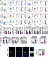 Figure 3
Atf7 overexpression inhibits macrophage M1 polarization in diabetic wounds. Single-cell suspensions were prepared from skin wound tissues of db/db mice. (A) Schematic of the gating strategy for identifying macrophages for the detection of M1 macrophages (MHCII+F4/80+CD115+CD11b+). (B) Quantitative analysis of the percentage of M1 macrophages (MHCII+F4/80+CD115+CD11b+). (C) Schematic of the gating strategy for the detection of M1 macrophages (CD86+F4/80+CD115+CD11b+). (D) Quantitative analysis of the percentage of M1 macrophages (CD86+F4/80+CD115+CD11b+). (E) mRNA expression levels of M1 macrophage markers (Nos2, Cd86, and MHCII) in wound tissues were measured by qRT-PCR. (F) Levels of proinflammatory cytokines (TNF-α, IL-6, and IL-1B) in wound tissues were measured by ELISA. (G) Levels of M2 macrophage markers (Arg1 and Cd206) were measured by qRT-PCR. (H) Representative immunofluorescence images showing CD206 expression in wound tissues and quantitative analysis for CD206+ cells. Nuclei counterstained with DAPI. Scale bar: 50 μm (original magnification: ×400) (E, F, and H). Results are expressed as mean ± SD. **P < 0.01, ***P < 0.001 by 2-tailed, unpaired Student’s t test. Each experimental group consisted of 6 animals (n = 6).
ATF7 promotes the proliferation of keratinocytes under high-glucose conditions. To investigate the effect of ATF7 on keratinocyte function in vitro, HaCaT cells were infected with lentivirus expressing human ATF7 (LV-ATF7) to achieve ATF7 overexpression (Supplemental Figure 1C) or with lentivirus expressing shRNA to knock down ATF7 (Supplemental Figure 1D), followed by culture under high-glucose (HG) conditions. CCK-8 assay results showed that ATF7 overexpression enhanced the viability of HaCaT cells exposed to HG (Figure 4A). IF staining for Ki67 revealed an increase in Ki67-positive cells in ATF7-overexpressing cells (Figure 4B), while ATF7 knockdown decreased Ki67-positive cells (Figure 4C). Furthermore, a wound healing assay revealed that ATF7 overexpression promoted the migration of HaCaT cells in HG medium, whereas ATF7 knockdown impaired cell migration (Figure 4D). Collectively, these results indicate that ATF7 ameliorates HG-induced damage to keratinocytes by promoting their proliferation and migration.
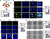 Figure 4
ATF7 promotes the proliferation of keratinocytes under high-glucose conditions. HaCaT cells were infected with lentivirus expressing ATF7 (LV-ATF7) or shATF7 (LV-shATF7) for 48 hours, followed by culture in 25 mM high-glucose (HG) medium for 24 hours. (A) Cell viability was assessed by CCK-8 assay. (B and C) Representative immunofluorescence images of Ki67 expression and quantitative analysis for Ki67+ cells. Nuclei counterstained with DAPI. Scale bar: 50 μm (original magnification: ×400). (D) Cell migration ability was analyzed by the scratch wound healing assay. Left: Representative images at 0 hours and 24 hours after scratching. Scale bars: 200 μm. Right: Quantification of wound closure percentage. Results are expressed as mean ± SD. **P < 0.01; *** P < 0.001 by 1-way ANOVA with Bonferroni’s post hoc test. All cell experiments were performed with n = 3 replicates.
ATF7 overexpression inhibits macrophage M1 polarization. To investigate the effect of ATF7-overexpressing keratinocytes on macrophage polarization, a coculture system was employed. Human monocytic THP-1 cells were differentiated into M0 macrophages (THP-1-M0) and cocultured with HaCaT cells using a Transwell system (Figure 5A). Following coculture, macrophages in the lower chamber were harvested for analysis. Notably, ATF7 overexpression in HaCaT cells suppressed the expression of M1 macrophage markers (NOS2, CD86, MHCII) in the cocultured macrophages (Figure 5B). Furthermore, flow cytometric analysis revealed a decrease in the percentage of MHCII+ and CD86+ cells within the macrophage population (Figure 5, C and D). The expression of TNFA, IL6, and IL1B was downregulated in these macrophages (Figure 5E). To test whether the effects of ATF7 on keratinocytes depend on its ability to suppress M1 polarization, we established a coculture system with a rescue design. HaCaT cells were infected with negative control lentivirus (LV-NC) or LV-ATF7, and cocultured with THP-1 under HG conditions. In the rescue group, macrophages were treated with LPS plus IFN-γ. We then assessed both macrophage polarization and keratinocyte function. qPCR and flow cytometry exhibited that in the rescue group, the expression of CD86 and MHCII in macrophages was restored compared with the LV-ATF7 group (Figure 5, F and G). CCK-8, IF, and wound healing assays revealed that while ATF7 overexpression enhanced HaCaT viability, proliferation, and migration, these beneficial effects were attenuated when macrophages were forced to remain in the M1 state (Figure 5, H–K). This in vitro rescue experiment showed that the ability of ATF7 to suppress M1 polarization is critical for its protective effects on keratinocytes.
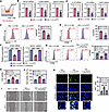 Figure 5
ATF7 overexpression inhibits macrophage M1 polarization. (A) Experimental workflow: Human monocytic THP-1 cells were differentiated into M0 macrophages (THP-1-M0) using 20 nM PMA for 36 hours. HaCaT cells were infected with lentivirus for 48 hours, followed by coculture with THP-1-M0 macrophages in a Transwell system for 24 hours (upper chamber, HaCaT; lower chamber, macrophages). (B) mRNA expression of M1 markers (NOS2, CD86, and MHCII) in cocultured macrophages was measured by qRT-PCR. (C) Flow cytometric analysis of MHCII+ cells in cocultured macrophages. (D) Flow cytometric analysis of CD86+ cells in cocultured macrophages. (E) mRNA expression of proinflammatory cytokines (TNFA, IL6, and IL1B) in cocultured macrophages were measured by qRT-PCR. HaCaT cells were infected with LV-NC or LV-ATF7 and cocultured with THP-1–derived macrophages under HG conditions. In the rescue group (LV-ATF7 + LPS/IFN-γ), macrophages were additionally treated with LPS and IFN-γ during coculture to force M1 polarization. (F) mRNA expression of M1 markers (CD86 and MHCII) in macrophages was measured by qRT-PCR. (G) Flow cytometric analysis of CD86+ cells in cocultured macrophages. (H) Cell viability was assessed by the CCK-8 assay. (I and J) Cell migration ability was analyzed by the scratch wound healing assay. Scale bar: 200 μm. (K) Representative immunofluorescence images of Ki67 expression and quantitative analysis for Ki67+ cells. Nuclei counterstained with DAPI. Scale bar: 50 μm (original magnification: ×400). Results are expressed as mean ± SD. **P < 0.01; ***P < 0.001 by 2-tailed, unpaired Student’s t test (B–E) or 1-way ANOVA with Bonferroni’s post hoc test (F–H, J, and K). All cell experiments were performed with n = 3 replicates.
ATF7-overexpressing keratinocyte-derived exosomes promote diabetic wound healing in vivo. Having established that ATF7 overexpression in keratinocytes suppresses M1 macrophage polarization in vitro, we next asked whether exosomes derived from ATF7-overexpressing keratinocytes are sufficient to promote diabetic wound healing in vivo. To this end, we purified exosomes from HaCaT cells infected with LV-NC (ExoLV-NC) or LV-ATF7 (ExoLV-ATF7), and injected them into full-thickness skin wounds of db/db mice. As shown in Figure 6, A and B, ExoLV-ATF7 treatment accelerated wound closure compared with ExoLV-NC treatment. Histological analysis by H&E staining revealed enhanced re-epithelialization, as evidenced by a reduced epithelial gap and an increased re-epithelialization ratio in ExoLV-ATF7–treated wounds (Figure 6C). IF staining for Ki67 showed an increase in proliferative keratinocytes in the ExoLV-ATF7 group (Figure 6D). Furthermore, flow cytometric analysis of wound tissues showed that ExoLV-ATF7 treatment decreased the percentage of M1 macrophages (MHCII+ and CD86+ populations) (Figure 6, E and F). Collectively, these results indicate that exosomes derived from ATF7-overexpressing keratinocytes promote healing in db/db mice.
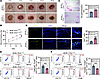 Figure 6
ATF7-overexpressing keratinocyte-derived exosomes promote diabetic wound healing in vivo. Exosomes were purified from conditioned media of HaCaT cells infected with LV-NC (ExoLV-NC) or LV-ATF7 (ExoLV-ATF7). Full-thickness skin wounds were created on the dorsum of db/db mice, followed by injection of exosomes. (A) Representative wound photographs at indicated days after wounding (days 0, 3, 7, 11, and 14). (B) Quantitative analysis of wound closure rate. (C) Representative H&E-stained sections of wound tissues and quantification of re-epithelialization ratio from H&E-stained sections. Scale bar: 500 μm. (D) Representative immunofluorescence images showing Ki67 expression in wound tissues and quantitative analysis for Ki67+ cells. Nuclei counterstained with DAPI. Scale bar: 50 μm (original magnification: ×400). (E) Quantitative analysis of the percentage of M1 macrophages (MHCII+F4/80+CD115+CD11b+). (F) Quantitative analysis of the percentage of M1 macrophages (CD86+F4/80+CD115+CD11b+). Results are expressed as mean ± SD. ***P < 0.001 by 2-way ANOVA with Bonferroni’s post hoc test (B) or 2-tailed, unpaired Student’s t test (C–F). Each experimental group consisted of n = 6 animals.
High-throughput omics screening identifies NOTCH1 as a downstream target of ATF7. To identify downstream molecular targets regulated by ATF7, HaCaT cells were infected with lentivirus and subsequently treated with HG. Transcriptome sequencing (RNA-seq) was then performed on these cells (Figure 7A). Principal component analysis (PCA) demonstrated clear separation between the experimental groups. Heatmap and volcano plot analysis visualized all differentially expressed genes (DEGs) identified by the RNA-seq. Gene Ontology (GO) enrichment analysis of upregulated and downregulated DEGs revealed enrichment in biological processes crucial for wound healing, including extracellular exosome, establishment of skin barrier, and regulation of wound healing. Similarly, Kyoto Encyclopedia of Genes and Genomes (KEGG) pathway enrichment analysis highlighted pathways implicated in wound healing, such as FoxO signaling pathway and Notch signaling pathway. In parallel, exosomes were purified from HG-treated, ATF7-overexpressing HaCaT cells and subjected to label-free quantitative proteomics (Figure 7B). This analysis yielded a set of differentially downregulated factors. Subsequently, we intersected the downregulated factors from label-free quantitative proteomics with the genes enriched in KEGG pathways identified by RNA-seq. This intersection yielded 3 candidate molecules: NOTCH1, YWHAH, and CTNNA1 (Figure 7C). Given the extensively documented role of the NOTCH1 signaling pathway in diabetic wound healing, including reports demonstrating its contribution to delayed healing in diabetic mice (13), NOTCH1 was selected for further investigation.
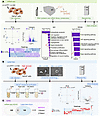 Figure 7
High-throughput omics screening identifies NOTCH1 as a downstream target of ATF7. (A) Transcriptomic analysis: HaCaT cells infected with LV-ATF7 or LV-NC lentivirus (48 hours) were cultured under HG (24 hours) for RNA sequencing. Left to right: Principal component analysis (PCA); volcano plot of differentially expressed genes (DEGs); and cluster heatmap of all DEGs. GO and KEGG pathway enrichment analysis of upregulated and downregulated DEGs are shown. (B) Exosomal proteomics: Exosomes purified from HG-treated HaCaT cells were characterized by transmission electron microscopy. Label-free quantitative proteomics was performed. Left to right: PCA; volcano plot of differentially expressed proteins (DEPs); and cluster heatmap of all DEPs. (C) Target identification: Intersection analysis between factors enriched in the top 5 KEGG pathways from transcriptomics (A) and downregulated proteins from exosomal proteomics (B), yielding 3 overlapping molecules (NOTCH1, YWHAH, and CTNNA1). NOTCH1 was prioritized for validation.
ATF7 transcriptionally represses NOTCH1 and reduces exosomal N1ICD expression. Our investigations revealed that ATF7 overexpression in HaCaT cells downregulated NOTCH1 expression (Figure 8A). Subsequently, exosomes were purified from these HaCaT cells, and analysis revealed a decrease in the expression of the NOTCH1 intracellular domain (N1ICD) within the exosomes (Figure 8B). Analysis using the ChIP-Atlas database predicted the binding for ATF7 within the NOTCH1 promoter region and identified enrichment of H3K9me3 at this promoter (Figure 8C). To validate these predictions, we performed a co-IP assay, which confirmed an interaction between ATF7 and the histone H3K9–specific tri-methyltransferase Suv39h1 (Figure 8D). Furthermore, a chromatin immunoprecipitation–qPCR (ChIP-qPCR) assay exhibited ATF7 binding to the NOTCH1 promoter in HaCaT cells (Figure 8E). Critically, ChIP-qPCR analysis further revealed an increase in H3K9me3 enrichment at the NOTCH1 promoter following ATF7 overexpression (Figure 8F). To extend these mechanistic findings to the in vivo setting, we analyzed wound tissues from db/db mice. Western blotting revealed that NOTCH1 protein levels were reduced in ATF7-overexpressing wounds (Figure 8G). Co-IP using wound tissues showed that ATF7 interacted with Suv39h1 in vivo (Figure 8H). Moreover, IF staining exhibited an increased H3K9me3 signal within tissues of ATF7-overexpressing wounds (Supplemental Figure 1F). To further test the functional relevance of NOTCH1, HaCaT cells were infected with a lentivirus overexpressing N1ICD (Supplemental Figure 1E) and then cultured under HG conditions. N1ICD overexpression impaired cell migration in a wound healing assay (Figure 8I) and decreased the number of Ki67-positive cells (Figure 8J). Altogether, these results imply that ATF7 recruits the histone methyltransferase Suv39h1 to the NOTCH1 promoter, leading to increased H3K9me3 deposition, transcriptional repression of NOTCH1, and consequently reduced levels of both cellular NOTCH1 and exosomal N1ICD.
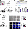 Figure 8
ATF7 transcriptionally represses NOTCH1 and reduces exosomal N1ICD expression. (A) NOTCH1 mRNA and NOTCH1 protein expression in HG-treated (25 mM, 24 hours) HaCaT cells infected with LV-ATF7 or LV-NC lentivirus (48 hours), analyzed by qRT-PCR and Western blotting. (B) N1ICD protein levels in exosomes purified from cells, detected by Western blotting. (C) Predicted binding sites of ATF7 and enrichment of H3K9me3 at the NOTCH1 promoter region through the ChIP-Atlas database. (D) Co-IP analysis of ATF7 interaction with histone H3K9 methyltransferase Suv39h1 in HG-treated HaCaT cells (25 mM, 24 hours). IgG: negative control. (E) ChIP-qPCR confirming ATF7 binding to the NOTCH1 promoter in LV-ATF7–infected, HG-treated HaCaT cells. (F) ChIP-qPCR analysis of H3K9me3 enrichment at the NOTCH1 promoter. (G) Western blot analysis of NOTCH1 protein levels in wound tissues from db/db mice with or without keratinocyte-specific ATF7 overexpression. (H) Co-IP analysis of ATF7 interaction with histone H3K9 methyltransferase Suv39h1 in wound tissues from db/db mice. HaCaT cells were infected with LV-NC or LV-N1ICD and cultured under HG conditions. (I) Cell migration ability was analyzed by the scratch wound healing assay. Scale bar: 200 μm. (J) Representative immunofluorescence images of Ki67 expression and quantitative analysis for Ki67+ cells. Nuclei counterstained with DAPI. Scale bar: 50 μm (original magnification: ×400). Results are expressed as mean ± SD. **P < 0.01, ***P < 0.001 by 2-tailed unpaired Student’s t test (A, E, and F) or 1-way ANOVA with Bonferroni’s post hoc test (I and J). Each experimental group consisted of n = 6 animals. All cell experiments were performed with n = 3 replicates.
Keratinocyte ATF7 inhibits macrophage M1 polarization by reducing exosomal N1ICD secretion. To elucidate the mechanism of exosome-mediated communication, exosomes derived from HaCaT cells were labeled with the green fluorescent dye PKH67 and subsequently coincubated with THP-1-M0 macrophages (Figure 9A). Microscopy analysis revealed an increase in exosome uptake by macrophages after 6 hours of incubation (Figure 9B). Concomitantly, the expression of N1ICD within these macrophages was also elevated (Figure 9C). Exosomes purified from these cells exhibited increased levels of N1ICD protein (Figure 9D). We then purified exosomes from HaCaT cells that were infected with lentivirus and treated with HG. Analysis showed that ATF7 overexpression in keratinocytes reduced exosomal N1ICD levels. This ATF7-mediated reduction in exosomal N1ICD was reversed by overexpression of N1ICD (Figure 9E). These results suggest that the transfer of keratinocyte-derived exosomal N1ICD to macrophages is regulated by ATF7. To assess the impact on macrophage polarization, THP-1-M0 macrophages were cocultured with HaCaT cells coinfected with the relevant lentiviruses and treated with HG (Figure 9F). Flow cytometric analysis revealed that ATF7 overexpression in keratinocytes decreased the percentage of CD86+ macrophages. Notably, this ATF7-induced decrease was partially reversed when N1ICD was co-overexpressed (Figure 9G). Furthermore, the ATF7 overexpression–mediated downregulation of NOS2 and TNFA expression in cocultured macrophages was also counteracted by N1ICD co-overexpression (Figure 9H). These findings indicate that ATF7 in keratinocytes inhibits macrophage M1 polarization by reducing the secretion of N1ICD within exosomes.
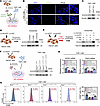 Figure 9
Keratinocyte ATF7 inhibits macrophage M1 polarization by reducing exosomal N1ICD secretion. (A) HaCaT cells and THP-1-M0 macrophages were pretreated with 25 mM HG (24 hours). Exosomes purified from HaCaT cells were labeled with PKH67 (green) and co-incubated with macrophages (6 hours). (B) Confocal microscopy images showing PKH67+ exosome internalization by macrophages (green, exosomes; blue, DAPI). Scale bar: 50 μm (original magnification: ×400). (C) N1ICD protein levels in macrophages after exosome incubation, detected by Western blotting. (D) HaCaT cells were infected with N1ICD-overexpressing lentivirus (LV-N1ICD, 48 hours). Exosomal N1ICD levels were analyzed by Western blotting. (E) HaCaT cells co-infected with LV-ATF7 or LV-N1ICD (48 hours) and treated with HG (24 hours). Exosomal N1ICD levels were analyzed by Western blotting. (F) Macrophages were incubated with exosomes, and recipient macrophage N1ICD levels were detected by Western blotting. (G) Flow cytometric analysis of CD86+ macrophages. (H) mRNA expression of NOS2 and TNFA in macrophages. Results are expressed as mean ± SD. *P < 0.05, ***P < 0.001 by 1-way ANOVA with Bonferroni’s post hoc test. All cell experiments were performed with n = 3 replicates.
-
Discussion
This study reveals an important role for ATF7 in diabetic wound healing. We demonstrate that Atf7 overexpression promotes wound closure in diabetic db/db mice. ATF7 achieves this function by acting as a transcriptional repressor; it recruits the histone methyltransferase Suv39h1 to the NOTCH1 promoter. This recruitment facilitates trimethylation of H3K9me3, leading to transcriptional silencing of NOTCH1 in keratinocytes. A key functional consequence of this ATF7-mediated NOTCH1 suppression is a reduction in the levels of N1ICD within keratinocyte-derived exosomes. Consequently, the delivery of this signaling molecule via exosomes to wound macrophages is diminished. This reduction in exosomal N1ICD transfer inhibits proinflammatory M1 polarization (Figure 10). These findings position ATF7 as a critical transcriptional hub in diabetic wound repair and highlight the therapeutic potential of modulating the ATF7/NOTCH1/exosome axis.
 Figure 10
Proposed mechanism. ATF7 promotes diabetic wound healing by repressing NOTCH1 transcription via H3K9me3, thereby reducing exosomal N1ICD secretion from keratinocytes and inhibiting macrophage M1 polarization.
We identify ATF7 as a beneficial regulator in the diabetic wound milieu. Its previously established roles in cellular stress responses and NF-κB pathway suppression (10), along with its pro-proliferative and migratory capacities (11, 12), are crucial for tissue repair. We observed an upregulation of Atf7 in wounds of db/db mice, which likely represents a physiological compensatory response to the diabetic stress within the wound environment. However, this endogenous upregulation appears insufficient to ameliorate the pathological state. Subsequent in vivo and in vitro overexpression of ATF7 confirmed our hypothesis that augmenting ATF7 levels promotes wound healing. Furthermore, utilizing transcriptomic analysis combined with exosomal proteomic screening, we identified NOTCH1 as a potential downstream target of ATF7.
NOTCH1, a member of the NOTCH protein family, is involved in diverse processes including cell proliferation, migration, and angiogenesis (14). The NOTCH receptor family comprises 4 members (NOTCH1–4) located on the cell membrane, activated by binding to ligands (such as JAG1, -2, or Delta-like 1, 3, and 4) on neighboring cells (15). Upon activation, the NOTCH1 receptor undergoes cleavage, releasing N1ICD, which translocates to the nucleus to regulate gene expression (16, 17). The role of NOTCH signaling in diabetic wound healing is well documented. For instance, Shao et al. demonstrated that NOTCH1 activation (knockin) inhibits fibroblast proliferation and migration, delaying wound healing in diabetic mice (13). Conversely, the NOTCH1 inhibitor DAPT promotes collagen accumulation and accelerates diabetic wound healing (18). In keratinocytes, HG treatment activates the NOTCH1 pathway, and both DAPT treatment and keratinocyte-specific NOTCH1 knockout promote skin wound healing in diabetic mice (19). Our findings demonstrate that ATF7 suppresses NOTCH1 transcription via H3K9me3 modification. Crucially, the therapeutic benefits of ATF7 overexpression were reversed by forced expression of N1ICD, unequivocally confirming the inhibitory effect of ATF7 on NOTCH1 signaling in the context of diabetic wound healing.
Moreover, multiple studies report that the NOTCH1 pathway promotes macrophage M1 polarization (20, 21). During diabetic wound healing, blocking the NOTCH1 pathway suppresses M1 polarization and mitigates the detrimental effects of angiogenesis inhibitors on wound repair (22). Evidence suggests that glioma stem cell–derived exosomes are enriched in NOTCH1 protein. Upon uptake by recipient cells, these exosomes release the N1ICD, influencing downstream gene transcription and target cell function (23). In our study, ATF7 overexpression suppressed markers of M1 polarization in wound macrophages, achieved through NOTCH1 blockade, and reduced exosomal N1ICD transfer. This shift in macrophage polarization phenotype is central to ATF7’s therapeutic effect, as sustained M1 inflammation is a major driver of chronicity in diabetic wounds, complementing prior findings.
Therapeutically, elucidating the ATF7/NOTCH1/exosome axis reveals multiple promising intervention points. Our rescue experiments provide genetic validation for targeting this pathway. Potential strategies include developing pharmacological agonists of ATF7, inhibiting NOTCH1 signaling using inhibitors like peptide inhibitor TAT-ANK (24) and the clinically relevant γ-secretase inhibitor DAPT (25). Alternatively, exosomes have been engineered to specifically deplete N1ICD cargo, analogous to promising applications of stem cell–derived exosomes (26). Targeting ATF7 or its downstream effectors offers potential advantages over growth factor therapies (VEGF and FGF) by acting upstream at the level of a transcriptional regulator, potentially yielding broader and more sustained effects while circumventing issues of protein instability or short half-life encountered by some growth factors in the harsh wound environment (27, 28).
Several limitations of this study should be acknowledged. First, while our experiments in db/db mice demonstrate that keratinocyte-specific Atf7 overexpression accelerates diabetic wound healing, and complementary in vitro experiments show that ATF7 knockdown impairs keratinocyte proliferation and migration, in vivo evidence from keratinocyte-specific Atf7-deficient mice is lacking. We recognize that the availability of such evidence would provide more proof of the important role of ATF7 in diabetic healing. Second, validation of our findings in human diabetic wounds is still at a preliminary stage. We collected human diabetic foot ulcer (DFU) tissues (n = 6) and non-diabetic normal skin tissues (n = 2). Western blot analysis revealed that both ATF7 and NOTCH1 proteins were elevated in DFU samples compared with controls (Supplemental Figure 1A). We interpret this co-upregulation as an endogenous protective attempt that is insufficient to overcome the complex chronic wound microenvironment. Consequently, NOTCH1 remains elevated and healing fails. This interpretation aligns with the literature showing that NOTCH1 impairs diabetic wound healing and underscores the therapeutic rationale for exogenously enhancing ATF7 activity — exactly as we validated in our mouse model. However, due to the small sample size of normal skin, definitive conclusions regarding ATF7 and NOTCH1 expression levels in human diabetic wounds await confirmation in larger cohorts. We are actively continuing sample collection and will examine ATF7, NOTCH1, and posttranslational regulators in future studies once sufficient tissue is obtained.
In conclusion, this study advances our understanding of the pathogenesis and repair processes in diabetic wounds by identifying ATF7 as a critical pro-healing regulator. It provides insights for developing innovative strategies to restore healing in chronic diabetic wounds.


