
Research ArticleCell biologyDermatology
Open Access |  10.1172/jci.insight.197838
Multiomics analysis reveals dermokine as a regulator of keratinocyte differentiation and adhesion
Vahap Canbay,1 Till Wüstemann,2 Weihua Tian,1 Tobias A. Beyer,3 Camilla Reiter Elbæk,1 Michael Stumpe,4 Gaetana Restivo,5 Chatpakorn Christiansen,1 Anabel Migenda Herranz,3 Susanne Mailand,3 Jürg Hafner,5 Rune Busk Damgaard,1 Steffen Goletz,1 Jörn Dengjel,6 Ulrich auf dem Keller,1 and Chiara Francavilla1,4
1Department for Biotechnology and Biomedicine, Technical University of Denmark, Lyngby, Denmark.
2Department of Biology, Swiss Federal Institute of Technology Zurich, Zurich, Switzerland.
3Cytosurge AG, Glattbrugg, Zurich, Switzerland.
4Division of Molecular and Cellular Function, Faculty of Biology, Medicine and Health, The University of Manchester, Manchester, United Kingdom.
5Department of Dermatology, University Hospital Zurich, Zurich, Switzerland.
6Department of Biology, University of Fribourg, Fribourg, Switzerland.
Address correspondence to: Chiara Francavilla, Department for Biotechnology and Biomedicine, Technical University of Denmark, Søltofts Plads, 2800 Kongens Lyngby, Denmark. Phone: 4593511641; Email: chiafra@dtu.dk.
Authorship note: UADK is deceased.
Find articles by Canbay, V. in: PubMed | Google Scholar
1Department for Biotechnology and Biomedicine, Technical University of Denmark, Lyngby, Denmark.
2Department of Biology, Swiss Federal Institute of Technology Zurich, Zurich, Switzerland.
3Cytosurge AG, Glattbrugg, Zurich, Switzerland.
4Division of Molecular and Cellular Function, Faculty of Biology, Medicine and Health, The University of Manchester, Manchester, United Kingdom.
5Department of Dermatology, University Hospital Zurich, Zurich, Switzerland.
6Department of Biology, University of Fribourg, Fribourg, Switzerland.
Address correspondence to: Chiara Francavilla, Department for Biotechnology and Biomedicine, Technical University of Denmark, Søltofts Plads, 2800 Kongens Lyngby, Denmark. Phone: 4593511641; Email: chiafra@dtu.dk.
Authorship note: UADK is deceased.
Find articles by Wüstemann, T. in: PubMed | Google Scholar
1Department for Biotechnology and Biomedicine, Technical University of Denmark, Lyngby, Denmark.
2Department of Biology, Swiss Federal Institute of Technology Zurich, Zurich, Switzerland.
3Cytosurge AG, Glattbrugg, Zurich, Switzerland.
4Division of Molecular and Cellular Function, Faculty of Biology, Medicine and Health, The University of Manchester, Manchester, United Kingdom.
5Department of Dermatology, University Hospital Zurich, Zurich, Switzerland.
6Department of Biology, University of Fribourg, Fribourg, Switzerland.
Address correspondence to: Chiara Francavilla, Department for Biotechnology and Biomedicine, Technical University of Denmark, Søltofts Plads, 2800 Kongens Lyngby, Denmark. Phone: 4593511641; Email: chiafra@dtu.dk.
Authorship note: UADK is deceased.
Find articles by Tian, W. in: PubMed | Google Scholar
1Department for Biotechnology and Biomedicine, Technical University of Denmark, Lyngby, Denmark.
2Department of Biology, Swiss Federal Institute of Technology Zurich, Zurich, Switzerland.
3Cytosurge AG, Glattbrugg, Zurich, Switzerland.
4Division of Molecular and Cellular Function, Faculty of Biology, Medicine and Health, The University of Manchester, Manchester, United Kingdom.
5Department of Dermatology, University Hospital Zurich, Zurich, Switzerland.
6Department of Biology, University of Fribourg, Fribourg, Switzerland.
Address correspondence to: Chiara Francavilla, Department for Biotechnology and Biomedicine, Technical University of Denmark, Søltofts Plads, 2800 Kongens Lyngby, Denmark. Phone: 4593511641; Email: chiafra@dtu.dk.
Authorship note: UADK is deceased.
Find articles by Beyer, T. in: PubMed | Google Scholar
1Department for Biotechnology and Biomedicine, Technical University of Denmark, Lyngby, Denmark.
2Department of Biology, Swiss Federal Institute of Technology Zurich, Zurich, Switzerland.
3Cytosurge AG, Glattbrugg, Zurich, Switzerland.
4Division of Molecular and Cellular Function, Faculty of Biology, Medicine and Health, The University of Manchester, Manchester, United Kingdom.
5Department of Dermatology, University Hospital Zurich, Zurich, Switzerland.
6Department of Biology, University of Fribourg, Fribourg, Switzerland.
Address correspondence to: Chiara Francavilla, Department for Biotechnology and Biomedicine, Technical University of Denmark, Søltofts Plads, 2800 Kongens Lyngby, Denmark. Phone: 4593511641; Email: chiafra@dtu.dk.
Authorship note: UADK is deceased.
Find articles by Elbæk, C. in: PubMed | Google Scholar
1Department for Biotechnology and Biomedicine, Technical University of Denmark, Lyngby, Denmark.
2Department of Biology, Swiss Federal Institute of Technology Zurich, Zurich, Switzerland.
3Cytosurge AG, Glattbrugg, Zurich, Switzerland.
4Division of Molecular and Cellular Function, Faculty of Biology, Medicine and Health, The University of Manchester, Manchester, United Kingdom.
5Department of Dermatology, University Hospital Zurich, Zurich, Switzerland.
6Department of Biology, University of Fribourg, Fribourg, Switzerland.
Address correspondence to: Chiara Francavilla, Department for Biotechnology and Biomedicine, Technical University of Denmark, Søltofts Plads, 2800 Kongens Lyngby, Denmark. Phone: 4593511641; Email: chiafra@dtu.dk.
Authorship note: UADK is deceased.
Find articles by Stumpe, M. in: PubMed | Google Scholar
1Department for Biotechnology and Biomedicine, Technical University of Denmark, Lyngby, Denmark.
2Department of Biology, Swiss Federal Institute of Technology Zurich, Zurich, Switzerland.
3Cytosurge AG, Glattbrugg, Zurich, Switzerland.
4Division of Molecular and Cellular Function, Faculty of Biology, Medicine and Health, The University of Manchester, Manchester, United Kingdom.
5Department of Dermatology, University Hospital Zurich, Zurich, Switzerland.
6Department of Biology, University of Fribourg, Fribourg, Switzerland.
Address correspondence to: Chiara Francavilla, Department for Biotechnology and Biomedicine, Technical University of Denmark, Søltofts Plads, 2800 Kongens Lyngby, Denmark. Phone: 4593511641; Email: chiafra@dtu.dk.
Authorship note: UADK is deceased.
Find articles by Restivo, G. in: PubMed | Google Scholar
1Department for Biotechnology and Biomedicine, Technical University of Denmark, Lyngby, Denmark.
2Department of Biology, Swiss Federal Institute of Technology Zurich, Zurich, Switzerland.
3Cytosurge AG, Glattbrugg, Zurich, Switzerland.
4Division of Molecular and Cellular Function, Faculty of Biology, Medicine and Health, The University of Manchester, Manchester, United Kingdom.
5Department of Dermatology, University Hospital Zurich, Zurich, Switzerland.
6Department of Biology, University of Fribourg, Fribourg, Switzerland.
Address correspondence to: Chiara Francavilla, Department for Biotechnology and Biomedicine, Technical University of Denmark, Søltofts Plads, 2800 Kongens Lyngby, Denmark. Phone: 4593511641; Email: chiafra@dtu.dk.
Authorship note: UADK is deceased.
Find articles by Christiansen, C. in: PubMed | Google Scholar
1Department for Biotechnology and Biomedicine, Technical University of Denmark, Lyngby, Denmark.
2Department of Biology, Swiss Federal Institute of Technology Zurich, Zurich, Switzerland.
3Cytosurge AG, Glattbrugg, Zurich, Switzerland.
4Division of Molecular and Cellular Function, Faculty of Biology, Medicine and Health, The University of Manchester, Manchester, United Kingdom.
5Department of Dermatology, University Hospital Zurich, Zurich, Switzerland.
6Department of Biology, University of Fribourg, Fribourg, Switzerland.
Address correspondence to: Chiara Francavilla, Department for Biotechnology and Biomedicine, Technical University of Denmark, Søltofts Plads, 2800 Kongens Lyngby, Denmark. Phone: 4593511641; Email: chiafra@dtu.dk.
Authorship note: UADK is deceased.
Find articles by Migenda Herranz, A. in: PubMed | Google Scholar
1Department for Biotechnology and Biomedicine, Technical University of Denmark, Lyngby, Denmark.
2Department of Biology, Swiss Federal Institute of Technology Zurich, Zurich, Switzerland.
3Cytosurge AG, Glattbrugg, Zurich, Switzerland.
4Division of Molecular and Cellular Function, Faculty of Biology, Medicine and Health, The University of Manchester, Manchester, United Kingdom.
5Department of Dermatology, University Hospital Zurich, Zurich, Switzerland.
6Department of Biology, University of Fribourg, Fribourg, Switzerland.
Address correspondence to: Chiara Francavilla, Department for Biotechnology and Biomedicine, Technical University of Denmark, Søltofts Plads, 2800 Kongens Lyngby, Denmark. Phone: 4593511641; Email: chiafra@dtu.dk.
Authorship note: UADK is deceased.
Find articles by Mailand, S. in: PubMed | Google Scholar
1Department for Biotechnology and Biomedicine, Technical University of Denmark, Lyngby, Denmark.
2Department of Biology, Swiss Federal Institute of Technology Zurich, Zurich, Switzerland.
3Cytosurge AG, Glattbrugg, Zurich, Switzerland.
4Division of Molecular and Cellular Function, Faculty of Biology, Medicine and Health, The University of Manchester, Manchester, United Kingdom.
5Department of Dermatology, University Hospital Zurich, Zurich, Switzerland.
6Department of Biology, University of Fribourg, Fribourg, Switzerland.
Address correspondence to: Chiara Francavilla, Department for Biotechnology and Biomedicine, Technical University of Denmark, Søltofts Plads, 2800 Kongens Lyngby, Denmark. Phone: 4593511641; Email: chiafra@dtu.dk.
Authorship note: UADK is deceased.
Find articles by Hafner, J. in: PubMed | Google Scholar
1Department for Biotechnology and Biomedicine, Technical University of Denmark, Lyngby, Denmark.
2Department of Biology, Swiss Federal Institute of Technology Zurich, Zurich, Switzerland.
3Cytosurge AG, Glattbrugg, Zurich, Switzerland.
4Division of Molecular and Cellular Function, Faculty of Biology, Medicine and Health, The University of Manchester, Manchester, United Kingdom.
5Department of Dermatology, University Hospital Zurich, Zurich, Switzerland.
6Department of Biology, University of Fribourg, Fribourg, Switzerland.
Address correspondence to: Chiara Francavilla, Department for Biotechnology and Biomedicine, Technical University of Denmark, Søltofts Plads, 2800 Kongens Lyngby, Denmark. Phone: 4593511641; Email: chiafra@dtu.dk.
Authorship note: UADK is deceased.
Find articles by Damgaard, R. in: PubMed | Google Scholar
1Department for Biotechnology and Biomedicine, Technical University of Denmark, Lyngby, Denmark.
2Department of Biology, Swiss Federal Institute of Technology Zurich, Zurich, Switzerland.
3Cytosurge AG, Glattbrugg, Zurich, Switzerland.
4Division of Molecular and Cellular Function, Faculty of Biology, Medicine and Health, The University of Manchester, Manchester, United Kingdom.
5Department of Dermatology, University Hospital Zurich, Zurich, Switzerland.
6Department of Biology, University of Fribourg, Fribourg, Switzerland.
Address correspondence to: Chiara Francavilla, Department for Biotechnology and Biomedicine, Technical University of Denmark, Søltofts Plads, 2800 Kongens Lyngby, Denmark. Phone: 4593511641; Email: chiafra@dtu.dk.
Authorship note: UADK is deceased.
Find articles by Goletz, S. in: PubMed | Google Scholar
1Department for Biotechnology and Biomedicine, Technical University of Denmark, Lyngby, Denmark.
2Department of Biology, Swiss Federal Institute of Technology Zurich, Zurich, Switzerland.
3Cytosurge AG, Glattbrugg, Zurich, Switzerland.
4Division of Molecular and Cellular Function, Faculty of Biology, Medicine and Health, The University of Manchester, Manchester, United Kingdom.
5Department of Dermatology, University Hospital Zurich, Zurich, Switzerland.
6Department of Biology, University of Fribourg, Fribourg, Switzerland.
Address correspondence to: Chiara Francavilla, Department for Biotechnology and Biomedicine, Technical University of Denmark, Søltofts Plads, 2800 Kongens Lyngby, Denmark. Phone: 4593511641; Email: chiafra@dtu.dk.
Authorship note: UADK is deceased.
Find articles by Dengjel, J. in: PubMed | Google Scholar
1Department for Biotechnology and Biomedicine, Technical University of Denmark, Lyngby, Denmark.
2Department of Biology, Swiss Federal Institute of Technology Zurich, Zurich, Switzerland.
3Cytosurge AG, Glattbrugg, Zurich, Switzerland.
4Division of Molecular and Cellular Function, Faculty of Biology, Medicine and Health, The University of Manchester, Manchester, United Kingdom.
5Department of Dermatology, University Hospital Zurich, Zurich, Switzerland.
6Department of Biology, University of Fribourg, Fribourg, Switzerland.
Address correspondence to: Chiara Francavilla, Department for Biotechnology and Biomedicine, Technical University of Denmark, Søltofts Plads, 2800 Kongens Lyngby, Denmark. Phone: 4593511641; Email: chiafra@dtu.dk.
Authorship note: UADK is deceased.
Find articles by auf dem Keller, U. in: PubMed | Google Scholar
1Department for Biotechnology and Biomedicine, Technical University of Denmark, Lyngby, Denmark.
2Department of Biology, Swiss Federal Institute of Technology Zurich, Zurich, Switzerland.
3Cytosurge AG, Glattbrugg, Zurich, Switzerland.
4Division of Molecular and Cellular Function, Faculty of Biology, Medicine and Health, The University of Manchester, Manchester, United Kingdom.
5Department of Dermatology, University Hospital Zurich, Zurich, Switzerland.
6Department of Biology, University of Fribourg, Fribourg, Switzerland.
Address correspondence to: Chiara Francavilla, Department for Biotechnology and Biomedicine, Technical University of Denmark, Søltofts Plads, 2800 Kongens Lyngby, Denmark. Phone: 4593511641; Email: chiafra@dtu.dk.
Authorship note: UADK is deceased.
Find articles by Francavilla, C. in: PubMed | Google Scholar
Published April 23, 2026 - More info
JCI Insight. 2026;11(13):e197838. https://doi.org/10.1172/jci.insight.197838.
© 2026 Canbay et al. This work is licensed under the Creative Commons Attribution 4.0 International License. To view a copy of this license, visit http://creativecommons.org/licenses/by/4.0/.
Received: July 10, 2025; Accepted: April 20, 2026


-
Results
Dermokine is important for early keratinocyte differentiation in 3D skin equivalents. To study a possible role of dermokine in keratinocyte differentiation (12), we edited the genomic sites coding for either dermokine-β/γ or -α/β (DMKN βγ or αβ) in human keratinocytes by two different CRISPR/Cas9 methods (19, 24) (Figure 1A). The immortalized but non-tumorigenic N/TERT keratinocyte cell line was used for this purpose, because it undergoes normal differentiation and stratification in 3D skin equivalents and allows the generation of clonally expanded lines (25). The first method, chemical transfection, which is based on fluorescence-activated cell sorting (FACS) of single-cell clones and clonal expansion, generated the DMKN βγ–/– cell line (26) (Figure 1B). The second method, the FluidFM CRISPR approach, which was based on coinjection of CRISPR ribonucleoprotein (RNP) and green fluorescent protein (GFP) mRNA into keratinocytes, generated the DMKN αβ–/– cell line (27) (Figure 1C). We confirmed the lack of expression of DMKN generated with either method by Sanger sequencing (28) (Supplemental Figure 1, A–D; supplemental material available online with this article; https://doi.org/10.1172/jci.insight.197838DS1). We then established 3D organotypic skin cultures by plating either WT or DMKN βγ–/– or DMKN αβ–/– keratinocytes on top of primary human fibroblasts incorporated in Matrigel, as previously described (19) (Figure 1, B and C). Under these conditions, keratinocytes form a multilayered, stratified epidermis, resembling human skin (19). We evaluated whether changing the calcium concentration in our experimental conditions from 1 mM to 5 mM would affect cell proliferation and differentiation by staining for keratin 14 (KRT14), a marker for non-differentiated keratinocytes, and for the early-differentiation marker KRT10 (Supplemental Figure 1E). Quantification of immunofluorescence stainings for KRT10 and KRT14 in varying calcium concentrations showed that the differentiation marker KRT10 did not change, while KRT14 levels decreased in response to elevated calcium (Supplemental Figure 1E). These results suggest that cells undergo the differentiation program in 3D organotypic skin culture, giving rise to a stratified, epidermis-like structure. To confirm changes in the expression of dermokine isoforms in this model system at the protein level, we could not use immunoblotting because of the lack of antibodies recognizing the specific isoforms. Instead, we used targeted proteomics and synthetic reference peptides (29, 30). We selected 5 proteotypic peptides spread across the entire dermokine sequence, including 2 near the N-terminus, 2 after the keratin-like domain, and 1 at the C-terminus (Figure 1D and Supplemental Table 1). As the C-terminal peptide GGVSPSSSASR is shared by both dermokine-β and -α (Figure 1D), deletion of the sole dermokine-β/γ isoform would still give a signal in the mass spectrometer because of the expression of the α isoform. Indeed, only a slight reduction in the peptide peak area was observed when only isoforms β and γ were genetically deleted (Figure 1E, Supplemental Figure 1F, and Supplemental Table 1). However, removing the dermokine-α and -β isoforms simultaneously resulted in a substantial decrease in the abundance of such a shared C-terminal peptide (Figure 1F, Supplemental Figure 1F, and Supplemental Table 1). Among the remaining 4 peptides, the N-terminal peptide VGEAAHALGNTGHEIGR showed the highest decrease in abundance upon depletion of both DMKN βγ and DMKN αβ in 3D organotypic skin cultures (Supplemental Figure 1, F–J, and Supplemental Table 1). Then, we analyzed WT and DMKN-KO 3D cultures by immunohistochemical (IHC) staining and showed the strong reduction of dermokine expression in both DMKN βγ–/– and DMKN αβ–/– compared with WT cultures (Figure 2). Hematoxylin and eosin as well as IHC staining revealed the lack of suprabasal layers beyond the spinous layer in the DMKN-KO compared with the WT samples (Figure 2). This observation was consistent with the known dermokine expression pattern in keratinocytes (13). Together, these data suggest that the expression of DMKN isoforms is indeed reduced at the protein level in our model system, which seems to affect keratinocyte differentiation.
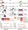 Figure 1
Targeted proteomics validates the knockout of dermokine in human keratinocytes. (A) Schematic of the genetic loci encoding the different dermokine isoforms and locations of guide RNAs (gRNA 1 and gRNA 2) targeting isoform-specific exons. (B and C) Methodologies used to delete dermokine in keratinocytes through either chemical transfection (B) or the FluidFM CRISPR approach (C). After transfection and clonal expansion, WT, DMKN αβ–/–, or DMKN βγ–/– keratinocytes were used for the generation of 3D organotypic skin cultures. (D) Schematic of the protein domains of the dermokine isoforms and of the 5 isoform-specific and proteotypic peptides used for targeted proteomics analysis. (E and F) Proteotypic dermokine peptide abundances are log2-transformed fold changes of summed transition areas from DMKN βγ–/– (E) or DMKN αβ–/– (F) relative to WT keratinocytes analyzed by targeted proteomics. N = 3 different 3D organotypic skin cultures. Statistical significance was calculated using 2-sided Student’s t tests on log2-transformed intensities, with multiple-testing correction performed using the Benjamini-Hochberg false discovery rate.
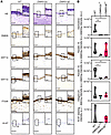 Figure 2
Staining of DMKN-KO and WT 3D organotypic skin cultures shows changes in epidermal marker proteins. (A) H&E and IHC staining with the indicated antibodies of WT and DMKN αβ–/– or DMKN βγ–/– keratinocyte 3D organotypic skin cultures. N = 3. Scale bars: 100 μm; and 25 μm in magnified images. (B) Truncated violin plots showing the semiquantitative IHC staining intensity with the indicated antibodies. N = 3 different 3D organotypic skin cultures. *P < 0.05 (1-way ANOVA and Fisher’s least significant difference test). a.u., arbitrary units.
Next, we evaluated whether the lack of dermokine affected the early-stage differentiation of keratinocytes by staining for KRT14 and KRT10 (1). The expression of KRT14 was comparable in WT and in both DMKN-KO cultures, whereas the expression of KRT10 decreased in the KOs compared with the WT samples (1) (Figure 2). These data suggest that dermokine is important for early keratinocyte differentiation. Expression of integrin α6 (ITGα6), a marker of basal keratinocytes (31), and expression of the proliferation marker Ki-67 were similar in WT and DMKN-KO keratinocytes, suggesting that hemidesmosome formation and proliferation do not change upon dermokine depletion (1) (Figure 2). Altogether, the IHC analysis suggests that the DMKN knockout reduces the amounts of differentiation-specific proteins.
Knockout of dermokine variants alters the keratinocyte proteome in organotypic skin cultures. To characterize the phenotype of DMKN-KO samples in an unbiased manner, we analyzed changes in the proteome of the total 3D organotypic skin cultures grown from either WT or DMKN-KO keratinocytes via quantitative liquid chromatography/mass spectrometry–based (LC-MS–based) proteomics. In total, we quantified 3,753 proteins in WT, DMKN βγ–/–, and DMKN αβ–/– 3D cultures with a coefficient of variation (CV) of less than 15% between the replicates (Supplemental Figure 2A and Supplemental Table 2). The Pearson’s correlation analysis of WT, DMKN βγ–/–, and DMKN αβ–/– 3D cultures revealed high correlation among replicates for each experimental condition (Supplemental Figure 2B), in line with previous publications (32, 33). Finally, principal component analysis (PCA) demonstrated clear differences in the proteome of cultures with the 3 different genotypes (Supplemental Figure 2C). Hierarchical clustering identified clusters including proteins that are more abundant in the WT (cluster 2), in both the KO (cluster 4), or only in one of the two KO samples (clusters 1 and 3) (Supplemental Figure 2D), suggesting individual but also overlapping functions of the two dermokine isoforms in keratinocytes. Gene Ontology (GO) term enrichment analysis showed that proteins in cluster 2 were enriched in terms like “formation of the cornified envelope” and proteins in cluster 4 were associated with signaling pathways (Supplemental Figure 2E). The differential abundance analysis, visualized by volcano plot, revealed 312 and 231 significantly more abundant proteins in the DMKN βγ–/– and DMKN αβ–/– 3D cultures, respectively, when compared with WT (Figure 3A and Supplemental Table 2). We also identified 217 and 230 significantly less abundant proteins in the respective DMKN βγ–/– and DMKN αβ–/– keratinocytes in comparison with the WT (Figure 3B and Supplemental Table 2). The protein abundance of dermokine and KRT10 was reduced in both DMKN-KO 3D cultures, confirming the results of the IHC staining (Figure 2 and Figure 3, A and B). KRT14 and ITGα6 showed no differential abundance in the DMKN βγ–/– clone, as seen in the IHC analysis, but were slightly increased in the DMKN αβ–/– clone (Figure 2 and Figure 3, A and B). Along with dermokine, the abundance of suprabasin (SBSN) and keratinocyte differentiation–associated protein (KRTDAP), which are all members of the stratified epithelium–secreted peptides complex (SSC) expressed in the suprabasal layers of the stratified epithelium (34), was also reduced (Figure 3, A and B). GO enrichment analysis of significantly less abundant proteins from both the DMKN KOs compared with WT clones showed the enrichment of pathways such as “keratinocyte differentiation” and “formation of the cornified envelope” (Figure 3C), whereas the GO enrichment analysis of significantly more abundant proteins showed the enrichment of “integrin binding,” “positive regulation of locomotion,” and “growth factor binding” pathways (Figure 3D).
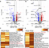 Figure 3
Proteomic analysis of dermokine-ablated 3D organotypic skin cultures shows phenotypic differences. (A and B) Volcano plots showing the differential protein abundance between DMKN αβ–/– (A) or DMKN βγ–/– (B) and WT keratinocytes in 3D organotypic skin cultures. Proteins characteristic of different epidermal strata are highlighted (1). Values are log2-transformed fold changes and –log10-transformed P values (2-sided moderated t test [empirical Bayes]) of 3 different (3D) organotypic skin cultures. (C and D) Gene Ontology analysis of significantly decreased (C) or increased (D) proteins from DMKN αβ–/– (A) or DMKN βγ–/– (B) relative to the WT proteome.
In conclusion, dermokine regulates the abundance of proteins of the suprabasal layers of the epidermis in human skin equivalents.
Phosphoproteomics analysis reveals that dermokine regulates cell-cell adhesion proteins. To uncover how loss of dermokine may affect keratinocyte differentiation, we studied changes in cellular signaling by deep quantitative proteomics and phosphoproteomics analyses of WT, DMKN αβ–/–, and DMKN βγ–/– keratinocytes in monocultures. In addition, we tested whether treatment of the KO samples with recombinant dermokine-β (35) rescues the alterations in the proteome and phosphoproteome.
We first focused on the proteome and found 5,118 unique quantified proteins (Supplemental Table 3) with the median CV of the protein groups being below 20% for all samples and the correlation analysis showing high correlation between replicates, but clear differences between the different genotypes in the PCA analysis (Supplemental Figure 3, B–D). The DMKN αβ–/– or DMKN βγ–/– proteomes differed from the WT proteome, as shown by hierarchical clustering (Supplemental Figure 4A), and this difference was not rescued by treatment with recombinant dermokine. When we subjected the significantly differentially abundant proteins in the different samples to GO enrichment analysis, we found several enriched GO terms, including the term “cadherin binding”, reciprocally enriched in 2 conditions: when comparing the most abundant proteins from non-treated DMKN αβ–/– and DMKN βγ–/– cells with those from WT keratinocytes (Supplemental Figure 4B) and when comparing the significantly less abundant proteins from the dermokine-treated DMKN αβ–/– and DMKN βγ–/– cell lines with those from WT keratinocytes (Supplemental Figure 4C). These 2D results confirm that dermokine knockout changes the keratinocyte proteome (Figure 3).
Next, we analyzed changes in the phosphoproteome of non-treated and dermokine-treated DMKN βγ–/– and DMKN αβ–/– cell lines compared with WT keratinocytes (Supplemental Figure 3A and Supplemental Table 4). We enriched phosphorylated peptides using automated iron(III)–nitrilotriacetic acid [Fe(III)-NTA] and used a modified high-resolution MS–data-independent acquisition (HRMS1-DIA) strategy covering a 400 to 1,400 m/z mass range (36–38) (Supplemental Figure 5A). In total, we identified 11,778 phosphorylation sites after filtering for localization site probability (≥0.75) and 4,453 fully quantifiable phosphorylated sites across all conditions on 2,695 proteins (Supplemental Figure 5B and Supplemental Table 4). All replicates showed high correlation scores (Supplemental Figure 5C), and we found 10,027 singly, 1,367 doubly, and 384 multiply phosphorylated sites (Supplemental Figure 5D) and 10,315, 1,164, and 299 phosphorylated serine, threonine, and tyrosine amino acids, respectively (Supplemental Figure 5E). Overall, the quality of the phosphoproteomics datasets was consistent with previous publications (38). We observed significant differences between the phosphoproteome of DMKN βγ–/– and DMKN αβ–/– cell lines compared with the WT cell phosphoproteome (4,453 phosphorylated sites) by hierarchical clustering (Supplemental Figure 5F). The phosphoproteome of dermokine-treated DMKN βγ–/– and DMKN αβ–/– keratinocytes was similar to the phosphoproteome of WT cells, suggesting a considerable reversal of the abnormal phosphorylation pattern of the KO keratinocytes upon treatment with recombinant dermokine (Supplemental Figure 5F). To study how dermokine affects cellular signaling, we applied a global kinase-substrate prediction approach (39), which reveals the likelihood of a kinase modifying a phosphorylated site by linking kinase recognition sequence motifs and known in vivo and in vitro phosphorylation profiles (39) to the 4,453 phosphorylated sites identified in the WT and the KO samples. This approach identified 94 putative kinases (Supplemental Table 5) whose known downstream phosphorylated sites were visualized in a heatmap (Figure 4A). Among the 94 kinases and based on the kinase activity score, we identified 30 kinases with a high change and 15 kinases that showed significantly high changes between the endogenous WT and all the DMKN βγ–/– and DMKN αβ–/– samples (Figure 4B and Supplemental Table 5). Four of the latter kinases were cytoplasmic tyrosine kinases (DYRK1A, PTK2B, SRC, and SYK), of which both SRC and SYK showed decreased activity scores in the DMKN βγ–/– and DMKN αβ–/– keratinocytes compared with WT (Figure 4B). Similarly, our data showed significantly higher activity scores of ROCK1, a key regulator of the cytoskeleton (40, 41), in KO keratinocytes relative to WT cells (Figure 4B). Next, we used the kinase-substrate annotations generated by the global kinase-substrate prediction approach to cluster substrates sharing similar kinase profiles and regulation (39), and we identified 8 clusters (Supplemental Figure 6 and Supplemental Table 5). Interestingly, each cluster had a different distribution of phosphorylated serine, threonine, and tyrosine residues, ranging from clusters 2, 3, 4, and 6 not containing any phosphorylated tyrosines, to cluster 8 containing only phosphorylated serines, and clusters 5 and 7 mainly containing phosphorylated tyrosine and threonine sites, with cluster 7 being associated with PTK2B, ROCK1, SRC, and SYK activity (Supplemental Figure 6). GO enrichment analysis of the 8 clusters revealed enrichment of terms related to RNA regulation in all clusters, specifically in cluster 7, which, together with cluster 2, also contained several proteins associated with “adherens junctions” and “cadherin binding” (Figure 4C). This finding is in line with SRC, a known player in cell-cell adhesion (42), being the most likely kinase regulating the proteins belonging to cluster 7 (Supplemental Figure 6) and with “cadherin binding” being the most strongly dysregulated pathway in the DMKN βγ–/– and DMKN αβ–/– keratinocytes compared with WT (Supplemental Figure 4). To identify potential dermokine-regulated phosphorylated sites, we counted the number of sites on each phosphorylated protein belonging to the pathways associated with “cadherin binding” and “adherens junctions” associated with clusters 2, 4, 5, and 7. We found several phosphorylated sites (between 8 and 12) on proteins associated with cell-cell adhesion, including on TNKS1BP1 (12), TJP1 (11), SCRIB (10), TJP2 (10), CTNNB1 (9), AFDN (8), and CTNND1 (or p120) (8) (Supplemental Figure 6 and Supplemental Table 5). Among these proteins, p120 was phosphorylated on S252, S268, S288, and T916, all sites playing crucial roles in modulating cell-cell adhesion complexes and cytoskeletal dynamics (23, 43, 44). To confirm that the regulated phosphorylated sites identified on p120 as well as other proteins were bona fide phosphorylated sites due to changes in kinase activity and independent of alterations in protein abundance (45), we normalized the phosphoproteome on the proteome (Supplemental Figure 7A). We found that the normalized phosphoproteome of WT differed from that of DMKN βγ–/– and DMKN αβ–/– keratinocytes. When the DMKN βγ–/– and DMKN αβ–/– keratinocytes were treated with recombinant dermokine, their phosphoproteome became similar to the phosphoproteome of WT keratinocytes (Supplemental Figure 7A). This finding is consistent with the result observed before normalization (Figure 5, A and B). The phosphorylation pattern on p120 also remained unchanged after normalization (Supplemental Table 4). We validated the phosphorylation of p120 on two of the identified phosphorylated sites, serine 252 (S252) and serine 268 (S268), for which antibodies and fully annotated MS/MS spectra were available (Supplemental Figure 7, B and C). Immunoblot analysis of lysates from DMKN βγ–/–, DMKN αβ–/–, and WT keratinocytes showed that the phosphorylation of both S252 and S268 on p120 increased in the absence of dermokine (Supplemental Figure 7D). Furthermore, we observed that p120 phosphorylation was similar to WT when recombinant dermokine was added to DMKN βγ–/–, DMKN αβ–/–, and WT keratinocytes (Supplemental Figure 7E). In summary, the phosphoproteomics analyses of dermokine KO samples identified differentially phosphorylated proteins, including p120.
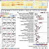 Figure 4
Absence of dermokine results in phosphorylation changes in adhesion proteins. (A) The kinase-substrate prediction approach (39) matched quantified phosphorylated sites in the WT and the two KO samples with the 94 identified putative kinases (Supplemental Table 5). The heatmap shows the combined score of the in vivo or in vitro kinases and sequence recognition motifs (39). The vertical axis shows substrates, and the horizontal axis represents kinases. Families of kinases are color-coded on top of the graph. (B) The kinase activity score for DMKN αβ–/–, DMKN βγ–/–, and WT conditions, treated with recombinant dermokine or vehicle, is shown for 15 significantly regulated kinases out of the 94 identified kinases. Values are kinase activity scores from N = 3 technical replicates from the different genotypes and treatments. One-way ANOVA test determined changes among all genotypes. (C) Gene Ontology analysis from proteins whose phosphorylated sites were annotated to kinases identified in clusters shown in Supplemental Figure 6. BP, biological process; CC, cellular component; MF, molecular function.
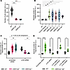 Figure 5
Cell-cell adhesion decreases in dermokine-depleted keratinocytes. (A) Quantification of 4 or more cell clusters from the cell-cell adhesion assay using DMKN αβ–/–, DMKN βγ–/–, and WT keratinocytes. Cell clusters (≥4) were counted, and each replicate was visualized as a dot in the violin plot. N = 3 biological replicates. ***P < 0.001 (1-way ANOVA and Fisher’s least significant difference test). (B) Quantification of dispase dissociation assay data using independent clones of human DMKN αβ–/–, DMKN βγ–/–, DMKN αβγ–/–, and WT keratinocytes transfected for 72 hours with p120 or scrambled siRNA. Values are normalized to total fragment size. N = 4 biological replicates. *P < 0.05, **P < 0.01 (1-way ANOVA and Fisher’s least significant difference test). (C) Quantification of dispase dissociation assay data using DMKN αβ–/–, DMKN βγ–/–, and WT keratinocytes transfected for 72 hours with p120 (1, 2, and 3) or scrambled siRNA. N = 4 biological replicates. All conditions are significant (P < 0.05) unless indicated otherwise (ns: P > 0.05) (2-way ANOVA and Fisher’s least significant difference test). (D) Quantification of dispase dissociation assay data of DMKN αβ–/–, DMKN βγ–/–, and WT keratinocytes transfected for 48 hours with p120 or scrambled siRNA followed by transfection with expression vectors encoding p120 mutants with a siRNA-resistant mutation and either S252D or S268D mutations. N = 3 biological replicates. *P < 0.05, ****P < 0.0001 (2-way ANOVA and Fisher’s least significant difference test).
Dermokine regulates cell-cell adhesion in keratinocytes. As p120 is known to regulate cell-cell adhesion (44) and “cell-cell adhesion” was also one of the dysregulated GO terms in the proteomics and phosphoproteomics analysis of the KO samples (Figure 4C, Supplemental Figure 4, and Supplemental Table 5), we next tested whether dermokine regulates cell-cell adhesion of keratinocytes using 2 different cell-cell adhesion assays (46, 47). An EDTA-based cell-cell adhesion assay showed that dermokine-ablated keratinocytes had significantly fewer cell clusters containing more than 4 cells compared with WT keratinocytes after EDTA treatment (Figure 5A and Supplemental Figure 8A). This finding suggests impaired cell-cell adhesion in dermokine-depleted cells. To further test this possibility, we performed a dispase dissociation assay using independent DMKN βγ–/–, DMKN αβ–/–, and DMKN αβγ–/– clones (47), and found an increase in the fragmentation of all the KO cells after dispase treatment (Figure 5B). These results confirm the impaired cell-cell adhesion in the KO cells, regardless of dermokine isoforms and clones. As we did not observe a change in cadherin-1 (CDH1) abundance by proteomic analysis of 2D cultures and IHC staining of WT, DMKN βγ–/–, and DMKN αβ–/– 3D organotypic skin cultures (Figure 3, A and B, and Supplemental Figure 8, B and C), we tested whether p120 may be involved in the dermokine-mediated effect on cell-cell adhesion in keratinocytes. We performed the same cell-cell adhesion assays in keratinocytes depleted of p120 by small interfering RNA (siRNA). The efficient knockdown was confirmed by immunoblotting (Supplemental Figure 8D). As previously shown (44), knockdown of p120 significantly decreased cell-cell adhesion in WT cells (Figure 5C and Supplemental Figure 8, E–G). Surprisingly, however, depletion of p120 in DMKN αβ–/– and DMKN βγ–/– keratinocytes decreased fragmentation in the dispase dissociation assay and increased the number of cell clusters containing more than 4 cells in the EDTA-based cell-cell adhesion assay, reaching values similar to those seen with WT cells (Figure 5C and Supplemental Figure 8, E–H). These findings suggest that the increased phosphorylation of p120 observed in KO cells (Supplemental Figure 7, D and E) may have a negative effect on the adhesive properties of p120 downstream of dermokine, confirming observations in other model systems (23, 48, 49). To test this idea, we generated siRNA-resistant mutants of p120 in which aspartic acid (D) was substituted for S252 and S268 (S252D, S268D) to mimic constitutive phosphorylation (Supplemental Figure 8I) and used them in the dispase dissociation assay. We confirmed that p120 depletion reduced fragmentation in KO keratinocytes (Figure 5, C and D). Most importantly, upon expression of the 2 phosphomimetic p120 mutants in p120-knockdown cells, the fragmentation in KO keratinocytes returned to the level seen in unperturbed KO cells (Figure 5D). These data suggest that dermokine modulates cell-cell adhesion, at least in part, through p120 phosphorylation. Together, our data demonstrate that dermokine regulates cell-cell adhesion in keratinocytes, possibly by affecting phosphorylation of p120.
Dermokine is a regulator of ROCK signaling. To gain insight into the molecular mechanisms underlying the dermokine-p120 interplay, we transfected WT and dermokine-KO keratinocytes with p120 or control siRNA and analyzed changes in the proteome. Using HRMS1-DIA, we identified 101,288 precursors, 78,704 peptides, and 7,394 proteins across all conditions, with high Pearson’s correlations between biological replicates (Supplemental Figure 9, A and B, and Supplemental Table 6). Hierarchical clustering of differentially abundant proteins revealed 4 clusters with distinct abundance patterns (Figure 6A). For instance, cluster 1 contained proteins whose abundance decreased upon p120 perturbation, including p120 itself and proteins regulating cell adhesion, such as α- and β-catenin and cadherin (Figure 6, A and B, and Supplemental Table 7). Interestingly, cluster 4 included proteins whose abundance followed the pattern observed in the dispase and wound closure assays (Figure 5 and Figure 6, A and B). Pathway enrichment analysis across clusters highlighted overhead terms associated with RhoA signaling, signal transduction, transport, and adherens junctions (Figure 6C, Supplemental Figure 9C, and Supplemental Table 8). Specifically, cluster 4 was enriched for RhoA-associated proteins, like cluster 1, suggesting that p120-perturbed KO cells and WT unperturbed keratinocytes shared similar levels of proteins associated with Rho signaling (Figure 6C, Supplemental Figure 9C, and Supplemental Table 8). These findings align with the results of the kinase-substrate prediction approach of phosphoproteomics data, which indicated significantly higher activity score of ROCK1 in KO cells relative to WT keratinocytes (Figure 4B and Supplemental Table 5). Based on these data, we hypothesized that RhoA signaling and its target ROCK1 (50) modulate the adhesion phenotype downstream of p120 in dermokine-KO cells. We functionally tested this possibility using the ROCK inhibitor (ROCKi) Y-27632 in dispase assays (Figure 6D). In WT and KO cells transfected with control siRNA, we did not observe a difference between ROCKi- and DMSO-treated cells (Figure 6D). Upon p120 knockdown, both WT cells and KO cells showed significantly more fragments relative to unperturbed cells when we normalized ROCKi- to DMSO-treated cells (Figure 6D). Furthermore, the number of fragments, indicating the extent of adhesion, was higher in KO than in WT cells in the latter conditions (Figure 6D). Together with data presented in Figure 5, these findings support the idea that, upon dermokine ablation, the p120-dependent adhesion phenotype is more sensitive to ROCK1 inhibition than in p120-perturbed cells alone.
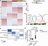 Figure 6
p120 perturbation and ROCK1 inhibition in keratinocytes regulate cell-cell adhesion. (A) Heatmap of the Z-scored differentially expressed proteins in the indicated genetic background show 4 distinct clusters. Genotypes and conditions are color-coded on top of the graph. (B) Line plot of scaled abundances of all proteins within clusters from A. Numbers indicate cluster number and total proteins for each cluster (in parentheses). (C) Pathway enrichment analysis of proteins in clusters 1, 2, 3, and 4. The color scale [–log10(qcombined)] represents the aggregated and multiple-testing-corrected significance of the module in each cluster. Shown are overhead terms, and all terms can be found in Supplemental Table 8. (D) Quantification of dispase dissociation assay using DMKN αβ–/–, DMKN βγ–/–, and WT keratinocytes transfected for 48 hours with p120 or scrambled siRNA and treated with ROCK inhibitor (ROCKi) for 24 hours. Data are normalized to DMSO-treated values. N = 3 biological replicates. **P < 0.01, ****P < 0.0001 (2-way ANOVA and Fisher’s least significant difference test).
Taken together, these data point to ROCK1 as a regulator of cellular adhesion downstream of the dermokine-p120 system in keratinocytes.
Dermokine expression is reduced in the hyperthickened epidermis of non-healing wounds. As abnormal dermokine expression has been previously associated with skin disorders (51), we stained skin biopsies from healthy and psoriatic skin and poorly and well-differentiated SCCs, as well as non-healing wounds (venous leg ulcers), for dermokine and p120 (Figure 7, Supplemental Figure 10, and Supplemental Table 9). We observed dermokine expression in suprabasal keratinocytes of healthy skin, psoriasis, and poorly and well-differentiated SCCs (Supplemental Figure 10, A–C), but a much weaker staining intensity in the hyperthickened epidermis of non-healing wounds (Figure 7, A and B, and Supplemental Figure 10D). We quantified dermokine expression relative to the distance to the wound bed in chronic wounds and found that keratinocytes proximal to the wound edge showed significantly lower dermokine expression across the epidermal profile, whereas, within the same biopsy, keratinocytes further away from the wound showed no change in dermokine expression (Figure 7C and Supplemental Figure 10E). To visualize the distance that keratinocytes express dermokine relative to the wounded tissue, we fitted a nonlinear regression curve previously used to explain differences in patient data from non-healing wounds and healthy skin (52). This analysis revealed that the average rate [K(fast)] at which dermokine expression decreases was higher in non-healing wound-edge keratinocytes [K(fast) = 4.4 × 10⁻³] compared with wound-edge distal keratinocytes in the respective biopsies [K(fast) = 5.7 × 10⁻⁸] (Figure 7C). Therefore, our data show less dermokine expression only in keratinocytes adjacent to the wound bed, which is consistent with the impaired differentiation of suprabasal keratinocytes in such wounds. Next, we evaluated changes in total and phosphorylated p120. For the latter, we used antibodies against phosphorylated S252 to recapitulate the immunoblotting findings and against phosphorylated T310 quantified in the phosphoproteomics dataset (Supplemental Figure 7, D and E, and Supplemental Table 4). After normalization of phosphorylated over total levels of p120, we found a significant increase in S252- and T310-phosphorylated p120 in non-healing wounds compared with healthy skin (Figure 7, D–F, and Supplemental Figure 10F).
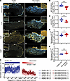 Figure 7
Dermokine expression decreases and p120 phosphorylation increases in keratinocytes of non-healing wounds. (A) Representative images of immunofluorescent staining of biopsies from healthy skin around the breast tissue from healthy individuals and non-healing wounds of venous leg ulcers (Supplemental Table 6). Scale bars: 400 μm for healthy skin, 1 mm for non-healing wounds, 50 μm for magnified images. (B and D–F) Quantification of immunofluorescent staining with the indicated antibodies from A. Anti-dermokine antibody values are the median Cy3 intensity values of N = 30 samples of 4 patients (non-healing wounds) and of 6 healthy individuals (healthy skin). Anti-p120 and anti–phosphorylated p120 antibody values are median Cy3 intensity values of N = 20 samples of 3 patients (non-healing wounds) and 3 patients (healthy skin). *P < 0.05, ***P < 0.001, ****P < 0.0001 (1-tailed, unpaired t test). (C) Quantification of dermokine intensity across 770.25 μm from the wound edge toward healthy keratinocytes. The quantified area was measured in 3 horizontal rectangles across the epidermal profile (Supplemental Figure 9D). Values are Cy3 anti-dermokine intensities along 3 equally shaped rectangles placed within the same biological sample at 2 different anatomical sites.
In conclusion, as all the tested phosphorylated sites on p120 are known to inhibit cell-cell adhesion (23, 48, 49), our results demonstrate that reduction of dermokine expression correlates with increased p120 phosphorylation, suggesting that the results obtained in vitro are also relevant to diseased skin in vivo (Figure 8).
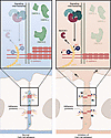 Figure 8
Model of dermokine function in human keratinocytes. Schematic of the putative relationship between dermokine, cell-cell adhesion, and p120 phosphorylation. Created in BioRender (biorender.com).


