Research ArticleNeurosciencePublic Health
Open Access | ![]() 10.1172/jci.insight.198059
10.1172/jci.insight.198059
Potentiation of fentanyl-induced respiratory depression by alcohol is not fully reversed by naloxone
Emma V. Frye,1 Lyndsay E. Hastings,1 Aniah N. Matthews,1 Adriana Gregory-Flores,1 Janaina C.M. Vendruscolo,1 Lindsay A. Kryszak,2 Shelley N. Jackson,2 Aidan J. Hampson,3 Nora D. Volkow,4 Leandro F. Vendruscolo,5 Renata C.N. Marchette,1 and George F. Koob1
1Neurobiology of Addiction Section and
2Translational Analytical Core, National Institute on Drug Abuse, Intramural Research Program, NIH, Baltimore, Maryland, USA.
3Division of Pharmacotherapeutic Development, National Institute on Drug Abuse, Rockville, Maryland, USA.
4Laboratory for Neuroimaging, National Institute on Alcohol Abuse and Alcoholism, Intramural Research Program, and
5Stress and Addiction Neuroscience Unit, National Institute on Drug Abuse and National Institute on Alcohol Abuse and Alcoholism, Intramural Research Programs, NIH, Baltimore, Maryland, USA.
Address correspondence to: Renata C.N. Marchette or George F. Koob, 251 Bayview Boulevard, BRC Room 08A727, Baltimore, Maryland 21224, USA. Phone: 667.312.5212; Email: renata.marchette@nih.gov (RCNM). Phone: 301.443.3885; Email: george.koob@nih.gov (GFK).
Authorship note: EVF and LEH have been designated as co–first authors.
Find articles by Frye, E. in: PubMed | Google Scholar
1Neurobiology of Addiction Section and
2Translational Analytical Core, National Institute on Drug Abuse, Intramural Research Program, NIH, Baltimore, Maryland, USA.
3Division of Pharmacotherapeutic Development, National Institute on Drug Abuse, Rockville, Maryland, USA.
4Laboratory for Neuroimaging, National Institute on Alcohol Abuse and Alcoholism, Intramural Research Program, and
5Stress and Addiction Neuroscience Unit, National Institute on Drug Abuse and National Institute on Alcohol Abuse and Alcoholism, Intramural Research Programs, NIH, Baltimore, Maryland, USA.
Address correspondence to: Renata C.N. Marchette or George F. Koob, 251 Bayview Boulevard, BRC Room 08A727, Baltimore, Maryland 21224, USA. Phone: 667.312.5212; Email: renata.marchette@nih.gov (RCNM). Phone: 301.443.3885; Email: george.koob@nih.gov (GFK).
Authorship note: EVF and LEH have been designated as co–first authors.
Find articles by Hastings, L. in: PubMed | Google Scholar
1Neurobiology of Addiction Section and
2Translational Analytical Core, National Institute on Drug Abuse, Intramural Research Program, NIH, Baltimore, Maryland, USA.
3Division of Pharmacotherapeutic Development, National Institute on Drug Abuse, Rockville, Maryland, USA.
4Laboratory for Neuroimaging, National Institute on Alcohol Abuse and Alcoholism, Intramural Research Program, and
5Stress and Addiction Neuroscience Unit, National Institute on Drug Abuse and National Institute on Alcohol Abuse and Alcoholism, Intramural Research Programs, NIH, Baltimore, Maryland, USA.
Address correspondence to: Renata C.N. Marchette or George F. Koob, 251 Bayview Boulevard, BRC Room 08A727, Baltimore, Maryland 21224, USA. Phone: 667.312.5212; Email: renata.marchette@nih.gov (RCNM). Phone: 301.443.3885; Email: george.koob@nih.gov (GFK).
Authorship note: EVF and LEH have been designated as co–first authors.
Find articles by Matthews, A. in: PubMed | Google Scholar
1Neurobiology of Addiction Section and
2Translational Analytical Core, National Institute on Drug Abuse, Intramural Research Program, NIH, Baltimore, Maryland, USA.
3Division of Pharmacotherapeutic Development, National Institute on Drug Abuse, Rockville, Maryland, USA.
4Laboratory for Neuroimaging, National Institute on Alcohol Abuse and Alcoholism, Intramural Research Program, and
5Stress and Addiction Neuroscience Unit, National Institute on Drug Abuse and National Institute on Alcohol Abuse and Alcoholism, Intramural Research Programs, NIH, Baltimore, Maryland, USA.
Address correspondence to: Renata C.N. Marchette or George F. Koob, 251 Bayview Boulevard, BRC Room 08A727, Baltimore, Maryland 21224, USA. Phone: 667.312.5212; Email: renata.marchette@nih.gov (RCNM). Phone: 301.443.3885; Email: george.koob@nih.gov (GFK).
Authorship note: EVF and LEH have been designated as co–first authors.
Find articles by Gregory-Flores, A. in: PubMed | Google Scholar
1Neurobiology of Addiction Section and
2Translational Analytical Core, National Institute on Drug Abuse, Intramural Research Program, NIH, Baltimore, Maryland, USA.
3Division of Pharmacotherapeutic Development, National Institute on Drug Abuse, Rockville, Maryland, USA.
4Laboratory for Neuroimaging, National Institute on Alcohol Abuse and Alcoholism, Intramural Research Program, and
5Stress and Addiction Neuroscience Unit, National Institute on Drug Abuse and National Institute on Alcohol Abuse and Alcoholism, Intramural Research Programs, NIH, Baltimore, Maryland, USA.
Address correspondence to: Renata C.N. Marchette or George F. Koob, 251 Bayview Boulevard, BRC Room 08A727, Baltimore, Maryland 21224, USA. Phone: 667.312.5212; Email: renata.marchette@nih.gov (RCNM). Phone: 301.443.3885; Email: george.koob@nih.gov (GFK).
Authorship note: EVF and LEH have been designated as co–first authors.
Find articles by Vendruscolo, J. in: PubMed | Google Scholar
1Neurobiology of Addiction Section and
2Translational Analytical Core, National Institute on Drug Abuse, Intramural Research Program, NIH, Baltimore, Maryland, USA.
3Division of Pharmacotherapeutic Development, National Institute on Drug Abuse, Rockville, Maryland, USA.
4Laboratory for Neuroimaging, National Institute on Alcohol Abuse and Alcoholism, Intramural Research Program, and
5Stress and Addiction Neuroscience Unit, National Institute on Drug Abuse and National Institute on Alcohol Abuse and Alcoholism, Intramural Research Programs, NIH, Baltimore, Maryland, USA.
Address correspondence to: Renata C.N. Marchette or George F. Koob, 251 Bayview Boulevard, BRC Room 08A727, Baltimore, Maryland 21224, USA. Phone: 667.312.5212; Email: renata.marchette@nih.gov (RCNM). Phone: 301.443.3885; Email: george.koob@nih.gov (GFK).
Authorship note: EVF and LEH have been designated as co–first authors.
Find articles by Kryszak, L. in: PubMed | Google Scholar
1Neurobiology of Addiction Section and
2Translational Analytical Core, National Institute on Drug Abuse, Intramural Research Program, NIH, Baltimore, Maryland, USA.
3Division of Pharmacotherapeutic Development, National Institute on Drug Abuse, Rockville, Maryland, USA.
4Laboratory for Neuroimaging, National Institute on Alcohol Abuse and Alcoholism, Intramural Research Program, and
5Stress and Addiction Neuroscience Unit, National Institute on Drug Abuse and National Institute on Alcohol Abuse and Alcoholism, Intramural Research Programs, NIH, Baltimore, Maryland, USA.
Address correspondence to: Renata C.N. Marchette or George F. Koob, 251 Bayview Boulevard, BRC Room 08A727, Baltimore, Maryland 21224, USA. Phone: 667.312.5212; Email: renata.marchette@nih.gov (RCNM). Phone: 301.443.3885; Email: george.koob@nih.gov (GFK).
Authorship note: EVF and LEH have been designated as co–first authors.
Find articles by Jackson, S. in: PubMed | Google Scholar
1Neurobiology of Addiction Section and
2Translational Analytical Core, National Institute on Drug Abuse, Intramural Research Program, NIH, Baltimore, Maryland, USA.
3Division of Pharmacotherapeutic Development, National Institute on Drug Abuse, Rockville, Maryland, USA.
4Laboratory for Neuroimaging, National Institute on Alcohol Abuse and Alcoholism, Intramural Research Program, and
5Stress and Addiction Neuroscience Unit, National Institute on Drug Abuse and National Institute on Alcohol Abuse and Alcoholism, Intramural Research Programs, NIH, Baltimore, Maryland, USA.
Address correspondence to: Renata C.N. Marchette or George F. Koob, 251 Bayview Boulevard, BRC Room 08A727, Baltimore, Maryland 21224, USA. Phone: 667.312.5212; Email: renata.marchette@nih.gov (RCNM). Phone: 301.443.3885; Email: george.koob@nih.gov (GFK).
Authorship note: EVF and LEH have been designated as co–first authors.
Find articles by Hampson, A. in: PubMed | Google Scholar
1Neurobiology of Addiction Section and
2Translational Analytical Core, National Institute on Drug Abuse, Intramural Research Program, NIH, Baltimore, Maryland, USA.
3Division of Pharmacotherapeutic Development, National Institute on Drug Abuse, Rockville, Maryland, USA.
4Laboratory for Neuroimaging, National Institute on Alcohol Abuse and Alcoholism, Intramural Research Program, and
5Stress and Addiction Neuroscience Unit, National Institute on Drug Abuse and National Institute on Alcohol Abuse and Alcoholism, Intramural Research Programs, NIH, Baltimore, Maryland, USA.
Address correspondence to: Renata C.N. Marchette or George F. Koob, 251 Bayview Boulevard, BRC Room 08A727, Baltimore, Maryland 21224, USA. Phone: 667.312.5212; Email: renata.marchette@nih.gov (RCNM). Phone: 301.443.3885; Email: george.koob@nih.gov (GFK).
Authorship note: EVF and LEH have been designated as co–first authors.
Find articles by Volkow, N. in: PubMed | Google Scholar
1Neurobiology of Addiction Section and
2Translational Analytical Core, National Institute on Drug Abuse, Intramural Research Program, NIH, Baltimore, Maryland, USA.
3Division of Pharmacotherapeutic Development, National Institute on Drug Abuse, Rockville, Maryland, USA.
4Laboratory for Neuroimaging, National Institute on Alcohol Abuse and Alcoholism, Intramural Research Program, and
5Stress and Addiction Neuroscience Unit, National Institute on Drug Abuse and National Institute on Alcohol Abuse and Alcoholism, Intramural Research Programs, NIH, Baltimore, Maryland, USA.
Address correspondence to: Renata C.N. Marchette or George F. Koob, 251 Bayview Boulevard, BRC Room 08A727, Baltimore, Maryland 21224, USA. Phone: 667.312.5212; Email: renata.marchette@nih.gov (RCNM). Phone: 301.443.3885; Email: george.koob@nih.gov (GFK).
Authorship note: EVF and LEH have been designated as co–first authors.
Find articles by
Vendruscolo, L.
in:
PubMed
|
Google Scholar
|

1Neurobiology of Addiction Section and
2Translational Analytical Core, National Institute on Drug Abuse, Intramural Research Program, NIH, Baltimore, Maryland, USA.
3Division of Pharmacotherapeutic Development, National Institute on Drug Abuse, Rockville, Maryland, USA.
4Laboratory for Neuroimaging, National Institute on Alcohol Abuse and Alcoholism, Intramural Research Program, and
5Stress and Addiction Neuroscience Unit, National Institute on Drug Abuse and National Institute on Alcohol Abuse and Alcoholism, Intramural Research Programs, NIH, Baltimore, Maryland, USA.
Address correspondence to: Renata C.N. Marchette or George F. Koob, 251 Bayview Boulevard, BRC Room 08A727, Baltimore, Maryland 21224, USA. Phone: 667.312.5212; Email: renata.marchette@nih.gov (RCNM). Phone: 301.443.3885; Email: george.koob@nih.gov (GFK).
Authorship note: EVF and LEH have been designated as co–first authors.
Find articles by
Marchette, R.
in:
PubMed
|
Google Scholar
|
1Neurobiology of Addiction Section and
2Translational Analytical Core, National Institute on Drug Abuse, Intramural Research Program, NIH, Baltimore, Maryland, USA.
3Division of Pharmacotherapeutic Development, National Institute on Drug Abuse, Rockville, Maryland, USA.
4Laboratory for Neuroimaging, National Institute on Alcohol Abuse and Alcoholism, Intramural Research Program, and
5Stress and Addiction Neuroscience Unit, National Institute on Drug Abuse and National Institute on Alcohol Abuse and Alcoholism, Intramural Research Programs, NIH, Baltimore, Maryland, USA.
Address correspondence to: Renata C.N. Marchette or George F. Koob, 251 Bayview Boulevard, BRC Room 08A727, Baltimore, Maryland 21224, USA. Phone: 667.312.5212; Email: renata.marchette@nih.gov (RCNM). Phone: 301.443.3885; Email: george.koob@nih.gov (GFK).
Authorship note: EVF and LEH have been designated as co–first authors.
Find articles by Koob, G. in: PubMed | Google Scholar
Published February 3, 2026 - More info
JCI Insight. 2026;11(6):e198059. https://doi.org/10.1172/jci.insight.198059.
© 2026 Frye et al. This work is licensed under the Creative Commons Attribution 4.0 International License. To view a copy of this license, visit http://creativecommons.org/licenses/by/4.0/.
Received: August 13, 2025; Accepted: January 29, 2026

-
Results
Additive effects of 25 μg/kg fentanyl and 1.18 g/kg alcohol on minute ventilation and apneic pauses
The first experiment sought to investigate whether a high, sedative-like dose of alcohol has additive effects to fentanyl in ventilatory parameters. The combination of 25 μg/kg fentanyl and the high dose of 1.18 g/kg alcohol resulted in high mortality (41.7% females, 33.3% males; Figure 1B and Supplemental Figure 2; supplemental material available online with this article; https://doi.org/10.1172/jci.insight.198059DS1), whereas neither alcohol nor fentanyl alone produced mortality. Figure 1 shows data from rats that completed all tests.
 Figure 1
Effects of 25 μg/kg fentanyl and 1.18 g/kg alcohol alone and combined on minute ventilation and apneic pauses. Rats received i.v. infusions of sterile water (5 mL/kg), fentanyl (25 μg/kg, 5 mL/kg), alcohol (1.18 g/kg, 30% vol/vol, 5 mL/kg), or a fentanyl+alcohol combination (25 μg/kg and 1.18 g/kg, respectively, 5 mL/kg) in a within-subjects Latin square design with each test separated by 1 week. (A) Timeline of each test. (B) Mortality for each drug. Data from the 12 rats that completed the experiment are shown in C–F. (C) Alcohol and fentanyl, alone and combined, decreased minute ventilation in a time-dependent manner. (D) Alcohol and fentanyl alone and combined increased apneic pauses in a time-dependent manner. The data are expressed as the mean ± SEM and were analyzed by 2-way RM-ANOVA followed by Duncan’s post hoc test. Filled symbols are different from water. fCombination different from fentanyl; ecombination different from alcohol (P < 0.05). (E) AUC of the first 15 min after infusion for minute ventilation. (F) AUC of the first 15 min after infusion for apneic pauses. The data are expressed as the mean ± SEM and were analyzed by 2-way RM-ANOVA followed by Šidák’s post hoc test when appropriate. Main effect of treatment: vP < 0.05, compared with water; eP < 0.05, compared with alcohol. n = 7 females, 5 males. (G) Representative raw plethysmography traces.
Analysis over time. The repeated-measures ANOVA (RM-ANOVA) for minute ventilation showed a main effect of time (F18,180 = 6.8939, P < 0.00001) and of treatment (F3,30 = 11.946, P = 0.00003) and a treatment × time interaction (F54,540 = 2.782, P < 0.0001; Figure 1C). Fentanyl decreased minute ventilation immediately upon injection (0 min) and increased ventilation at 20–30 and 85–90 min after infusion compared with vehicle. Alcohol decreased minute ventilation at 0–35 and 60–65 min after infusion, and fentanyl+alcohol decreased minute ventilation at 0–10 min after infusion compared with vehicle and at 10–30 min after infusion compared with fentanyl alone. There was no significant effect of sex, no sex × treatment interaction, no sex × time interaction, and no sex × treatment × time interaction.
The RM-ANOVA for apneic pause showed a main effect of time (F18,180 = 2.6952, P = 0.0004) and of treatment (F3,30 = 3.3695, P = 0.031) and a significant treatment × time interaction (F54,54 = 2.0224, P < 0.0001; Figure 1D). Fentanyl increased apneic pauses at 0–10 min after infusion, alcohol at 5–10, 45, and 60–70 min after infusion, and fentanyl+alcohol at 0–30, 40, 60, and 90 min after infusion compared with vehicle. Fentanyl+alcohol also increased apneic pauses at 0–20 and 70 min after infusion compared with alcohol alone, and at 5–45, 85, and 90 min after infusion compared with fentanyl alone. There was no significant effect of sex, no sex × treatment interaction, no sex × time interaction, and no sex × treatment × time interaction.
AUC. The AUC of minute ventilation for the first 15 min after infusion showed a main effect of treatment (F3,40 = 15.05, P < 0.0001). Fentanyl alone (P = 0.018), alcohol alone (P < 0.00001), and fentanyl+alcohol (P < 0.00001) induced a greater decrease in minute ventilation than vehicle (Figure 1E). Alcohol alone had a greater effect than fentanyl alone (P = 0.0423). Fentanyl+alcohol had similar effects to alcohol (P = 0.99) and fentanyl alone (P = 0.13). There was no effect of sex and no sex × treatment interaction.
The AUC of apneic pause for the first 15 min after infusion showed a main effect of treatment (F3,40 = 8.719, P = 0.0001). Šidák’s post hoc test showed that alcohol alone (P = 0.024) and fentanyl+alcohol (P < 0.00001) induced a greater increase in apneic pauses than vehicle, but fentanyl alone did not (P = 0.065; Figure 1F), whereas increases in apneic pauses between fentanyl+alcohol and alcohol did not differ (P = 0.27). There was no effect of sex and no sex × treatment interaction.
The AUCs for the other 9 parameters are shown in Table 1. Fentanyl+alcohol had additive effects on tidal volume, inspiratory time, peak expiratory flow, and end-inspiratory pause compared with fentanyl and alcohol alone. Altogether, these data indicate an additive effect of a sedative-like dose of alcohol with fentanyl on ventilatory parameters.
Additive effects of 25 μg/kg fentanyl and 0.59 g/kg alcohol on minute ventilation and apneic pauses
The second experiment sought to investigate whether a binge-like dose of alcohol has additive effects to fentanyl in ventilatory parameters.
Analysis over time. The RM-ANOVA for minute ventilation showed a main effect of time (F18,324 = 10.064, P < 0.00001) and of treatment (F3,54 = 6.512, P = 0.0011) and a significant treatment × time interaction (F54,972 = 5.910, P < 0.0001). Fentanyl alone decreased minute ventilation at 0 and 5 min and increased ventilation at 15–35 and 55–60 min after infusion (P < 0.05); alcohol alone decreased minute ventilation at 0–10 min after infusion (P < 0.05), whereas fentanyl+alcohol decreased minute ventilation at 0–10, 20, and 60 min after infusion compared with vehicle. Fentanyl+alcohol decreased minute ventilation at 25–45 min after infusion compared with fentanyl alone and at 0, 20, and 60 min after infusion compared with alcohol alone (P < 0.05; Figure 2A). There was no significant effect of sex, no sex × treatment interaction, no sex × time interaction, and no sex × treatment × time interaction.
 Figure 2
Effects of 25 μg/kg fentanyl and 0.59 g/kg alcohol alone and combined on minute ventilation and apneic pauses. Rats received i.v. infusions of sterile water (2.5 mL/kg), fentanyl (25 μg/kg, 2.5 mL/kg), alcohol (0.59 g/kg, 30% vol/vol, 2.5 mL/kg), or a fentanyl+alcohol combination (25 μg/kg and 0.59 g/kg, respectively, 2.5 mL/kg) in a within-subjects Latin square design with each test separated by 1 week. (A) Alcohol, fentanyl, and fentanyl+alcohol decreased minute ventilation in a time-dependent manner. (B) Alcohol, fentanyl, and fentanyl+alcohol increased apneic pauses in a time-dependent manner. The data are expressed as the mean ± SEM and were analyzed by 2-way RM-ANOVA followed by Duncan’s post hoc test when appropriate. Filled symbols are different from water. fCombination different from fentanyl; ecombination different from alcohol (P < 0.05). (C) AUC of the first 15 min after infusion for minute ventilation. *P < 0.05, drug × sex interactions. (D) AUC of the first 15 min after infusion for apneic pauses. The data are expressed as the mean ± SEM and were analyzed by 2-way RM-ANOVA followed by Šidák’s post hoc test when appropriate. Main treatment effect: vP < 0.05, compared with water; fP < 0.05, compared with fentanyl; eP < 0.05, compared with alcohol. n = 7–8 females, 10–11 males. (E) Representative raw plethysmography traces.
The RM-ANOVA for apneic pauses showed a main effect of time (F18,324 = 5.920, P <0.0001), no main effect of treatment (F3,54=1.115, P = 0.351), and a significant treatment × time interaction (F54,972 = 5.396, P < 0.0001; Figure 2B). Fentanyl increased apneic pauses at 0–5 min after infusion (P < 0.05), alcohol at 50 min after infusion (P < 0.05), and fentanyl+alcohol at 0–15 and 60 min after infusion compared with vehicle. Fentanyl+alcohol also increased apneic pauses at 0–15 min after infusion compared with fentanyl alone, and at 0–15, 50, and 60 min after infusion compared with alcohol alone (P < 0.05). There was no main effect of sex, no sex × treatment interaction, and no sex × treatment × time interaction. There was a significant sex × time interaction (F18,324 = 2.712, P = 0.0002), with males and females differing at 0 and 5 min.
AUC. For minute ventilation the AUC for the first 15 min after infusion showed a main effect of treatment (F3,68 = 23.81, P < 0.0001; Figure 2C). There was a significant effect of sex (F1,68 = 8.133, P = 0.0058; females > males) and a significant sex × treatment interaction (F3,68 = 4.672, P = 0.0050). Females that received fentanyl exhibited a greater decrease in minute ventilation than males (P = 0.0001). In females, fentanyl (P < 0.0001) and fentanyl+alcohol (P < 0.0001) but not alcohol (P = 0.217) induced a greater decrease than vehicle. In males, fentanyl (P = 0.024), alcohol (P = 0.044), and fentanyl+alcohol (P = 0.0001) produced a greater decrease than vehicle.
For apneic pauses, the AUC for the first 15 min after infusion showed a main effect of treatment (F3,60 = 34.26, P < 0.0001). Fentanyl (P = 0.0002) and fentanyl+alcohol (P < 0.00001) caused a greater increase than vehicle (Figure 2D), and fentanyl+alcohol induced a greater effect than fentanyl (P < 0.0001) and alcohol (P < 0.0001). There was a trend toward an effect of sex (F1,60 = 3.900, P = 0.053; females > males) but no sex × treatment interaction (F3,60 = 1.456, P = 0.236).
The AUCs for the other 9 parameters are shown in Table 2. All treatments increased AUC of all 9 parameters. Females had a more pronounced effect of fentanyl on inspiratory time, peak expiratory flow, and end-expiratory pause and a more pronounced effect of fentanyl+alcohol on tidal volume and a blunted effect on inspiratory time compared with males. Altogether, these data indicate an additive effect of a binge-like dose of alcohol with fentanyl on ventilatory parameters.
A low dose of fentanyl (3.125 μg/kg) did not have additive effects with 0.59 g/kg alcohol
The third experiment investigated whether a binge-like dose of alcohol had additive effects with a low dose of fentanyl in ventilatory parameters (Supplemental Figure 4 and Supplemental Table 1). Altogether, the data indicate no additive effect of a binge-like dose of alcohol with a low dose of fentanyl on ventilatory parameters.
Naloxone injected 5 min after fentanyl+alcohol administration reversed the effect of fentanyl+alcohol (25 μg/kg + 0.59 g/kg) on minute ventilation but not on apneic pauses
The fourth experiment investigated whether naloxone could rescue or prevent the effects of fentanyl+alcohol on ventilatory parameters.
Naloxone (100 μg/kg): analysis over time. For minute ventilation, the 2-way RM-ANOVA revealed a main effect of time (F18,270 = 4.790, P < 0.0001), a main effect of naloxone treatment (F1,15 = 8.683, P = 0.010), and a treatment × time interaction (F18,270 = 1.860, P = 0.019). Naloxone restored minute ventilation upon injection at 0 min, but at 10–30 and 40 min, ventilation was inhibited to a greater degree when naloxone was present than in its absence compared with saline (Figure 3B). For apneic pauses, the 2-way RM-ANOVA revealed a main effect of time (F18,23 = 7.303, P < 0.0001), no main effect of treatment (naloxone) (F1,13 = 0.862, P = 0.370), but no treatment × time interaction (F18,234 = 0.997, P = 0.4645; Figure 3C). There was no effect of sex, no sex × drug interaction, and no sex × drug × time interaction for either measure.
.") Figure 3
Figure 3Effect of naloxone on respiratory depression induced by fentanyl+alcohol (25 μg/kg + 0.59 g/kg). Rats received an i.v. infusion of a fentanyl+alcohol combination (25 μg/kg and 0.59 g/kg, 2.5 mL/kg), followed by an injection of naloxone (0, 100, 300, and 1,000 μg/kg) 5 min later. (A)Timeline of each test. (B) Naloxone (100 μg/kg) transiently reversed the fentanyl+alcohol–induced decrease in minute ventilation. (C) Naloxone (100 μg/kg) did not change the fentanyl+alcohol–induced increase in apneic pauses. (D) Naloxone (300 and 1,000 μg/kg) reversed fentanyl+alcohol–induced decreases in minute ventilation. (E) Naloxone (300 and 1,000 μg/kg) did not change the fentanyl+alcohol–induced increase in apneic pauses. The data are expressed as the mean ± SEM and were analyzed by 2-way RM-ANOVA followed by Duncan’s post hoc test when appropriate. (F) AUC of the first 15 min after naloxone infusion for minute ventilation. (G) AUC of the first 15 min after naloxone infusion for apneic pauses. (H) AUC of the first 15 min after naloxone infusion for minute ventilation. (I) AUC of the first 15 min after naloxone infusion for apneic pauses. The data are expressed as the mean ± SEM and were analyzed by 2-way RM-ANOVA followed by Šidák’s post hoc test when appropriate. n = 7–10 females, 7 males. (J) Representative raw plethysmography traces.
Naloxone (100 μg/kg): AUC. For AUC of minute ventilation, the 2-way RM-ANOVA revealed no main effect of sex (F1,15 = 1.272, P = 0.28) nor of naloxone dose (F1,15 = 0.6353, P = 0.44) and no sex × dose interaction (F1,15 = 0.0004, P = 0.9841; Figure 3F). For AUC of apneic pause, the 2-way RM-ANOVA revealed no main effect of sex (F1,12 = 2.030, P = 0.18) nor of naloxone dose (F1,12 = 4.210, P = 0.06) and no sex × dose interaction (F1,12 = 0.057, P = 0.81; Figure 3G).
The AUCs for the other 9 parameters are shown in Table 3. Treatment with naloxone 100 μg/kg blunted the effects of fentanyl+alcohol on frequency of breathing, inspiratory time, peak inspiratory flow, and end-expiratory pause, while it potentiated its effects on tidal volume, peak expiratory flow, and end-inspiratory pause.
Naloxone (300 and 1,000 μg/kg): analysis over time. For minute ventilation with the higher naloxone doses, the 2-way RM-ANOVA revealed a main effect of time (F18,216 = 17.8438, P < 0.0001), no main effect of treatment (naloxone) (F2,2 = 0.0720, P = 0.9308), and a treatment × time interaction (F36,432 = 7.4079, P < 0.0001). Naloxone at 300 and 1,000 μg/kg increased minute ventilation upon injection at 0–5 min compared with saline (Figure 3D). There was no effect of sex, no sex × drug interaction, and no sex × drug × time interaction.
For apneic pauses, the 2-way RM-ANOVA revealed a main effect of time (F18,216 = 11.118, P < 0.0001), no main effect of treatment (naloxone) (F2,24 = 0.397, P = 0.6765), and no treatment × time interaction (F36,432 = 1.374, P = 0.0777; Figure 3E). There was no effect of sex or sex × dose × time interaction, but there was a significant sex × naloxone dose interaction (F2,24 = 3.582, P = 0.044). Females had higher apneic pauses after 1,000 μg/kg naloxone than males (P = 0.022).
Naloxone (300 and 1,000 μg/kg): AUC. The 2-way RM-ANOVA of the minute ventilation AUC did not reveal a main effect of sex (F1,35 = 0.184, P = 0.67), a main effect of naloxone dose (F2,35 = 1.781, P = 0.18), or a sex × dose interaction (F2,35 = 0.273, P = 0.76; Figure 3H). The 2-way RM-ANOVA of the apneic pause AUC did not reveal a main effect of sex (F1,12 = 1.135, P = 0.31), a main effect of naloxone dose (F2,23 = 0.268, P = 0.77), or a sex × dose interaction (F2,23 = 0.991, P = 0.39; Figure 3I).
The AUCs for the other 9 parameters are shown in Table 4. Naloxone at 300 and 1,000 μg/kg blunted the effects of fentanyl+alcohol on inspiratory time, peak inspiratory flow, and end-expiratory pause. Naloxone at 300 μg/kg potentiated fentanyl+alcohol effects on expiratory time, relaxation time, and end-inspiratory pause. Naloxone at 1,000 μg/kg potentiated fentanyl+alcohol effects on peak expiratory flow, relaxation time, and end-inspiratory pause.
Altogether, these data indicate that high doses of naloxone are necessary to partially rescue the ventilatory alterations caused by fentanyl+alcohol, but naloxone treatment worsens expiratory-related measures. Effects of 300 μg/kg naloxone on 25 μg/kg fentanyl alone and 0.59 g/kg alcohol alone are shown in Supplemental Figures 5–7.
Naloxone injected before fentanyl+alcohol did not fully prevent fentanyl+alcohol–induced impairment of minute ventilation or apneic pauses
Analysis over time. For minute ventilation, the 2-way RM-ANOVA revealed no main effect of time or of sex, no sex × group interaction, no time × sex interaction, and no sex × group × time interaction. However, there was a main effect of naloxone on fentanyl+alcohol–induced minute ventilation (F1,25 = 6.642, P = 0.0162), and there was a treatment × time interaction (F17,425 = 2.813, P = 0.0002). Naloxone increased minute ventilation at 0 and 15–25 min compared with saline (Figure 4B).
 on minute ventilation and apneic pauses.") Figure 4
Figure 4Effect of pretreatment with naloxone on effects of fentanyl+alcohol (25 μg/kg + 0.59 g/kg) on minute ventilation and apneic pauses. Rats received an injection of naloxone (0 and 300 μg/kg) followed by an i.v. infusion of fentanyl+alcohol (25 μg/kg + 0.59 g/kg, 2.5 mL/kg). (A) Timeline of each test. (B) Pretreatment with 300 μg/kg naloxone attenuated the fentanyl+alcohol–induced decrease in minute ventilation in a time-dependent manner. (C) Pretreatment with 300 μg/kg naloxone attenuated the fentanyl+alcohol–induced increase in apneic pauses in a time-dependent manner. The data are expressed as the mean ± SEM and were analyzed by 2-way RM-ANOVA followed by Duncan’s post hoc test when appropriate. Filled symbols are different from vehicle (P < 0.05). (D) AUC of the first 15 min after fentanyl+alcohol infusion for minute ventilation. (E) AUC of the first 15 min after fentanyl+alcohol infusion for apneic pauses. The data are expressed as the mean ± SEM and were analyzed by 2-way RM-ANOVA. Main treatment effect: *P < 0.05, naloxone vs. saline. n = 16 females, 12 males. (F) Representative raw plethysmography traces.
For apneic pauses, the 2-way RM-ANOVA revealed a main effect of time on fentanyl+alcohol–induced pauses (F17,435 = 9.228, P < 0.0001). There was no main effect of naloxone on fentanyl+alcohol–induced apnea compared with saline (F1,25 = 1.148, P = 0.294). There was a treatment × time interaction (F17,425 = 2.472, P = 0.001). Naloxone decreased apnea at 5–25 min (Figure 4C). There was no effect of sex, no sex × group interaction, no time × sex interaction, and no sex × group × time interaction.
AUC. For minute ventilation, the AUC analyses revealed no main effect of sex and no sex × treatment interaction. There was a main effect of naloxone in attenuating the fentanyl+alcohol–induced impairment of minute ventilation (F1,25 = 32.48, P < 0.0001; Figure 4D). For apneic pause, the AUC analysis revealed no effect of sex and no sex × treatment interaction (Figure 4E) but showed a main effect of naloxone (F1,25 = 15.41, P = 0.0006).
The AUCs for the other 9 parameters are shown in Table 5. Pretreatment with naloxone blunted the effects of fentanyl+alcohol (decreased the AUC) on frequency, inspiratory time, peak inspiratory flow, and end-expiratory pause and potentiated its effects on end-inspiratory pause. Altogether, these data indicate that naloxone is partially effective in preventing the ventilatory alterations caused by fentanyl+alcohol.
 Table 5
Effect of naloxone administration before fentanyl+alcohol administration on ventilation parameters
Effect of 25 μg/kg fentanyl in fentanyl- and alcohol-dependent rats
Next, we tested the effects of fentanyl, alcohol, and their combination in fentanyl- and alcohol-dependent groups compared with a nondependent group. Alcohol and fentanyl dependence had no effect on baseline ventilatory parameters (Supplemental Figure 8).
Analysis over time. For minute ventilation, the RM-ANOVA showed a main effect of time (F18,684 = 13.231, P < 0.0001) but no main effect of group (F2,38 = 3.063, P = 0.0568). There was a group × time interaction (F36,684 = 2.0408, P = 0.0004; Figure 5B). The fentanyl-dependent group exhibited a faster recovery of minute ventilation 10–20 min after infusion compared with the nondependent group (P < 0.05). For apneic pauses, the RM-ANOVA showed a main effect of time (F18,666 = 15.417, P < 0.0001), no main effect of group (F2,37 = 1.023, P = 0.369), and no group × time interaction effect (F36,666 = 1.065, P = 0.369; Figure 5C).
 Figure 5
Effect of 25 μg/kg fentanyl in fentanyl- and alcohol-dependent and nondependent rats. Rats received an i.v. infusion of fentanyl (25 μg/kg, 2.5 mL/kg). (A) Timeline of each test. CIAV, chronic, intermittent alcohol vapor. (B) Fentanyl decreased minute ventilation in a time-dependent manner. (C) Fentanyl increased apneic pauses in a time-dependent manner. The data are expressed as the mean ± SEM and were analyzed by 2-way RM-ANOVA followed by Duncan’s post hoc test when appropriate. Filled symbols are different from nondependent (P < 0.05). *P < 0.05, fentanyl-dependent vs. alcohol-dependent. (D) AUC of the first 15 min after infusion for minute ventilation. (E) AUC of the first 15 min after infusion for apneic pauses. The data are expressed as the mean ± SEM and were analyzed by 2-way RM-ANOVA followed by Šidák’s post hoc test when appropriate. n = 5–7 females, 5–7 males.
AUC. For minute ventilation, the AUC analyses for the first 15 min after fentanyl infusion showed a main effect of sex (F1,35 = 11.22, P = 0.002) with females showing a greater decrease than males. There was no effect of group (F2,35 = 1.409, P = 0.26) and no sex × group interaction (F2,35 = 0.207, P = 0.81; Figure 5D). For apneic pauses, the area under analyses for the first 15 min after fentanyl infusion showed no effect of group or sex and no sex × group interaction (Figure 5E).
The AUCs for the other 9 parameters are shown in Table 6. Compared with the nondependent group, chronic exposure to alcohol and fentanyl led to lower AUC for (i.e., reduced effect on) frequency of breathing, expiratory time, and relaxation time in response to bolus fentanyl injection.
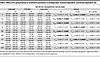 Table 6
Effect of 25 μg/kg fentanyl on ventilation parameters in nondependent, fentanyl-dependent, and alcohol-dependent rats
Effect of 0.59 g/kg alcohol in fentanyl- and alcohol-dependent rats
Analysis over time. The 2-way RM-ANOVA showed a main effect of time on minute ventilation (F18,684 = 13.231, P < 0.0001) but no main effect of group (F2,38 = 0.023, P = 0.977). There was no group × time interaction (F36,684 = 1.092, P = 0.329; Figure 6B). The 2-way RM-ANOVA of the effects of alcohol over time on apneic pauses showed no main effect of time (F18,684 = 0.824, P = 0.673) or main effect of group (F2,38 = 2.253, P = 0.119). However, there was a significant group × time interaction in response to alcohol (F36,684 = 1.610, P = 0.014; Figure 6C). Duncan’s post hoc test showed that the alcohol-dependent group exhibited an increase in apneic pauses at 10 and 15 min after infusion (P < 0.05) compared with the nondependent group.
 Figure 6
Effect of 0.59 g/kg alcohol in fentanyl- and alcohol-dependent and nondependent rats. Rats received an i.v. infusion of alcohol (0.59 g/kg, 2.5 mL/kg). (A) Timeline of each test. CIAV, chronic, intermittent alcohol vapor. (B) Alcohol did not affect minute ventilation. (C) Alcohol increased apneic pauses in a time-dependent manner. The data are expressed as the mean ± SEM and were analyzed by 2-way RM-ANOVA followed by Duncan’s post hoc test when appropriate. Filled symbols are different from nondependent (P < 0.05). *P < 0.05, fentanyl-dependent vs. alcohol-dependent. (D) AUC of the first 15 min after infusion for minute ventilation. (E) AUC of the first 15 min after infusion for apneic pauses. The data are expressed as the mean ± SEM and were analyzed by 2-way RM-ANOVA followed by Šidák’s post hoc test when appropriate. n = 5–7 females, 5–8 males.
AUC. For minute ventilation, the AUC for the first 15 min after alcohol infusion showed no main effect of group (F2,35 = 0.722, P = 0.49), no main effect of sex (F1,35 = 0.299, P = 0.59), and no sex × group interaction (F2,35 = 1.914, P = 0.16; Figure 6D). For apneic pauses, the AUC for the first 15 min after alcohol infusion showed a significant main effect of group (F2,35 = 3.486, P = 0.042; Figure 6E). Šidák’s post hoc test showed that the alcohol-dependent group exhibited a trend toward more severe apnea than the nondependent group (P = 0.057). There was no main effect of sex (F1,35 = 1.618, P = 0.21) and no sex × group interaction (F2,35 = 2.42, P = 0.104).
The AUCs for the other 9 parameters are shown in Table 7. Compared with the nondependent group, chronic exposure to alcohol led to higher AUC for (i.e., sensitization effect on) end-expiratory pause.
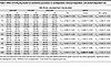 Table 7
Effect of 0.59 g/kg alcohol on ventilation parameters in nondependent, fentanyl-dependent, and alcohol-dependent rats
Effect of fentanyl+alcohol (25 μg/kg + 0.59 g/kg) in fentanyl- and alcohol-dependent rats
Analysis over time. For minute ventilation, the RM-ANOVA showed a main effect of time (F18,576 = 6.244, P < 0.0001), a main effect of group (F2,32 = 7.693, P = 0.002), and a significant group × time interaction (F36,544 = 2.361, P < 0.0001). Duncan’s post hoc test showed that the fentanyl-dependent group had higher minute ventilation at 0, 10, 15, 20, 45, and 75 min after infusion compared with the nondependent group. In contrast, the alcohol-dependent group exhibited higher minute ventilation at 20, 30, 40, 45, and 55 min after infusion (Figure 7B). For apneic pauses, the RM-ANOVA showed a main effect of time (F18,522 = 19.025, P < 0.0001), no main effect of group (F2,29 = 0.711, P = 0.499), and a significant group × time interaction (F36,522 = 1.557, P = 0.022; Figure 7C). Duncan’s post hoc test showed that the alcohol-dependent group exhibited an increase in apneic pauses at 0 min after infusion compared with the nondependent group (P < 0.05), whereas the fentanyl-dependent group exhibited a decrease in apneic pauses at 10 min after infusion.
 in fentanyl-dependent, alcohol-dependent, and nondependent rats.") Figure 7
Figure 7Effect of fentanyl+alcohol (25 μg/kg + 0.59 g/kg) in fentanyl-dependent, alcohol-dependent, and nondependent rats. Rats received an i.v. infusion of a fentanyl+alcohol combination (25 μg/kg and 0.59 g/kg, respectively, 2.5 mL/kg). (A) Timeline of each test. CIAV, chronic, intermittent alcohol vapor. (B) Fentanyl+alcohol decreased minute ventilation in a time-dependent manner. (C) Fentanyl+alcohol increased apneic pauses in a time-dependent manner. The data are expressed as the mean ± SEM and were analyzed by 2-way RM-ANOVA followed by Duncan’s post hoc test when appropriate. Filled symbols are different from nondependent (P < 0.05). *P < 0.05, fentanyl-dependent vs. alcohol-dependent. (D) AUC of the first 15 min after infusion for minute ventilation. (E) AUC of the first 15 min after infusion for apneic pauses. The data are expressed as the mean ± SEM and were analyzed by 2-way RM-ANOVA followed by Šidák’s post hoc test. n = 5–7 females, 5–7 males.
AUC. For minute ventilation, the AUC for the first 15 min after fentanyl+alcohol infusion showed no main effect of group (F2,30 = 0.800, P = 0.46) or sex (F1,30 = 0.061, P = 0.81) and no sex × group interaction (F2,30 = 0.963, P = 0.39; Figure 7D). For apneic pause, the AUC for the first 15 min after fentanyl+alcohol infusion showed no effect of group (F2,30 = 1.642, P = 0.21; Figure 7E), no effect of sex (F1,30 = 3.894, P = 0.058), and no sex × group interaction (F2,30 = 1.642, P = 0.21).
The AUCs for the other 9 parameters are shown in Table 8. Compared with the nondependent group, chronic exposure to fentanyl had a reduced effect on expiratory time and end-expiratory pause.
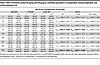 Table 8
Effect of fentanyl+alcohol (25 μg/kg and 0.59 g/kg) on ventilation parameters in nondependent, fentanyl-dependent, and alcohol-dependent rats
Blood gasometry and blood and brain drug concentrations following fentanyl, alcohol, and fentanyl+alcohol
Acute treatment with fentanyl, alcohol, and fentanyl+alcohol did not change arterial blood oxygen pressure (F3,15 = 0.6788, P = 0.5785; Figure 8A), carbon dioxide pressure (F3,15 = 0.9435, P = 0.4443; Figure 8B), or oxygen saturation (F3,15 = 0.8418, P = 0.492; Figure 8D). Fentanyl+alcohol decreased arterial blood pH (F3,15 = 5.825, P = 0.0076; Figure 8C) compared with vehicle (P < 0.01).
 Figure 8
Effects of 25 μg/kg fentanyl and 0.59 g/kg alcohol on arterial partial pressure gasometry and serum and brain fentanyl and alcohol levels. Rats received i.v. infusions of sterile water (2.5 mL/kg), fentanyl (25 μg/kg, 2.5 mL/kg), alcohol (30% vol/vol, 0.59 g/kg, 2.5 mL/kg), and fentanyl+alcohol (25 μg/kg + 0.59 g/kg, 2.5 mL/kg). Arterial blood was collected 5 min after the infusion, and trunk blood and brains were collected 10 min after infusion. (A–L) Effect of water, fentanyl, alcohol, and fentanyl+alcohol on partial pressure of oxygen (pO2) (A), partial pressure of carbon dioxide (pCO2) (B), hydrogen ion concentration (pH) (C), oxygen saturation (SO2) (D), alcohol concentration in blood (E), fentanyl concentration in blood (F), alcohol concentration in blood in nondependent and dependent rats (G), fentanyl concentration in blood nondependent and dependent rats (H), alcohol concentration in brain (I), fentanyl concentration in brain (J), alcohol concentration in brain in nondependent and dependent rats (K), and fentanyl concentration in brain in nondependent and dependent rats (L). The data are expressed as the mean ± SEM and were analyzed by 1-way ANOVA followed by Šidák’s post hoc test. **P < 0.01, ****P < 0.0001. n = 10 males, 9 females.
Acute treatment with alcohol and fentanyl+alcohol led to detectable serum alcohol levels (F3,16 = 145.4, P < 0.0001; Figure 8E). Rats that received alcohol and fentanyl+alcohol had measurable brain alcohol levels (F3,16 = 6168, P < 0.0001; Figure 8I). Rats that received fentanyl and fentanyl+alcohol had measurable blood (F3,15 = 7.250, P = 0.0031; Figure 8F) and brain fentanyl levels (F3,16 = 142.1, P < 0.0001; Figure 8J).
Acute treatment with fentanyl+alcohol led to similar levels of alcohol in blood (F2,14 = 0.1442, P = 0.867; Figure 8G) and brain (F2,13 = 1.202, P = 0.332; Figure 8K) in dependent and nondependent rats and similar fentanyl levels in blood (F2,13 = 3.186, P = 0.0748; Figure 8H) and brain (F2,13 = 0.3941, P = 0.682; Figure 8L) in dependent and nondependent rats.


.")


 on minute ventilation and apneic pauses.")


 in fentanyl-dependent, alcohol-dependent, and nondependent rats.")


-
Methods
Detailed methods are presented in Supplemental Methods and Results.
Sex as a biological variable. Male and female Long-Evans rats were used in all experiments (Table 9).
Animals. Seven-week-old Long-Evans rats (experiments 1, 2, 3, and 4) or 4-week-old Long-Evans rats (experiment 5) were obtained from Charles River (Table 9). Additionally, 9-week-old Long-Evans pre-catheterized rats (experiment 6) were obtained from Envigo.
Drugs. Fentanyl citrate was obtained from the National Institute on Drug Abuse Drug Supply Program (Research Triangle Institute, Research Triangle Park, North Carolina, USA) and dispensed by the National Institute on Drug Abuse Intramural Research Program Pharmacy (Baltimore, Maryland, USA). We also used alcohol (190-proof ethyl alcohol; Warner Graham Co.), naloxone hydrochloride (Tocris Bioscience, Bio-Techne), and 0.9% sterile saline (Hospira).
Intravenous catheter surgery. Rats were implanted with an indwelling silastic catheter (Dow Corning) in the right jugular vein under general anesthesia (2%–3% isoflurane in O2) as previously described (47).
Plethysmography apparatus. Ventilation was noninvasively monitored using 4 whole-body plethysmograph chambers (SCIREQ) as previously described (16). The ventilatory parameters listed in Table 10 were generated using IOX 2.10.0.40 software (emka TECHNOLOGIES).
Fentanyl dependence. In experiment 5, rats were made dependent on fentanyl by daily, subcutaneous injections of escalating doses of fentanyl as previously described (16).
Alcohol vapor exposure. In experiment 5, rats were made alcohol dependent by chronic, intermittent alcohol vapor exposure as previously described (48).
General procedure for plethysmography experiments. After intravenous (i.v.) catheter implantation and recovery, the rats were habituated to the plethysmography chambers in sessions in which ventilation was not monitored. During testing, the rats were acclimated to the chambers for 10 min, followed by 30 min of baseline data collection. Each rat then received all treatments in a within-subjects Latin square design, with tests 1 week apart. Ventilation was monitored for 90 min after infusion. If a rat’s catheter lost patency during testing, it was repaired or replaced, and the drug was readministered in a makeup test on the fifth week of testing. Detailed methods are described in Supplemental Methods and Results.
Experiment 1: 25 μg/kg fentanyl + 1.18 g/kg alcohol (“high, sedative-like” dose). Each rat received a 1 min i.v. infusion of sterile water (5 mL/kg), fentanyl (25 μg/kg, 5 mL/kg), alcohol (1.18 g/kg, 5 mL/kg), or a fentanyl+alcohol combination (25 μg/kg + 1.18 g/kg, 5 mL/kg; Figure 1A). Representative traces of raw ventilation curves in response to each drug are shown in Figure 1G. Alcohol dose was determined based on pilot experiment (Supplemental Figure 1).
Experiment 2: 25 μg/kg fentanyl + 0.59 g/kg alcohol (“binge-like” dose). In this experiment, half of the alcohol dose was used to achieve human binge-like blood alcohol levels (>80 mg/dL; n = 9–11 per sex per drug). Each rat received a 1 min i.v. infusion of sterile water (2.5 mL/kg), fentanyl (25 μg/kg, 2.5 mL/kg), alcohol (0.59 g/kg, 2.5 mL/kg), or a fentanyl+alcohol combination (25 μg/kg + 0.59 g/kg, 2.5 mL/kg; Figure 2A). Representative traces of raw ventilation curves in response to each drug are shown in Figure 2E.
Experiment 3: 3.125 μg/kg fentanyl + 0.59 g/kg alcohol. This experiment used a dose of fentanyl that does not cause respiratory depression (i.e., a subeffective dose). Each rat received a 1 min i.v. infusion of sterile water (2.5 mL/kg), fentanyl (3.125 μg/kg, 2.5 mL/kg), alcohol (0.59 g/kg, 2.5 mL/kg), or a fentanyl+alcohol combination (3.125 μg/kg + 0.59 g/kg, 2.5 mL/kg). The dose of fentanyl was based on a pilot experiment shown in Supplemental Figure 3.
Experiment 4: naloxone reversal of respiratory depression induced by fentanyl+alcohol. To test whether naloxone reverses respiratory depression caused by a fentanyl+alcohol combination, we tested 2 cohorts of rats with different naloxone doses. Each rat received a 1 min i.v. infusion of a fentanyl+alcohol combination (25 μg/kg + 0.59 g/kg, 2.5 mL/kg). In cohort 1, 5 min later, the rats received a bolus injection (1 mL/kg, i.v.) of naloxone (0 and 100 μg/kg). In cohort 2, the rats received a bolus injection of naloxone (0, 300, and 1,000 μg/kg; Figure 3A). Representative traces of raw ventilation curves in response to each drug are shown in Figure 3J.
Lastly, we tested whether 300 μg/kg naloxone prevents effects of a fentanyl+alcohol combination. Each rat received a bolus injection of naloxone (0 or 300 μg/kg, 1 mL/kg, i.v.), followed by a 1 min i.v. infusion of a fentanyl+alcohol combination (25 μg/kg + 0.59 g/kg, 2.5 mL/kg) 5 min later (Figure 4A). Representative traces of raw ventilation curves in response to each drug are shown in Figure 4F.
Experiment 5: fentanyl+alcohol in fentanyl- and alcohol-dependent rats. Rats were made dependent on alcohol as described above. Fentanyl-dependent rats were tested 4–6 hours into withdrawal. Nondependent rats were concomitantly tested. Each rat received a 1 min i.v. infusion of fentanyl (25 μg/kg, 2.5 mL/kg; Figure 5A), alcohol (0.59 g/kg, 2.5 mL/kg; Figure 6A), or a fentanyl+alcohol combination (25 μg/kg + 0.59 g/kg, 2.5 mL/kg; Figure 7A) in this order at 1-week intervals between tests.
Experiment 6: blood gasometry and blood and brain drug concentration measurements. Twenty rats (10 females, 10 males) with jugular vein and femoral artery catheters were used. The rats received a 1 min i.v. infusion of fentanyl (25 μg/kg, 2.5 mL/kg), alcohol (0.59 g/kg, 2.5 mL/kg), or a fentanyl+alcohol combination (25 μg/kg + 0.59 g/kg, 2.5 mL/kg). Five minutes after the infusion, arterial blood was collected for the measurement of blood gases. The rats were euthanized 10 min after infusion, and trunk blood and brains were collected.
To measure blood gas levels (Table 11), 100 μL of arterial blood was injected into CG8+ cartridges (Abbott Laboratories) and analyzed using an i-STAT 1 analyzer (Abbott Laboratories). Fentanyl and alcohol levels were measured in blood and brain (see Supplemental Methods and Results).
Statistics. A custom-made application (rvent_app) was used to import, bin, compile, plot, and export Microsoft Excel data sheets from the TXT files that were generated and calculated using IOX 2.10.0.40 software (emka TECHNOLOGIES). Prism 8 software (GraphPad) was used for figure preparation. Statistica 13 software (TIBCO) was used for statistical analyses. All data were aggregated into 1 min bins for analysis of the first 15 min post-infusion or in 5 min bins for the whole 90 min session analyses. The data are expressed as the mean ± SEM percentage of baseline values.
For statistical purposes, the analyses of treatment over time included the post-injection data but not the baseline data; we used 3-way repeated-measures analysis of variance (RM-ANOVA), with drug (treatment) and time as within-subjects factors and sex as a between-subjects factor. To determine the effect of treatments on the AUC, we used 2-way RM-ANOVA, with sex as a between-subjects factor and drug (treatment) as a within-subjects factor. Dunnett’s, Duncan’s, and Šidák’s post hoc tests were used when appropriate. Values of P < 0.05 were considered statistically significant.
Study approval. All procedures were performed according to the Guide for the Care and Use of Laboratory Animals (National Academies Press, 2011) and were approved by the National Institute on Drug Abuse, Intramural Research Program, Animal Care and Use Committee (protocol 23-INRB-13).
Data availability. All data presented in this article are available in the Supporting Data Values file.


