
Endocrinology
Abstract
BACKGROUND. Body weight and insulin resistance in women increase in midlife. It is not known whether this is driven by the menopause transition or solely by chronological aging. We aimed to determine the role of the menopause transition and menopause-related BMI changes in the development of insulin resistance in midlife women. METHODS. Mixed effects, linear spline modeling of longitudinal data in 1315 women, 42- to 52-years-old, pre- or early perimenopausal in 1995 to 1996 with up to 16 follow up visits by 2018. Homeostatic-model-assessed-insulin-resistance (HOMA-IR) was measured up to 12 times. Date of final menstrual period (FMP) was prospectively determined. RESULTS. HOMA-IR increased faster over a 4-year interval extending from 1.5 years prior to 2.5 years after the FMP, than in the years before or after it; p value for both slope changes < 0.0001. In the referent woman (White, age 52 years at FMP, non-smoker, body mass index [BMI] 25.6 kg/m2 at baseline, and not on sex hormone therapy or statins), the average annualized rate of HOMA-IR increase, adjusted for covariates including changes in BMI, was 1.2%, 4.3%, and 0.9% respectively, before, during, and after the 4-year transition. CONCLUSION. Consistent with an independent effect of the menopause transition, insulin resistance increased significantly faster during the transition (from 1.5 years before the to 2.5 years after the FMP) than in the years preceding or following the transition. Future studies need to investigate mechanisms underpinning this observed association and determine the impact of lifestyle on dampening menopause-associated increases in insulin resistance. FUNDING. National Institutes of Health.
Authors
Arun S. Karlamangla, Wei Juan Han, Preethi Srikanthan, Albert Shieh, Duncan Thomas, Barbara Sternfeld, Monique M. Hedderson, Gail A. Greendale
Abstract
The hypothalamic-pituitary-adrenal (HPA) axis integrates central neural processing and responses to stress with endocrine and metabolic responses. We utilized a monoclonal antibody binding corticotrophin-releasing factor (anti-CRF) to achieve durable HPA axis modulation. In mice, anti-CRF treatment decreased plasma corticosterone, food-intake, body weight and fat mass, but preserved lean mass and improved metabolic parameters including insulin sensitization. These body composition effects were observed in middle aged mice on normal chow or high fat diet, aged mice, and Mc4r-/- and ob/ob mice. Single-dose studies demonstrated impacts on weight gain and body composition lasting beyond the period of pharmacological target engagement. Multi-organ transcriptomic and proteomic analyses revealed organ-specific effects of anti-CRF and are consistent with altered glucocorticoid receptor (GR) signaling as a mediator of the observed phenotypes. These studies support anti-CRF-mediated HPA-axis modulation as a metabolic and anti-obesity therapeutic approach distinct from incretins.
Authors
Zachary A. Krumm, Tariful Islam, Caroline M. Watson, Yong Ran, Yona Levites, Hunter S. Futch, Luming Yin, Edgardo Rodriguez-Lebron, Xuefei Liu, Kazi Farhana Afroz, Ariella Goloborodsky, Thomas B. Ladd, Kristy D. Dillon, Danny Ryu, Jennifer L. Bizon, Jada Lewis, Karen A. Scott, Annette de Kloet, Nicholas T. Seyfried, Eric G. Krause, Karen N. McFarland, Todd E. Golde
Abstract
Prader-Willi syndrome (PWS) is a complex genetic disorder resulting from the deficiency of several maternally imprinted genes, including SNORD116, in the 15q11-q13 region. Loss of Snord116 in mice recapitulates some of the most salient clinical features of PWS, including growth hormone (GH) deficiency and hypogonadism. This study explored the impact of Snord116 deficiency on early postnatal pituitary development and growth in Snord116-KO mice. Snord116 was found to be expressed in both the anterior and posterior pituitary. Pituitary transcriptomes of Snord116-KO and WT mice at 2 developmental stages, P0 and 4 weeks of age, were interrogated and related to ex vivo analyses of GH secretion in the pituitaries of 5-week-old mice. Significant differences in pituitary transcriptomes were detected between Snord116-KO and WT mice at 4 weeks of age but not at P0. The differentially expressed genes and affected molecular pathways play important roles in regulating embryonic and postnatal pituitary development. Our results suggested that PWS GH deficiency was mainly due to pituitary hypoplasia and decreased GH production but not to reduced GH secretory function per se, implicating Snord116 in the specific molecular/cellular pathways that account for impaired postnatal pituitary development and GH deficiency in PWS.
Authors
Gabriel F. Batzli, Kaiying Guo, Fahrünisa Meryem Betül Erol, Charles A. LeDuc, Lisa C. Burnett, Rudolph L. Leibel, Yiying Zhang
Abstract
Activation of the mechanistic target of rapamycin (mTOR) complex1 (mTORC1) promotes muscle protein synthesis, mass, and function. Muscle mTORC1 can be activated by feeding and contraction. Here, muscle mTORC1 signaling, protein synthesis, mass, and function are characterized in a genetic mouse model that separates these two major modes of muscle mTORC1 regulation. AKT signaling is required for feeding-induced muscle mTORC1 signaling and protein synthesis, and mice expressing a mutant of tuberous sclerosis complex 2 (TSC2) that cannot be phosphorylated by AKT specifically in skeletal muscle (SkM-TSC2-5A) attenuate these effects of feeding. Despite this loss of postprandial protein synthesis, SkM-TSC2-5A mice have similar muscle and myofiber size compared to SkM-TSC2-WT mice. SkM-TSC2-5A mice maintain normal muscle mTORC1 activation in response to contraction and exhibit no differences in atrophy-related gene expression or ribosomal content. SkM-TSC2-5A mice exhibit improved maximal endurance capacity without changes in muscle contractile function. This phenotype occurs without alterations in muscle glycogen content or myofiber type but does coincide with a modest increase in muscle mitochondrial content. Therefore, AKT-mediated phosphorylation of TSC2 is required for postprandial mTORC1 activation and the induction of protein synthesis; however, these are dispensable for the development and maintenance of muscle mass in sedentary mice.
Authors
Samuel C. Lapp, Krystle C. Kalafut, Madi Y. Cissé, Khaled Tighanimine, Dean M. Rosenthal, Will Doxsey, Sheng Hui, Karen E. Inouye, Claire E. Morrow, Yann Cormerais, Brendan D. Manning
Abstract
Estrogen can promote aggressive tumor phenotypes in estrogen receptor–positive (ER+) breast cancer; however, ER– cell lines are not widely considered estrogen responsive. Noncanonical estrogen-stimulated pathways such as the membrane-bound G protein–coupled estrogen receptor (GPR30) can mediate migratory and proliferative phenotypes in breast cancer and are postulated to promote resistance to aromatase therapies. Moreover, dysregulation of UDP-glucose 6-dehydrogenase (UGDH), a ubiquitously expressed enzyme critical to the metabolism of UDP-glucuronic acid into extracellular matrix precursors and hormone regulation, is associated with tumorigenesis. Here, we illustrated the impact of estrogen stimulation on tumor phenotypes in ER+ and ER– cell models in vitro and in vivo. We then demonstrated UGDH’s association with metastatic breast cancer via single-cell sequencing of patient specimens. Genetic knockdown of UGDH blunted estrogen-stimulated tumor phenotypes in vitro, ex vivo, and in vivo using both ER+ and ER– breast cancer lines. Finally, we demonstrated that UGDH knockdown blunted noncanonical estrogen stimulation through GPR30. Ultimately, our study validated prior studies demonstrating estrogen-responsive malignant phenotypes in ER– breast cancer and demonstrated that estrogen-stimulated breast cancer progression can be mediated through noncanonical pathways (e.g., UGDH/GPR30), regardless of ER status.
Authors
Meghan J. Price, Annee D. Nguyen, Corinne H. Strawser, Trupti Trivedi, Cesar C.D. Baeta, Catherine Lavau, Jovita K. Byemerwa, Debarati Mukherjee, Suzanne E. Wardell, Sandeep Artham, Vardhman Kumar, Shyni Varghese, C. Rory. Goodwin
Abstract
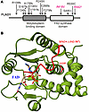
The FLAD1 gene codes for flavin adenine dinucleotide (FAD) synthase. FAD is a cofactor for many redox enzymes involved in vital processes from respiration to signal transduction. In this work, we described a clinical case of 2 siblings carrying compound heterozygous mutations in the FLAD1 gene resulting in the substitutions A418V and R542* at the protein level. The patients demonstrate adrenal insufficiency, which has not previously been associated with FLAD1 protein defects. To verify that adrenal insufficiency is caused by FLAD1 mutations, we created a personalized mouse model carrying the mutations found in the patients. The mutation in the FLAD1 gene, leading to the A418V substitution, appeared viable in the homozygous state, with minimal difference from the WT. The FLAD1 gene mutation leading to the R542* truncation is lethal when homozygous. The mouse model of the compound heterozygous FLAD1A418V/R542* mutations recapitulated the physiological, biochemical, and endocrine manifestations of FLAD1 mutations in patients. The mouse model created demonstrates the causal effect of FLAD1 mutations on the described pathology and potentially paves the way for understanding the disease’s molecular mechanism and developing better therapies.
Authors
Olga A. Averina, Natalia Yu. Kalinchenko, Vitaly A. Ioutsi, Andrey V. Pirogov, Alexander V. Bogachev, Oleg A. Permyakov, Vitaly S. Buev, Ekaterina A. Guseva, Anastasia V. Priymak, Olga A. Bazhanova, Mariia A. Emelianova, Olga O. Grigoryeva, Galina V. Baydakova, Maxim A. Abakumov, Vasily N. Manskikh, Olga A. Dontsova, Petr V. Sergiev, Anatoly N. Tiulpakov
Abstract
Autoimmune Addison’s disease (AD) is a rare but life-threatening disorder caused by immune-mediated destruction of the adrenal cortex, and progress in therapy has been limited by insufficient mechanistic insight. Here, we establish a model of Experimental Autoimmune Adrenalitis (EAA) that recapitulates key features of AD and reveals sex-dependent differences in disease manifestation within the model. Immunization with peptides derived from the adrenal self-antigen CYP11A1 induces corticosterone insufficiency. We show that autoimmune adrenalitis is driven by IFNG produced by self-reactive CD4+ T cells, promoting granulomatous inflammation in the adrenal cortex. Together, these findings identify IFNG as a central effector of autoimmune adrenalitis and suggest that targeting the IFNG pathway may represent a potential therapeutic strategy for AD.
Authors
Arina Andreyeva, Juraj Michalik, Veronika Niederlova, Veronika Cimermanova, Ales Drobek, Radislav Sedlacek, Jan Prochazka, Juraj Labaj, Olha Fedosieieva, Waldemar Kanczkowski, Peter Draber, André Sulen, Ondrej Stepanek, Aleš Neuwirth
Abstract
Locally produced nonpituitary growth hormone (npGH) promotes DNA damage accumulation and epithelial-mesenchymal transition (EMT) in aging human colon epithelium. GH receptor (GHR) and npGH are expressed in normal human prostate and benign prostatic hyperplasia (BPH) prevalence increases with age. We hypothesized that local prostate GH action may promote EMT and contribute to BPH pathogenesis. We show here that the number of patients expressing npGH increases >10-fold after age 60, concordant with increased γH2AX, a marker of DNA damage, and EMT activation. GH-treated human primary prostate epithelial cells, normal prostate cells, and primary cell cultures derived from resected BPH specimens exhibited enhanced DNA damage and activated EMT, with induced TWIST2 and suppressed E-cadherin, as well as increased Ki67, cell motility, and proliferation. In mice, prostate tissue adjacent to allografted GH-expressing fibroblasts showed increased γH2AX, TWIST2, and Ki67 compared to controls, along with morphological changes consistent with BPH. While GH and GH-induced IGF-1 both activated EMT, GH triggered DNA damage independent of IGF-1. These results elucidate a novel role for local npGH in aging prostate tissue, whereby npGH increases DNA damage and promotes EMT to enable a microenvironment favoring BPH development. Prostate GHR signaling may be an attractive therapeutic target for BPH.
Authors
Masaki Ryuzaki, Svetlana Zonis, Neil A. Bhowmick, Sandrine Billet, Saravana Kumar Kailasam Mani, Stephen J. Freedland, Hyung L. Kim, Vera Chesnokova, Shlomo Melmed
Abstract
The hypothalamic changes that occur after the loss of ovarian estrogen remain poorly characterized. Here, we performed a comprehensive temporal characterization of the mouse hypothalamus following ovariectomy (OVX), combining physiological measurements with bulk RNA-sequencing of the posterior hypothalamus (PH) and preoptic area (POA) at short-term (14 days) and long-term (4 months) post-OVX. Serum LH levels rose progressively and then declined, while core temperature peaked early and subsequently normalized, recapitulating the endocrine and thermoregulatory dynamics of reproductive aging in humans. Transcriptomic analysis revealed time-dependent activation of inflammatory pathways, glial markers, and KNDy neuron-related gene networks, with the most pronounced changes emerging at 4 months post-OVX, particularly in the PH. Immunofluorescence confirmed increased NKB release, declining KNDy neuronal activity, and heightened astrocytic reactivity in the arcuate nucleus after prolonged estrogen withdrawal. To contextualize these findings, we analyzed publicly available human hypothalamic RNA-seq data across chronological age. Age-related transcriptomic patterns in women, including progressive inflammatory signaling, glial activation, and altered KNDy gene expression, showed significant correlation with the OVX mouse model, particularly at the pathway level. These findings establish a temporal framework for hypothalamic molecular changes after estrogen withdrawal, identify conserved neuroinflammatory signatures across species, and provide a preclinical platform for testing interventions targeting menopausal-associated hypothalamic dysfunction.
Authors
Jordana C.B. Bloom, Encarnación Torres, Sidney A. Pereira, Liliana Arvizu-Sanchez, Audrey N. Fontes, Hadine Joffe, David C. Page, Victor M. Navarro
Abstract
Adults with type 2 diabetes mellitus (T2DM) are at increased risk for stroke, myocardial infarction, and cardiovascular death, yet individual risk is heterogeneous and incompletely captured by clinical models. In the Exenatide Study of Cardiovascular Event Lowering (EXSCEL), adults with T2DM were randomized to a GLP-1 RA (exenatide) or placebo and followed longitudinally for major adverse cardiovascular events (MACE). High-throughoput discovery proteomics was done in plasma collected at baseline and 12-months. Proteins associated with time-to-MACE were identified using multivariable regression and incorporated into supervised machine learning models. A multi-protein score was developed and externally validated in two independent population-based and trial cohorts, Cardiovascular Health Study and the Prospective Multicentre Imaging Study for Evaluation of Chest Pain (PROMISE). The proteomic score showed incremental improvement in cardiovascular risk discrimination beyond clinical factors alone, and several proteins were consistently prioritized across modeling approaches. The protein score and a top-ranked protein, tetranectin, were modified by GLP-1 RA treatment, and a decrease in the protein score was associated with improved outcomes, supporting modifiability of MACE risk. External validation confirmed generalizability across cohorts with and without diabetes. Together, these findings demonstrate that plasma proteomic signatures can enhance cardiovascular risk stratification and identify treatment-responsive biomarkers in T2DM, supporting their potential role in precision prevention strategies.
Authors
Kristin M. Corey, Maggie Nguyen, Michael Y. Mi, Megan E. Ramaker, Ilya Zhbannikov, Harald Sourij, G. Michael Felker, Naveed Sattar, Jennifer B. Green, Pamela S. Douglas, Robert E. Gerszten, Robert J. Mentz, Adrian F. Hernandez, Rury R. Holman, Bruce M. Psaty, James S. Floyd, Svati H. Shah
No posts were found with this tag.


