

Abstract
Type 2 (T2) immune cells dominate the airways of patients with mild-moderate asthma (MMA) with a more complex type 1 (T1)-T2 mixed immune response evident in treatment-refractory severe asthma (SA). We hypothesized that comparing the transcriptomes of the airway epithelium of patients with SA and MMA would reveal molecular signatures associated with more severe disease in the context of a complex immune response. Using our interpretable machine learning tool, SLIDE, meaningful latent factors (context-specific gene co-expression networks) were revealed that distinguished SA from MMA. Unexpectedly, an aberrant high expression of normally host-protective, membrane-tethered, and IFN-inducible mucins, MUC1 and MUC4, was identified in SA. Gene networks in the significant latent factors discriminating SA from MMA corresponded to enrichment of a keratinization program in SA airways. Keratinization was marked by increased expression of the stress keratin KRT16, signifying squamous metaplasia suggesting adaptive reprogramming of the airway epithelium in response to chronic stress. These mucins and KRT16 were inversely associated with lung function in 2 separate asthma cohorts. Imaging of endobronchial biopsies revealed significantly higher KRT16 protein expression in SA compared with MMA that strongly correlated with MUC1 protein expression. Our study identifies dysregulated host-protective and maladaptive repair responses in SA distinguishing from MMA.
Authors
Sagar L. Kale, Augusta Vincent, Mark A. Ross, Isha Mehta, Michael J. Calderon, Richard P. Ramonell, Himanshu Setya, Jessica C. McCreary-Partyka, Huijuan Yuan, Stephanie A. Christenson, Prescott G. Woodruff, Mario Castro, Kaharu Sumino, Nizar N. Jarjour, Loren C. Denlinger, Benjamin Gaston, Eugene R. Bleecker, Deborah A. Meyers, Wendy C. Moore, Elliot Israel, Bruce D. Levy, David Mauger, Serpil Erzurum, Anthony Newbrough, Taylor J. Nee, Prabir Ray, Claudette M. St. Croix, Sally E. Wenzel, Jishnu Das, Anuradha Ray, Marc C. Gauthier

Abstract
Chronic graft-versus-host disease (cGVHD) remains a major cause of morbidity and mortality after allogeneic hematopoietic transplantation. cGVHD pathophysiology involves cooperation between T follicular helper cells (TFHs) and germinal center B cells (GCBs), allo- and autoantibody depositions in cGVHD tissues, and fibrosis. We evaluated human CD19–directed chimeric antigen receptor (CAR19) T cell therapy in a clinically relevant murine cGVHD model with bronchiolitis obliterans syndrome (BOS). Although CD8+ CAR19 T cells effectively reduced peripheral B cell and GCB frequencies, pulmonary function was unimproved. In contrast, a single infusion of CAR19 CD4+ regulatory T cells (Tregs) mitigated ongoing pulmonary disease and modulated germinal centers (GCs) associated with reduced TFH frequencies compared with control Tregs but without measurable B cell depletion. Compared with EGFR Treg infusion, mice receiving CAR19 Tregs exhibited enhanced suppression of B cell activation and preserved splenic architecture, and CAR19 Treg infusion provided greater opportunities for interaction with CD19+ B cells at the B cell follicle boundary zones. Taken together with the absence of detectable B cell cytolysis, these findings were most consistent with GC suppression rather than B cell depletion as the dominant mechanism. Overall, our findings suggest that CAR19 Tregs represent a promising and safe cGVHD/BOS therapeutic strategy, offering immunosuppressive benefits and improved disease outcomes that may be more limited with CD8+ CAR19 T cell treatment.
Authors
Sujeong Jin, Michael C. Zaiken, Cameron McDonald-Hyman, Christina R. Hartigan, Sara Bolivar-Wagers, Jemma H. Larson, Yiyun Peng, Sophia Hani, Megan Riddle, Asim Saha, Angela Panoskaltsis-Mortari, Eun Ko, Yujie Zhao, Rocio Amaro Marquez, Pooja Shree Marri Baskar, Cindy R. Eide, William J. Murphy, Keli L. Hippen, Geoffrey R. Hill, Jakub Tolar, Peter T. Sage, Christopher A. Pennell, Leslie S. Kean, Bruce R. Blazar

Abstract
Undifferentiated pleomorphic sarcoma (UPS) is one of the most common adult soft-tissue sarcomas (STSs), yet therapeutic progress remains limited because of the absence of recurrent oncogenic driver mutations. To identify tumor suppressors contributing to UPS pathogenesis, we performed a customized in vivo CRISPR/Cas9 screen in mice. This approach identified BRCA1-associated protein 1 (BAP1) as a potent tumor suppressor in STS. Integrative analyses using RNA sequencing, multiplex immunohistochemistry, and flow cytometry revealed that Bap1-deficient sarcomas exhibited a markedly immunosuppressive tumor microenvironment. Consistent with these findings, BAP1 protein expression was reduced in human UPS, whereas polo-like kinase 1 (PLK1) expression was elevated. Functional studies demonstrated that PLK1 was required for the growth and survival of Bap1-deficient sarcomas. Pharmacologic inhibition of PLK1 with volasertib significantly suppressed tumor growth in both syngeneic and autochthonous mouse models. Moreover, combining PLK1 inhibition with anti–PD-1 therapy enhanced tumor control and improved survival compared with either treatment alone. Together, these results identify PLK1 as a potential therapeutic vulnerability in BAP1-deficient sarcomas and support further evaluation of combined PLK1 inhibition and immune checkpoint blockade as a treatment strategy for a subset of STSs.
Authors
Jianguo Huang, Xingliang Liu, Warren Floyd, William Haugh, Zhaoyu Sun, Melissa J. Kasiewicz, Yaping Wu, Brian Piening, John T. Welle, Wesley K. Rosales, Venkatesh Rajamanickam, So Young Kim, Eric S. Xu, Lixia Luo, Yan Ma, Rutulkumar Patel, Ziqiang Zhang, Brady Bernard, William L. Redmond, Walter J. Urba, R. Bryan Bell, David G. Kirsch
Abstract
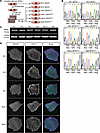
Charcot-Marie-Tooth disease (CMT) is a group of inherited progressive conditions affecting distal motor and sensory neurons, leading to muscle weakness, pain, and loss of sensation in limbs. CMT type 2A (CMT2A) is the most common form of axonal CMT and is associated with a more severe clinical manifestation. However, there are no treatments currently available. To investigate disease mechanisms and facilitate treatment discovery, we developed an in vitro model for CMT2A by introducing the patient-specific MFN2R94Q/+ variant into human embryonic stem cells (hESCs). Isogenic variant and wild-type hESCs differentiated into spinal motor neurons with similar efficiency and gave rise to functional motor neurons in vitro. However, MFN2R94Q/+ spinal motor neurons displayed impaired mitochondrial trafficking, resulting in altered distribution of mitochondria in axons. Unbiased quantitative proteomic profiling of the endogenous MFN2 interactome revealed dose-dependent remodelling by the R94Q variant across 412 proteins, highlighting candidate mechanisms in disease pathology. Importantly, we showed that mitochondrial trafficking defects could be alleviated by treatment with an HDAC6 inhibitor. Chemical inhibition of HDAC6 also rescued the motor phenotype in a zebrafish CMT2A model. Taken together, our study reveals a variant-specific insight into CMT2A disease mechanisms and confirms HDAC6 as a promising target for further therapeutic development.
Authors
Lydia H. Jestice, Larissa Butler, Rebecca A. Lea, Kathryn I. Adamson, Jonas Van Lent, Stuart L. Johnson, Hollie Weedon, Eldriena D’Silva, Gabriele Gelezauskaite, Bob Asselbergh, Eloise Brown, Owen Laing, Christopher J. Price, Dylan Stavish, Anestis Tsakiridis, Mark O. Collins, Vincent Timmerman, Kurt J. De Vos, Alison E. Twelvetrees, Andrew J. Grierson, Ivana Barbaric

Abstract
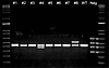
Single-cell RNA sequencing (scRNA-seq) of peripheral blood mononuclear cells (PBMCs) has enhanced our understanding of host immune mechanisms in small cohorts, particularly in diseases with complex and heterogeneous immune responses such as sepsis. However, standard PBMC isolation from blood requires technical expertise and over 2 hours of on-site processing using Ficoll density gradient separation (“Ficoll”) for scRNA-seq compatibility, precluding large-scale sample collection at most clinical sites. To minimize on-site processing, we developed cryopreservation with PBMC recovery offsite (Cryo-PRO), a method of immediate on-site whole-blood cryopreservation and subsequent batched PBMC isolation in a central laboratory prior to sequencing. We compared multimodal single-cell immune profiling results from samples processed using Cryo-PRO versus standard on-site Ficoll separation in 23 patients with sepsis. Key outputs, including cell substate fractions, marker genes, and surface protein expression were similar for each method across multiple cell types and substates, including an important monocyte substate enriched in patients with sepsis. Capture of T cell receptor transcripts was also comparable across both methods. Cryo-PRO reduced on-site sample processing time from more than 2 hours to less than 15 minutes and was reproducible across 2 enrollment sites, thus demonstrating potential for expanding multimodal single-cell analyses in multicenter studies of sepsis and other diseases.
Authors
Alyssa K. DuBois, Pierre O. Ankomah, Alexis C. Campbell, Renee Hua, Olivia K. Nelson, Christopher A. Zeuthen, M. Kartik Das, Shira Mann, Abigail Mauermann, Blair A. Parry, Nathan I. Shapiro, Michael R. Filbin, Roby P. Bhattacharyya
Abstract
Pulmonary arterial hypertension (PAH) is a rare vascular disorder characterized by elevated pressure in pulmonary arteries, eventually leading to right ventricular failure. Approximately 50% of pediatric disease and 20% of adult disease can be linked to a genetic mutation, with nearly 70% of these cases involving mutations in the bone morphogenetic protein receptor type 2 (BMPR2) locus. Investigations using rodent models have made substantial advances in our understanding of BMPR2 signaling; however, limited data exist regarding the onset and course of PAH, and etiologies for phenotypic expression in these patients remain unknown. In this work, we describe the development of an ovine model of heritable PAH. Because homozygous disruption of BMPR2 is embryonic lethal, we developed heterozygous BMPR2-edited (BMPR2+/–) sheep by using a PAM-disrupting synonymous single-stranded oligodeoxyribonucleotide alongside a single guide RNA and Cas9-mediated gene editing strategy. The resulting BMPR2+/– lambs demonstrated cardiac and pulmonary vascular pathology that are consistent with BMPR2 mutation–driven PAH observed in humans. Given the genetic and physiological similarities of BMPR2+/– sheep to humans with heritable PAH, this large animal model will serve as a vital platform for mechanistic molecular studies and will provide a much-needed preclinical model for extensive treatment evaluations.
Authors
Sanjeev A. Datar, Nicholas Werry, Austin R. Brown, Devon S. Fitzpatrick, Oluwafemi Falade, Josephine F. Trott, Rachel Hutchings, Elena K. Amin, Jessica M. Morgan, Hythem Nawaytou, Gail H. Deutsch, Eric G. Johnson, Omar A. Gonzales Viera, Thomas F. Bishop, Tara Urbano Beach, Bret R. McNabb, Eric D. Austin, Jeffery R. Fineman, Alison L. Van Eenennaam

Abstract
Vaccine development for tuberculosis (TB) is a global priority. Our studies using Collaborative Cross (CC) mice show that genetic diversity influences the efficacy of BCG, the most widely used TB vaccine. BCG vaccination of CC042 mice reduced their lung bacillary burden and increased their survival following low-dose aerosol Mycobacterium tuberculosis infection (MTBI), despite impaired T cell trafficking due to a defective Itgal gene. BCG vaccination conferred early bacillary control that appeared to be independent of B cell or T cell recall responses following MTBI. In contrast, long-term survival of BCG-vaccinated CC042 mice after MTBI required T cells. Thus, CC042 mice reveal two phases of immunity induced by BCG: an early phase mediated by innate immunity or innate-like T cells and a later phase mediated by conventional memory CD4+ and/or CD8+ T cells. Although measurement of vaccine-induced protection 30 days after MTBI is a standard measure of vaccine efficacy in the TB model, this time point might be independent of memory T cells in CC042 mice. Our results suggest that vaccine-elicited innate/innate-like responses could have a larger role in protection than previously considered. The concordance between lung CFU, pathology, and survival makes CC042 mice useful for mechanistic studies on vaccine-induced immunity.
Authors
Abiola F. Ogunsola, Rocky Lai, Kelly Cavallo, Anthony V. Tran, Gillian L. Beamer, Samuel M. Behar
Abstract
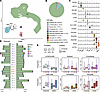
Systemic sclerosis (SSc) is a rare autoimmune disease characterized by vasculopathy and fibrosis of the skin and internal organs. Individuals with SSc often suffer from chronic acid reflux and dysphagia due to loss of esophageal motility. To determine whether distinct changes in esophageal epithelial cells contribute to esophageal involvement in SSc, we investigated the stratified squamous esophageal epithelium from proximal and distal biopsies using single-cell RNA sequencing in individuals with SSc compared with those with gastroesophageal reflux disease (GERD) and healthy controls. Cellular and molecular changes in SSc were highly correlated with those seen in GERD, indicating they were secondary to reflux; however, their magnitudes were more pronounced in the proximal esophagus, suggesting that esophageal dysmotility leads to greater proximal acid exposure, which may contribute to aspiration. SSc-specific gene dysregulation implicated immunoregulatory pathways likely pertinent to pathogenic mechanisms. Ligand-receptor interaction analysis revealed enhanced profibrotic signaling between fibroblasts and epithelial cells in SSc. Cell type localization and SSc-specific changes were confirmed by spatial molecular imaging. By offering a comprehensive view of transcriptional dysregulation at single-cell resolution in human esophageal epithelial cells in SSc compared with GERD and healthy tissue, this work clarifies the state of epithelial cells in SSc-induced esophageal dysfunction.
Authors
Matthew Dapas, Margarette H. Clevenger, Hadijat-Kubura M. Makinde, Tyler Therron, Dustin A. Carlson, Mary Carns, Kathleen Aren, Cenfu Wei, Kainat Mian, Lutfiyya N. Muhammad, Carrie Richardson, Parambir S. Dulai, Monique Hinchcliff, John Pandolfino, Harris Perlman, Deborah R. Winter, Marie-Pier Tetreault

Microbiome-derived metabolites shape CD4+ T cell differentiation and immune aging in HIV-1 infection
Abstract
The role of aromatic gut-derived bacterial metabolites (GDBMs) in shaping immune cell metabolism and function remains poorly explored. Using ex vivo metabolomic profiling of paired plasma and CD4+ T cells from people living with HIV-1 (PLWH), we identified a network of aromatic GDBMs whose cell-associated abundance, rather than systemic levels, was linked to broad alterations in CD4+ T cell metabolic and functional states. Among these, p-cresol sulfate (PCS) emerged as a mechanistic prototype. Ex vivo flow cytometry and scRNA-seq of CD4+ T cells stratified by cell-associated PCS levels revealed dose-dependent enrichment of transcriptional programs associated with impaired differentiation, regulatory-like identity, and cellular senescence. In vitro transcriptomic and proteomic analyses of PCS-exposed CD4+ T cells demonstrated induction of cell-cycle arrest, mitochondrial dysfunction, and senescence-associated programs, including upregulation of p16 and p21. Integration of these immunometabolic findings with HIV-1 reservoir measurements revealed that CD4+ T cell states defined by cell-associated GDBMs track with intact proviral DNA levels in vivo. These findings define a microbiome-derived axis that reshapes CD4+ T cell metabolism and fate, promotes immune aging in PLWH, and may foster immunometabolic states linked to long-term HIV-1 reservoir persistence.
Authors
Amanda Cabral Da Silva, Luke Flantzer, Jaclyn Weinberg, Shuya Kyu, Lisa P. Daley-Bauer, Anyce Godoy, Ana Carolina Santana, Aarthi Talla, Amber Lynn Rittgers, Sarah Welbourn, David Ezra Gordon, Jeffery Alan Tomalka, Vincent C. Marconi, Dean P. Jones, Souheil-Antoine Younes
Abstract
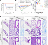
Post-acute sequelae of SARS-CoV-2 (PASC) occurs in subsets of individuals, including those with preexisting lung disease. To investigate PASC pathogenesis and therapeutics in a chronic bronchitis mouse model (Scnn1b-Tg), Scnn1b-Tg and WT mice were inoculated with a mouse-adapted SARS-CoV-2 virus (SARS-CoV-2 MA10) and followed for 60 days. Viral titer, histology, immunohistochemistry, single-cell RNA sequencing, RNA in situ hybridization, and spatial transcriptomic profiling characterized disease pathologies. Scnn1b-Tg mice inoculated with SARS-CoV-2 MA10 exhibited lower viral titers and less weight loss than WT mice. Airway epithelia of Scnn1b-Tg mice were less infected than epithelia of WT mice, reflecting increased airway mucus and enhanced epithelial antiviral activities in Scnn1b-Tg mice. However, Scnn1b-Tg mice subsequently exhibited heterogeneous airway and parenchymal disease with elevated Il33 expression characteristic of human eosinophilic pneumonia. Cohorts of infected mice were given a monoclonal antibody targeting the IL-33 receptor (ST2) or enteral prednisone. Administration of an anti-ST2 monoclonal antibody mitigated development of eosinophilic pneumonia, while enteral prednisone suppressed IL-33 expression and disease. The eosinophilic pneumonia in Scnn1b-Tg mice after SARS-CoV-2 MA10 infection mimics reports of eosinophilic pneumonia in humans after SARS-CoV-2, suggesting that targeting of IL-33 may be beneficial in treating post-viral eosinophilic pneumonia in humans.
Authors
Padraig E. Hawkins, Sarah R. Leist, Hong Dang, Minako Saito, Lisa C. Morton, Jesse B. Hines, Rodney C. Gilmore, Stephen A. Schworer, Ella F. Burns, Jason R. Rock, Robert S. Hagan, James J. Pestka, Alexandra Schäfer, Kenichi Okuda, Lauren K. Heine, Jack R. Harkema, Wanda K. O’Neal, Alessandra Livraghi-Butrico, Raymond J. Pickles, Ralph S. Baric, Richard C. Boucher
No posts were found with this tag.


