
Research ArticleAIDS/HIVImmunologyInflammation
Open Access |  10.1172/jci.insight.201258
Dietary indoles influence the AHR-RORγt axis and mucosal immune homeostasis in ART-treated SIV infection
Siva Thirugnanam,1 Alison R. Van Zandt,1 Alexandra B. McNally,1,2 Victoria A. Hart,1 Isabelle Berthelot,1 Cecily C. Midkiff,1 Lara A. Doyle-Meyers,1 David A. Welsh,3 Robert V. Blair,1 Andrew G. MacLean,1,2,4,5 and Namita Rout1,2,5
1Tulane National Biomedical Research Center, Covington, Louisiana, USA.
2Department of Microbiology and Immunology, Tulane University School of Medicine, New Orleans, Louisiana, USA.
3Department of Microbiology, Immunology and Parasitology, Louisiana State University School of Medicine, New Orleans, Louisiana, USA.
4Louisiana Cancer Research Center, New Orleans, Louisiana, USA.
5Tulane Center for Aging, Tulane University School of Medicine, New Orleans, Louisiana, USA.
Address correspondence to: Namita Rout, Tulane National Biomedical Research Center, 18703 Three Rivers Road, Covington, Louisiana 70433, USA. Phone: 985.871.6361; Email: nrout@tulane.edu.
Find articles by Thirugnanam, S. in: PubMed | Google Scholar
1Tulane National Biomedical Research Center, Covington, Louisiana, USA.
2Department of Microbiology and Immunology, Tulane University School of Medicine, New Orleans, Louisiana, USA.
3Department of Microbiology, Immunology and Parasitology, Louisiana State University School of Medicine, New Orleans, Louisiana, USA.
4Louisiana Cancer Research Center, New Orleans, Louisiana, USA.
5Tulane Center for Aging, Tulane University School of Medicine, New Orleans, Louisiana, USA.
Address correspondence to: Namita Rout, Tulane National Biomedical Research Center, 18703 Three Rivers Road, Covington, Louisiana 70433, USA. Phone: 985.871.6361; Email: nrout@tulane.edu.
Find articles by Van Zandt, A. in: PubMed | Google Scholar
1Tulane National Biomedical Research Center, Covington, Louisiana, USA.
2Department of Microbiology and Immunology, Tulane University School of Medicine, New Orleans, Louisiana, USA.
3Department of Microbiology, Immunology and Parasitology, Louisiana State University School of Medicine, New Orleans, Louisiana, USA.
4Louisiana Cancer Research Center, New Orleans, Louisiana, USA.
5Tulane Center for Aging, Tulane University School of Medicine, New Orleans, Louisiana, USA.
Address correspondence to: Namita Rout, Tulane National Biomedical Research Center, 18703 Three Rivers Road, Covington, Louisiana 70433, USA. Phone: 985.871.6361; Email: nrout@tulane.edu.
Find articles by McNally, A. in: PubMed | Google Scholar
1Tulane National Biomedical Research Center, Covington, Louisiana, USA.
2Department of Microbiology and Immunology, Tulane University School of Medicine, New Orleans, Louisiana, USA.
3Department of Microbiology, Immunology and Parasitology, Louisiana State University School of Medicine, New Orleans, Louisiana, USA.
4Louisiana Cancer Research Center, New Orleans, Louisiana, USA.
5Tulane Center for Aging, Tulane University School of Medicine, New Orleans, Louisiana, USA.
Address correspondence to: Namita Rout, Tulane National Biomedical Research Center, 18703 Three Rivers Road, Covington, Louisiana 70433, USA. Phone: 985.871.6361; Email: nrout@tulane.edu.
Find articles by Hart, V. in: PubMed | Google Scholar
1Tulane National Biomedical Research Center, Covington, Louisiana, USA.
2Department of Microbiology and Immunology, Tulane University School of Medicine, New Orleans, Louisiana, USA.
3Department of Microbiology, Immunology and Parasitology, Louisiana State University School of Medicine, New Orleans, Louisiana, USA.
4Louisiana Cancer Research Center, New Orleans, Louisiana, USA.
5Tulane Center for Aging, Tulane University School of Medicine, New Orleans, Louisiana, USA.
Address correspondence to: Namita Rout, Tulane National Biomedical Research Center, 18703 Three Rivers Road, Covington, Louisiana 70433, USA. Phone: 985.871.6361; Email: nrout@tulane.edu.
Find articles by Berthelot, I. in: PubMed | Google Scholar
1Tulane National Biomedical Research Center, Covington, Louisiana, USA.
2Department of Microbiology and Immunology, Tulane University School of Medicine, New Orleans, Louisiana, USA.
3Department of Microbiology, Immunology and Parasitology, Louisiana State University School of Medicine, New Orleans, Louisiana, USA.
4Louisiana Cancer Research Center, New Orleans, Louisiana, USA.
5Tulane Center for Aging, Tulane University School of Medicine, New Orleans, Louisiana, USA.
Address correspondence to: Namita Rout, Tulane National Biomedical Research Center, 18703 Three Rivers Road, Covington, Louisiana 70433, USA. Phone: 985.871.6361; Email: nrout@tulane.edu.
Find articles by Midkiff, C. in: PubMed | Google Scholar
1Tulane National Biomedical Research Center, Covington, Louisiana, USA.
2Department of Microbiology and Immunology, Tulane University School of Medicine, New Orleans, Louisiana, USA.
3Department of Microbiology, Immunology and Parasitology, Louisiana State University School of Medicine, New Orleans, Louisiana, USA.
4Louisiana Cancer Research Center, New Orleans, Louisiana, USA.
5Tulane Center for Aging, Tulane University School of Medicine, New Orleans, Louisiana, USA.
Address correspondence to: Namita Rout, Tulane National Biomedical Research Center, 18703 Three Rivers Road, Covington, Louisiana 70433, USA. Phone: 985.871.6361; Email: nrout@tulane.edu.
Find articles by
Doyle-Meyers, L.
in:
PubMed
|
Google Scholar
|

1Tulane National Biomedical Research Center, Covington, Louisiana, USA.
2Department of Microbiology and Immunology, Tulane University School of Medicine, New Orleans, Louisiana, USA.
3Department of Microbiology, Immunology and Parasitology, Louisiana State University School of Medicine, New Orleans, Louisiana, USA.
4Louisiana Cancer Research Center, New Orleans, Louisiana, USA.
5Tulane Center for Aging, Tulane University School of Medicine, New Orleans, Louisiana, USA.
Address correspondence to: Namita Rout, Tulane National Biomedical Research Center, 18703 Three Rivers Road, Covington, Louisiana 70433, USA. Phone: 985.871.6361; Email: nrout@tulane.edu.
Find articles by Welsh, D. in: PubMed | Google Scholar
1Tulane National Biomedical Research Center, Covington, Louisiana, USA.
2Department of Microbiology and Immunology, Tulane University School of Medicine, New Orleans, Louisiana, USA.
3Department of Microbiology, Immunology and Parasitology, Louisiana State University School of Medicine, New Orleans, Louisiana, USA.
4Louisiana Cancer Research Center, New Orleans, Louisiana, USA.
5Tulane Center for Aging, Tulane University School of Medicine, New Orleans, Louisiana, USA.
Address correspondence to: Namita Rout, Tulane National Biomedical Research Center, 18703 Three Rivers Road, Covington, Louisiana 70433, USA. Phone: 985.871.6361; Email: nrout@tulane.edu.
Find articles by
Blair, R.
in:
PubMed
|
Google Scholar
|
1Tulane National Biomedical Research Center, Covington, Louisiana, USA.
2Department of Microbiology and Immunology, Tulane University School of Medicine, New Orleans, Louisiana, USA.
3Department of Microbiology, Immunology and Parasitology, Louisiana State University School of Medicine, New Orleans, Louisiana, USA.
4Louisiana Cancer Research Center, New Orleans, Louisiana, USA.
5Tulane Center for Aging, Tulane University School of Medicine, New Orleans, Louisiana, USA.
Address correspondence to: Namita Rout, Tulane National Biomedical Research Center, 18703 Three Rivers Road, Covington, Louisiana 70433, USA. Phone: 985.871.6361; Email: nrout@tulane.edu.
Find articles by MacLean, A. in: PubMed | Google Scholar
1Tulane National Biomedical Research Center, Covington, Louisiana, USA.
2Department of Microbiology and Immunology, Tulane University School of Medicine, New Orleans, Louisiana, USA.
3Department of Microbiology, Immunology and Parasitology, Louisiana State University School of Medicine, New Orleans, Louisiana, USA.
4Louisiana Cancer Research Center, New Orleans, Louisiana, USA.
5Tulane Center for Aging, Tulane University School of Medicine, New Orleans, Louisiana, USA.
Address correspondence to: Namita Rout, Tulane National Biomedical Research Center, 18703 Three Rivers Road, Covington, Louisiana 70433, USA. Phone: 985.871.6361; Email: nrout@tulane.edu.
Find articles by Rout, N. in: PubMed | Google Scholar
Published April 2, 2026 - More info
JCI Insight. 2026;11(10):e201258. https://doi.org/10.1172/jci.insight.201258.
© 2026 Thirugnanam et al. This work is licensed under the Creative Commons Attribution 4.0 International License. To view a copy of this license, visit http://creativecommons.org/licenses/by/4.0/.
Received: October 13, 2025; Accepted: March 27, 2026

-
Results
iFABP recovery during ART parallels decline in SIV-induced proinflammatory cytokines. Two cohorts of rhesus macaques were infected with SIV and, following peak viremia, were subsequently treated with daily ART and followed for more than 6 months, with one group of 5 macaques receiving broccoli-based dietary AHR ligands to target gut inflammation (Figure 1A). Data points represent a combination of animals from a previously described cohort (9) and the current dietary intervention cohort to demonstrate model concordance. To ensure a robust baseline, we first confirmed the virological and immunological concordance between our current and previous control cohorts (see Methods), allowing for pooled analysis in Figures 1 and 2. Plasma viremia peaked at 2 weeks (7.16 log10 copies/mL) and ranged between 3 and 6.4 log10 copies/mL at 4 weeks (Figure 1B). Daily ART (tenofovir disoproxil fumarate [TDF], emtricitabine [FTC], and dolutegravir [DTG]) began at 6 weeks after infection, achieving viral suppression below the assay limit of detection within 6 weeks. Plasma inflammatory and IEBD biomarkers were measured at baseline (pre-SIV), acute infection (day 30 after SIV), early ART (3 months after SIV+ART), and late chronic ART phases (>5 months of ART).
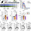 Figure 1
Plasma biomarkers of epithelial barrier disruption, microbial translocation, and inflammatory cytokines during SIV infection and ART. (A) Study design. SIV-infected rhesus macaques (n = 11) received daily ART (TDF, FTC, DTG). Baseline blood and gut biopsy samples were collected at week –2 and day 0 SIV challenge, and ART was initiated at 6 weeks after SIV infection. (B) Plasma SIV RNA levels over 30 weeks. Dashed line marks ART initiation, blue symbols represent animals in the DS group, red symbols represent control group, and green line shows average viral loads (VL) for all animals. (C) Plasma iFABP, zonulin, and sCD14 concentrations at pre-SIV baseline, day 30 after SIV infection (acute SIV), and 12 weeks after SIV+ART (post-ART). (D) Plasma CXCL13, MCP-1, IL-18, eotaxin, and IL-1RA levels at pre-SIV baseline, acute SIV (day 30), and 12 weeks post-ART time points. Filled symbols represent control group, and open symbols represent DS group animals. Data are shown as mean ± SEM and P values using repeated-measures 1-way ANOVA.
iFABP, a marker of enterocyte damage, rose significantly during acute infection (4 weeks, P = 0.019) and remained elevated despite ART, whereas zonulin increased later, 3 months after ART (P = 0.015), suggesting that early epithelial injury triggers tight junction disruption (Figure 1C). Soluble CD14 (sCD14), a marker of microbial translocation, remained unchanged. Circulating proinflammatory cytokines (CXCL13, IL-18, eotaxin, IL-1RA) surged at 4 weeks after SIV and partially declined with ART (Figure 1D). These findings indicate that SIV-induced IEBD occurs early, with epithelial damage leading to zonulin-mediated tight junction disruption and inflammation, and although ART partially normalizes iFABP and reduces inflammation, zonulin remains elevated beyond baseline, likely contributing to persistent microbial translocation and inflammation despite effective viral suppression.
Depletion of circulating Vδ2 T cells and ILC3s during acute SIV infection is associated with peak viremia and elevated integrin α4β7 expression. Extensive HIV/SIV replication in gut-associated lymphoid tissue depletes CCR5+ and Th17 CD4+ T cells, disrupting the epithelial barrier and promoting microbial translocation. Compensatory mucosal populations, including γδ T cells and ILC3s, support barrier integrity via IL-17 and IL-22 production (9, 13). We longitudinally tracked CD3+Vδ1+ T cells, CD3+Vδ2+ T cells, and lineage–CD8α–CD127+CD161+ ILC3 populations during acute SIV infection and subsequent ART. Despite differences in CD4 expression, Vδ1 and Vδ2 T cells both expressed high CD95, consistent with a memory/activated phenotype (Supplemental Figure 1; supplemental material available online with this article; https://doi.org/10.1172/jci.insight.201258DS1), and showed similar IL-17 and IL-22 production at steady state. A trend toward increased CD8+ T cell frequencies was observed at day 30 in response to acute SIV infection, while the CD4+ T cell compartment exhibited a significant decline following peak viremia and returned to baseline at 3 months after ART (Figure 2B). Vδ2 γδ T cells underwent a rapid, transient expansion during early acute infection (day 7) before returning to baseline levels by day 30 (Figure 2C), corroborating our earlier findings (9). This transient expansion reflects an acute proliferative response, which was followed by a significant contraction that coincided with peak viral replication. On the other hand, a rapid and progressive decline in ILC3s beginning during peak viremia was observed (Figure 2D). Despite their divergent kinetics at 1 week after infection, the decline of both peripheral Vδ2 T cells and ILC3s between weeks 1 and 4 was associated with viral dynamics, displaying an inverse relationship with plasma viral loads (Figure 2, E and F). Notably, the depletion of these circulating populations was accompanied by a significant upregulation of the gut-homing integrin α4β7 on Vδ1, Vδ2, and ILC3 subsets (Figure 2G). These data suggest that the loss of these critical innate-like populations from the circulation during peak viremia is likely driven by enhanced trafficking to the inflamed intestinal mucosa.
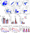 Figure 2
Temporal dynamics of Vδ2 T cell and ILC3 depletion during acute SIV infection and their relationship to peak viremia. (A) Representative gating schematic for Vδ1 T cells, Vδ2 T cells, and ILC3s in PBMCs. (B–D) Frequencies of CD4+ and CD8+ T cells (B), Vδ1 and Vδ2 γδ T cells (C), and ILC3s (D) in peripheral blood of study animals at baseline (2 weeks before SIV infection), acute SIV infection (day 7 and day 30 after SIV), and 12 weeks after SIV+ART (post-ART). Filled symbols represent control group, and open symbols represent DS group animals. (E and F) Circulating immune cell frequencies (blue, left y axis) and SIV viral load in log scale (red, right y axis) at each sampling time point for Vδ2 T cells (E) and ILC3s (F). (G) Frequencies of PBMC Vδ1 T cells, Vδ2 T cells, and ILC3s expressing integrin α4β7 at indicated time points. Data are shown as mean ± SEM by paired ANOVA with mixed-effects analysis using Tukey’s test for integrin α4β7.
SIV-driven Th1 polarization persists in circulating γδ T cells and ILC3s during ART, marked by T-bet upregulation and RORγt/AHR downregulation. The shift in Th1/Th17 balance in mucosal tissues is a well-documented hallmark of HIV/SIV infections (19, 20). To examine changes in functional immunophenotype of circulating ILC3s and γδ T cells, changes in their transcription factor profiles were evaluated at baseline (Figure 3, A and B), following SIV infection, and during the immune reconstitution phase of ART (Figure 3, C–E). At baseline, γδ T cells and ILC3s exhibited higher frequencies of cells expressing the Th17-associated transcription factors AHR and RORγt compared with those expressing the Th1-associated factor T-bet (Figure 3B). Notably, among the 3 subpopulations, ILC3s displayed significantly higher RORγt mean fluorescence intensity (MFI) relative to Vδ1 T cells and lower T-bet MFI in comparison with both Vδ1 and Vδ2 T cells, suggesting a predominant Th17-type functional phenotype (Supplemental Figure 2).
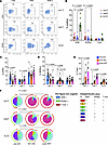 Figure 3
Transcription factor expression in circulating type 3 immune cells during SIV infection and ART. (A) Representative flow cytometry plots showing expression of the transcription factors AHR, RORγt, and T-bet in Vδ1 T cells, Vδ2 T cells, and ILC3s. (B) Frequencies of AHR, RORγt, and T-bet expression in PBMC Vδ1 T, Vδ2 T, and ILC3 subsets at baseline. (C–E) Longitudinal changes in the frequencies of AHR+ (C), RORγt+ (D), and T-bet+ (E) Vδ1 T, Vδ2 T, and ILC3 populations evaluated before SIV, at acute SIV infection (day 30 after SIV), and 12 weeks after SIV+ART (post-ART). Data are shown as mean ± SEM and paired ANOVA test P values. Filled symbols represent control group, and open symbols represent DS group animals. (F) Pie charts depicting the relative expression of various combinations of AHR, RORγt, and T-bet by Vδ1 T, Vδ2 T, and ILC3 populations from pre-SIV baseline through SIV infection and post-ART. Pie arcs represent expression of individual transcription factors, and pie slices represent the proportion of coexpressed transcription factors under each category. Significant differences between each time point for the overall phenotype were evaluated by SPICE permutation test.
Following SIV infection, AHR+ Vδ1 T cells transiently declined but normalized with ART (Figure 3C). Vδ2 T cells, by contrast, showed no significant change in AHR expression during acute infection but exhibited increased AHR expression during ART, indicating enhanced AHR signaling in the setting of viral control. Notably, Vδ2 T cells also showed an early increase in RORγt expression after infection, which returned to baseline after 2 months on ART, coinciding with an upregulation of T-bet (Figure 3, D and E). These findings suggest that ART restores the frequency of certain Th17-associated transcriptional profiles, such as AHR+ Vδ2 T cells and RORγt+ Vδ1 T cells, but may also promote a shift in type 3 immune cells from a Th17-like phenotype (AHR+RORγt+) toward a Th1-like phenotype (T-bet+) during acute infection and viral suppression (Figure 3F). This dynamic may reflect immune reprogramming associated with treated SIV infection, where ART partially restores immune homeostasis but may also skew innate-like T cell subsets toward Th1-type responses.
Compartmentalized γδ T cell and ILC3 dynamics in colonic epithelium versus lamina propria during SIV and ART. To further understand the gut-specific impacts of SIV infection and the influence of viral suppression with ART on γδ T cells and ILC3s, using the gating strategy shown in Figure 2A, we next examined their frequencies and immunophenotype in colonic lamina propria lymphocyte (LPL) and intraepithelial lymphocyte (IEL) compartments. Direct comparison between peripheral and mucosal compartments was restricted to the day 0, day 30, and week 12 (ART) time points to maintain a standardized sampling interval across all subjects and prioritize animal safety during the acute phase. As expected, a significant loss of CD4+ T cells was observed in colonic LPLs (P < 0.0001) and IELs (P < 0.01) by 6 weeks after SIV infection, resulting in near-complete depletion of CD4+ T cells in this gut compartment around the time of set-point viremia (Figure 4, A and D). Three months of daily ART partially restored CD4+ T cells in both IEL and LPL compartments (P < 0.05; Figure 4, A and D). However, CD4+ T cell frequencies in the LPLs remained significantly below baseline levels (P = 0.0058), indicating that viral suppression by ART promotes differential recovery of CD4+ T cells in the colonic lamina propria versus the epithelium.
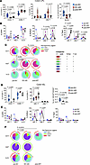 Figure 4
Frequencies of colonic mucosal γδ T cells and ILC3s and expression of Th1/Th17-associated transcription factors during acute SIV infection and ART. (A) Longitudinal frequencies of CD4+ T, CD8+ T, Vδ1 T, Vδ2 T, and ILC3 populations in the LPLs from colon biopsies at indicated time points using the gating strategy described in Figure 2A. (B) AHR, RORγt, and T-bet expression by colonic LPL Vδ1 T, Vδ2 T, and ILC3 populations. (C) Pie charts depicting the relative expression of AHR, RORγt, and T-bet by Vδ1 T, Vδ2 T, and ILC3 populations in colonic LPLs isolated from pre-SIV baseline through SIV infection and ART. (D) Longitudinal frequencies of CD4+ T, CD8+ T, Vδ1 T, Vδ2 T, and ILC3 populations in colonic IELs at indicated time points. (E) AHR, RORγt, and T-bet expression by colonic IEL Vδ1 T, Vδ2 T, and ILC3 populations. (F) Pie charts depicting the relative expression of AHR, RORγt, and T-bet by Vδ1 T, Vδ2 T, and ILC3 populations in colonic IEL fraction from pre-SIV baseline through SIV infection and ART. Pie arcs represent expression of individual transcription factors, and pie slices represent the number of coexpressed transcription factors. Note that IEL data represent n = 5–6 animals because of insufficient cell yields from cryopreserved colon biopsies for these specific assays. The rare frequencies of IEL Vδ2 T and ILC3 populations precluded a meaningful analysis of their combinatorial transcription factor profiles at the post-ART time point. Data are shown as mean ± SEM and paired ANOVA test P values.
In LPLs, Vδ2 T cells increased during acute infection and returned to baseline by 3 months after ART (P = 0.017), while Vδ1 T cells remained stable (Figure 4A). However, ILC3s mirrored CD4+ T cell kinetics, sharply declining at 1 month after infection (P < 0.0001) with partial ART-mediated restoration (Figure 4A). Notably, a transient Th17-type skewing, reflected by elevated RORγt, occurred in Vδ2 T cells and ILC3s during acute infection (P ≤ 0.012), whereas AHR remained unchanged across all 3 cell populations during acute SIV infection and after ART (Figure 4B). Progressive T-bet upregulation was noted in γδ T cells, most pronounced in Vδ1 (P = 0.0004), while T-bet+ ILC3s increased transiently during acute infection before returning to baseline (Figure 4B). These findings suggest that during acute SIV infection, Vδ2 T cells and ILC3s undergo transient Th17-type skewing in response to viral replication and CD4+ Th17 cell depletion in the gut (Figure 4C). Furthermore, the sustained T-bet upregulation in Vδ1 T cells, despite effective viral suppression with ART, points to a potential role for this subset in maintaining mucosal inflammation during chronic infection (Figure 4C).
The colonic IEL compartment exhibited a near-complete depletion of CD4+ T cells following SIV infection, accompanied by a reciprocal increase in CD8+ T cell frequencies (Figure 4D), highlighting a major shift in T cell composition at the mucosal barrier. Vδ2 T cells remained nearly undetectable at all time points, consistent with prior findings that Vδ1 T cells predominate among γδ T cells in the gut epithelium (21, 22). Similarly, lineage-negative CD127+ ILC3-like cells, a relatively rare subset in the IELs (23), showed a progressive and sustained decline after infection that was not reversed by ART (Figure 4D), suggesting poor restoration of this critical innate population in the epithelial niche. Interestingly, IEL-resident Vδ1 T cells increased significantly by 3 months after SIV+ART (P = 0.046), pointing to a selective expansion or enhanced retention of this subset in the epithelial compartment (Figure 4D). Functionally, RORγt-expressing Vδ1 T cells were significantly elevated during acute infection (P = 0.0025), mirroring the Th17-like skewing observed in Vδ2 T cells in the LPLs (Figure 4, E and F). However, this increase in RORγt expression was transient and declined to below baseline levels by 3 months after ART (Figure 4E). Because of the limited cellular yield from longitudinal colon biopsies, transcription factor analysis was prioritized for the diet-supplemented cohort (n = 5; Figure 4, B and E) to characterize the functional profile following intervention. The low abundance of ILC3s precluded a meaningful analysis of their transcription factor profiles in the IELs. Thus, compartmentalized γδ T cell and TCR-negative innate cell responses were observed in the colonic lamina propria and epithelium during SIV infection.
Th1/Th17 cytokine dysregulation and transcription factor correlates in colonic γδ T cells during ART-treated SIV infection. To assess how RORγt, AHR, and T-bet expression affects gut γδ T cell and ILC3 function, we measured cytokine production in colonic LPLs after ex vivo mitogen stimulation. Consistent with our earlier findings (9), ex vivo production of Th17-type cytokines, IL-17A and IL-22, was markedly impaired during SIV infection despite ART, whereas IFN-γ production in mitogen-stimulated cells remained largely intact, except for a significant decline in CD4+ T cells (Supplemental Figure 3D). In colonic LPLs, IL-22 was significantly reduced in CD4+ (P = 0.0002) and Vδ2 T cells (P = 0.002), with a trend in ILC3s (P = 0.059), and IL-17 was decreased in CD4+ (P = 0.005) and Vδ2 T cells (P = 0.007) (Figure 5, A and B).
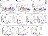 Figure 5
Cytokine production by colonic mucosal T cells and ILC3s during ART-suppressed SIV infection and association with lineage-defining transcription factors. Freshly isolated colonic LPLs were evaluated for cytokine production following ex vivo stimulation with PMA/calcium-ionomycin. (A–C) Intracellular expression of IL-22 (A), IL-17A (B), and IFN-γ (C) across γδ T cells, CD4+ T cells, CD8+ T cells, and ILC3s. Lines connect longitudinal samples from the same animal to illustrate shifts from pre-SIV baseline to ART-suppressed SIV infection. (D–F) Transcription factor correlations: correlation analysis between master regulators and cytokine output in colonic T cells and ILC3s. Transcription factor expression was measured in parallel and correlated with cytokine production. Correlations between RORγt (D) and AHR (E) expression and IL-17A or IL-22 production by colonic γδ T cell subsets are shown. Correlations between T-bet (F) and IFN-γ, IL-17A, or IL-22 production by colonic γδ T cells and ILC3s are presented. Spearman’s rank correlations were calculated using pooled samples from baseline and 30 days after infection to assess maintenance of these relationships during SIV-associated immune activation. For A–C, data represent mean ± SEM, with P values determined by Wilcoxon’s paired signed-rank test. For D–F, Spearman’s rank correlations were calculated using pooled longitudinal data to define the relationship between transcription factors and cytokines across healthy and SIV-associated immune activation.
Notably, IL-22 expression in ex vivo–stimulated Vδ2 T cells correlated positively with RORγt expression (P = 0.01; Figure 5D), whereas IL-17 production was associated with AHR expression in both Vδ1 (P = 0.017) and Vδ2 T cells (P = 0.007) (Figure 5E), highlighting distinct transcriptional dependencies for these cytokines. As expected, T-bet expression strongly correlated with IFN-γ production in colonic γδ T cells (P = 0.004 in Vδ2; P = 0.015 in Vδ1; Figure 5F). Strikingly, T-bet also negatively correlated with IL-22 in Vδ2 T cells and ILC3s (P = 0.019 and 0.038, respectively), consistent with the skewing toward an IFN-γ–dominant phenotype observed with increased T-bet and reduced RORγt in colonic LPLs (Figure 5C). Interestingly, IL-17 production in Vδ1 T cells correlated positively with T-bet, suggesting that IFN-γ/IL-17–coproducing Vδ1 T cells are regulated via integrated T-bet and AHR signaling. No significant correlation was observed between AHR or RORγt expression and cytokine production in ILC3s (Supplemental Figure 3). In summary, chronic SIV+ART infection reprograms mucosal γδ T cells and ILC3s toward a T-bet–driven, IFN-γ–biased phenotype with diminished IL-17 and IL-22 production.
In vitro effects of AHR ligands on mitigating HIV-induced damage to colonic epithelial monolayers. HIV proteins, including Tat, can disrupt intestinal epithelial integrity independent of viral replication (24). We treated differentiated human colonic epithelial Caco-2 monolayers with recombinant SIV Tat protein as an in vitro model. Tat exposure resulted in significantly reduced cell viability comparably to 5%–10% ethanol (Supplemental Figure 4), used as a well-established inducer of intestinal epithelial permeability. Further, SIV Tat decreased AHR expression (Figure 6, A and B), suggesting that Tat-mediated damage may occur, at least in part, via AHR suppression. Immunofluorescence staining for the tight junction protein ZO-1 revealed disrupted junctional morphology in Tat-treated Caco-2 monolayers, characterized by diminished ZO-1 signal intensity and visible intercellular gaps (Figure 6C, red arrows), indicative of compromised barrier integrity. Cotreatment with the AHR ligand 6-formylindolo[3,2-b]carbazole (FICZ) or indole-3-carbinol (I3C) preserved ZO-1 organization and restored the characteristic zigzag junctional pattern associated with well-polarized, mature epithelial monolayers (Figure 6C, bottom), with FICZ showing a stronger effect. Real-time cell analysis confirmed that Tat impaired barrier function (decreased impedance/cell index), which was significantly restored by FICZ and I3C, particularly FICZ (P < 0.0001), as evidenced by both real-time recovery kinetics and area under the curve analysis (Figure 6, D and E). Because cruciferous vegetables such as broccoli contain indole glucosinolates hydrolyzed into AHR agonists like indolocarbazole (ICZ) (25, 26), our subsequent experiments aimed to explore the in vivo effects of dietary indoles in modulating gut AHR signaling and intestinal epithelial barrier functions during ART-suppressed chronic SIV infection.
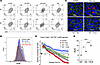 Figure 6
In vitro epithelial monolayer disruption and AHR downregulation by SIV Tat is reversed by ligand-specific activation through indoles. (A) Representative flow cytometry contour plots showing AHR expression in Caco-2 cells at 0 hours and 6 hours following exposure to control medium or medium supplemented with 5% EtOH, 10% EtOH, or 3 μM SIV Tat. (B) Representative histogram showing AHR expression in Caco-2 cells after 6 hours of exposure to EtOH- or Tat-containing medium compared with control conditions. (C) Representative immunofluorescence images of ZO-1 (green) and nuclei (DAPI, blue) in Caco-2 cells under control conditions (top left) or following 6 hours of treatment with Tat alone (top right), Tat plus i3C (bottom left), or Tat plus FICZ (bottom right). Scale bars: 50 μm. (D) Real-time cell analysis (RTCA) of Caco-2 monolayer integrity. Cells were seeded and allowed to form a confluent monolayer until the normalized cell index reached a stable plateau (50 hours after seeding), at which point HIV Tat alone or in combination with AHR agonists (i3C or FICZ) was added. Cell index was continuously monitored over the subsequent 6 hours and normalized to baseline at the time of treatment. Data are shown as mean ± SD of triplicate wells. (E) Area under the curve (AUC) analysis is shown as a single, quantitative value that denotes the cumulative cellular response over 6 hours, represented as mean ± SEM (**P < 0.005, ****P < 0.0001).
Broccoli-based AHR ligand supplementation enhances epithelial barrier integrity, Vδ2 T cell function, and mucosal immune subset distribution during chronic SIV infection and ART. We next assessed the effects of short-term broccoli-based dietary supplementation (DS) as a physiologically relevant means of augmenting AHR ligand availability in the gut, on AHR signaling and intestinal inflammation in chronically SIV-infected macaques on long-term ART. At 5 months after SIV+ART, animals received approximately 30 μmol sulforaphane/Avmacol (2 tablets/day) for 2 weeks, escalating to approximately 60 μmol sulforaphane (4 tablets/day) for 2 more weeks. Plasma biomarkers showed reduced iFABP by 2 weeks and LPS-binding protein (LBP) by 4 weeks (Figure 7, A and B), indicating improved barrier integrity and decreased microbial translocation. Plasma sCD14 concentrations declined in three of the five animals by the two-week time point (Figure 7C). Concomitantly, DS enhanced IL-17 responses in circulating Vδ2 T cells, as demonstrated by an increase in IL-17 spot-forming cells following HMBPP stimulation of PBMCs (Figure 7D). Furthermore, at 4 weeks after DS, there was a coordinated trend toward reduced systemic inflammation, including TNF-α, the chemokines MCP-1 and CXCL13, and eotaxin, suggesting a broad systemic antiinflammatory effect. IL-1RA also declined, reflecting dampened negative feedback, while SDF-1α, FGF-2, and VEGF-D levels decreased, indicating reduced inflammatory cell recruitment and pro-angiogenic signaling (Supplemental Figure 5).
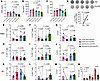 Figure 7
Effects of dietary indoles on plasma biomarkers and type 3 immune cell frequencies in chronically SIV-infected macaques on long-term ART. (A–C) Plasma iFABP (A), LBP (B), and sCD14 (C) at baseline and 2 and 4 weeks after supplementation and at baseline and 4-week-equivalent time points in control group. (D) IL-17+ PBMCs after Vδ2 T agonist stimulation at baseline and 4 weeks. (E–M) Frequencies of Vδ1 T cells, Vδ2 T cells, and ILC3s in PBMCs (E–G), LPLs (H–J), and IELs (K–M) from control versus DS groups. (N) Vδ1/Vδ2 ratios in PBMCs, LPLs, and IELs. For panel N, a Mann-Whitney test was used.
Flow cytometry showed a significant increase in circulating Vδ2 T cells at 4 weeks in the DS group, contrasting with significantly lower frequencies at the matching time point of 6 months after SIV in the control group (Figure 7F). Additionally, circulating ILC3 frequencies were maintained in the DS group, while there was a significant decline in the control group (Figure 7G). In colonic LPLs, although there were no significant changes in Vδ2 T cell frequencies both within and between groups, the DS group displayed higher ILC3 frequencies than the control group (Figure 7J). Notably, colonic IELs exhibited significantly reduced Vδ1 and increased Vδ2 frequencies (Figure 7, K and L) along with a trend of increased ILC3 frequencies (Figure 7M) in the DS group. Overall, these changes resulted in a lower Vδ1/Vδ2 ratio across all compartments, particularly in IELs (Figure 7N). Circulating Vδ1 T cell subsets (Figure 7E) and colonic LPL Vδ1 and Vδ2 T cell subsets (Figure 7, H and I) showed no significant differences in frequency. Classical T cell frequencies were unchanged in the colon (Supplemental Figure 6), but the DS group showed increased naïve CD4+ T cell frequencies (Figure 8A) and no change in CD8+ naïve cells (Figure 8D). Additionally, compared to the unchanged profiles of the control group, DS animals displayed significantly expanded effector memory (Figure 8C, F) and contracted central memory (Figure 8B, E) populations across both CD4+ and CD8+ T cell subsets.
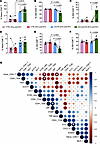 Figure 8
Impact of dietary indoles on colonic mucosal T cell memory distribution and its relationship to circulating inflammatory cytokines and barrier integrity. (A–F) Frequencies of CD95–CD28+ naive, CD95+CD28+ central memory (CM), and CD95+CD28– effector memory (EM) subsets in colonic lamina propria CD4+ T cells (A–C) and CD8+ T cells (D–F). Data are shown as mean ± SEM. Longitudinal comparisons within the DS group across 3 time points were analyzed using 1-way repeated-measures ANOVA with Bonferroni’s post hoc correction. Comparisons between control and DS groups over time were performed using 2-way repeated-measures ANOVA to evaluate the interaction between time and intervention. (G) Pearson’s correlations from –1 (red) to +1 (blue); white stars indicate significance.
Correlation analysis linked immune subsets with plasma biomarkers (Figure 8G). Naive CD4+ T cells negatively correlated with iFABP, consistent with their recovery after DS, and ILC3s tracked with CXCL13, suggesting a gut mucosal–B cell chemokine axis. MCP-1, CXCL13, and eotaxin clustered together, highlighting a shared inflammatory program spanning monocyte/macrophage activation, eosinophil recruitment, and lymphoid remodeling, which was dampened by DS (Supplemental Figure 5). Overall, DS improved epithelial barrier markers, enhanced IL-17–producing Vδ2 responses, and reshaped mucosal T cell and ILC3 subsets during chronic SIV+ART.
In colonic LPLs, DS significantly increased AHR in Vδ2 T cells (P = 0.01) with a trend in Vδ1 T cells (P = 0.06) but decreased AHR in ILC3s (P = 0.006) (Figure 9A). RORγt increased across all subsets, most prominently in ILC3s (P = 0.003) and Vδ1 T cells (P = 0.04), while T-bet declined in Vδ1 T cells (P = 0.004) (Figure 9, B and C). These shifts indicate a move toward a type 3 immune phenotype, supported by polyfunctional transcription factor expression profiles in Vδ1 T cells and ILC3s (Figure 9D). Ex vivo stimulation showed increased IL-22 response in Vδ1 (P = 0.0008) and Vδ2 (P = 0.007), reduced IFN-γ, and unchanged IL-17A (Figure 9, E–G), with no significant changes in polyfunctional cytokine profiles (Figure 9, H–K). ILC3 cytokines were unaffected despite significantly increased RORγt expression. CD4+ and CD8+ T cells exhibited no functional recovery following supplementation. Their IL-17 and IL-22 responses continued to decline under chronic SIV+ART, consistent with progressive loss of epithelial barrier–protective functions, and were accompanied by a marked reduction in IFN-γ production (Figure 9G and Supplemental Figure 7), indicative of dampened inflammatory capacity. Taken together, these findings demonstrate that DS reprograms colonic γδ T cells toward type 3 immunity characterized by increased IL-22 production, a shift likely to enhance mucosal barrier integrity and epithelial repair.
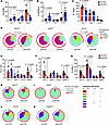 Figure 9
Effects of dietary indoles on transcription factor expression and cytokine-producing functions of colonic mucosal γδ T cells and ILC3s during chronic SIV+ART. Colonic LPLs were analyzed for transcription factor expression and cytokine production following PMA/ionomycin stimulation. (A–C) Frequencies of AHR (A), RORγt (B), and T-bet (C) expression were compared before and 1 month after dietary supplementation (DS) with indoles. (D) Pie charts depicting changes in the relative expression of AHR, RORγt, and T-bet by Vδ1 T, Vδ2 T, and ILC3 populations in colonic LPL fraction from pre- and post-DS. Pie arcs represent expression of individual transcription factors, and pie slices represent the number of coexpressed transcription factors. (E–K) Cytokine production of IL-22 (E), IL-17A (F), and IFN-γ (G) by Vδ1 T, Vδ2 T, ILC3, and CD4+ T cells was measured following mitogen stimulation of colonic LPLs, and combinatorial cytokine responses are shown in pie charts from before and after DS (H–K). Data are shown as mean ± SEM; paired t test values for comparisons between pre- and post-DS are shown.
To evaluate the localized impact of dietary indole supplementation on mucosal immunity, we used multiplex confocal immunohistochemistry (IHC) to quantify the density of AHR+RORγt+ cells within the colonic lamina propria. Representative images demonstrate that while SIV-naive animals maintained strong, continuous ZO-1 expression and abundant AHR+ and RORγt+ populations, chronic SIV infection resulted in the characteristic loss of apical ZO-1 and a depletion of these critical immune subsets (Figure 10A). Quantitative analysis revealed more than 2-fold higher mean density of dual-positive AHR+RORγt+ cells in the DS group compared with the control group (12.22 vs. 5.33 cells/mm2, respectively). While this comparison (Figure 10B) did not reach the threshold for traditional statistical significance (Mann-Whitney P = 0.0693), likely owing to the inherent constraints of the nonhuman primate cohort size (n = 5–6 per group), the greater AHR+RORγt+ cell density suggests a biological trend toward the enrichment of these regenerative immune subsets with the intervention. These data suggest that a higher prevalence of the AHR-RORγt axis may be a critical factor in the intestinal barrier restitution observed in the DS-treated cohort.
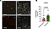 Figure 10
Enrichment of AHR/RORγt-coexpressing cells in dietary indole–supplemented animals during chronic SIV+ART. Fluorescent IHC demonstrating ZO-1, AHR, and RORγt expression in colon. (A) Representative colon sections from one SIV-naive (top) and one chronic SIV–positive macaque (bottom). The SIV-naive animal shows strong ZO-1 expression (green, arrows) along the apical margin of epithelial cells (red; top left), and abundant AHR-expressing (green, arrow) cells both with and without RORγt expression (red, arrowheads; top right). Inset highlights an AHR+RORγt+ cell in yellow. The chronically SIV-infected macaque demonstrates a loss of ZO-1 expression with only patchy, residual expression (green, arrows; bottom left), and a reduction in AHR-expressing cells within the mucosa and a depletion of single- and dual-positive RORγt-expressing cells (red, arrowhead; bottom right). Inset highlights an AHR+RORγt+ cell. Scale bars: 100 μm. Original magnification, ×200. White, DAPI (right); red, pan-cytokeratin (left), RORγt (right); green, ZO-1 (left), AHR (right). (B) AHR+RORγt+ cells/mm2 quantified using HALO software. Data show mean ± SEM and comparison between control and DS group by Mann-Whitney test.


