
Cell biology
Anti-Notch plus endocrine therapy in ER+ breast cancer inhibits cancer stem cells by increasing DAXX
Abstract
Resistance to endocrine therapy (ET) in ER+ breast cancer is mediated by Notch signaling, but the clinical application of anti-Notch therapy has been limited by the lack of predictive biomarkers. To identify Notch-regulated biomarkers, we conducted a pre-surgical window study evaluating ET combined with the γ-secretase inhibitor (GSI) MK-0752. RNA expressions in tumors were measured using an Affymetrix array and by real-time PCR. ET plus GSI showed more genes were decreased than ET alone. Specifically, DAXX, NOXA, and LFNG RNAs were increased, while fifteen additional transcripts were decreased. Mechanistically, GSI reduced Notch1 occupancy at CSL-binding elements within HES1, HEY2, HEYL, CCND1, MKI67, and DAXX genes, and inhibited cancer stem cells (CSCs) by 90% to 100%. This anti-CSC effect required DAXX, while GSI treatment or Notch1/4 knockdown increased DAXX expression, suggesting transcriptional repression by Notch. Using mouse tumor xenograft studies, ET plus MK-0752 resulted in complete regression of MCF-7 tumors, with DAXX-high tumors showing greater treatment sensitivity. Clinically, high DAXX expression was associated with improved recurrence-free and overall survival. This study found that anti-Notch plus ET in ER+ breast cancer inhibits cancer stem cells by increasing DAXX, a promising predictive biomarker. These findings support clinical evaluation of therapies that increase DAXX expression.
Authors
Kathy S. Albain, Debra Wyatt, Andrei Zlobin, Susan G. Hilsenbeck, Cheryl M. Czerlanis, Daniel S. Peiffer, Kyle R. Convington, Constantine Godellas, Shelly S. Lo, Patricia A. Robinson, Kathy Czaplicki, Barbara Busby, Davide Bova, Ping Tang, Patrick J. Stiff, Suzanne A.W. Fuqua, Lucio Miele, Clodia Osipo
Abstract
ADAMTS9 mutations cause the ciliopathies nephronophthisis and Joubert syndrome. Here we demonstrated that deletion of ADAMTS9 in the proximal nephron led to polycystic kidney development in mice. In males, Adamts9 deletion caused kidneys to become highly cystic while remaining small without undergoing enlargement. In contrast, female mice developed cystic kidneys at a slower rate. ADAMTS9 deletion disrupted ciliogenesis through the loss of cleavage of the ciliary transition zone (TZ) protein TMEM67, which led to loss of the MKS/B9 module – a key component of the ciliary gate. Functional analysis of all eight ciliopathy patient variants of ADAMTS9 identified to date showed TMEM67 C-terminus failed to localize to the TZ, thus disrupting a key regulatory mechanism in patient renal ciliogenesis. Modeling ADAMTS9-mediated TMEM67 cleavage utilizing TMEM67-cleavage deficient mice revealed loss of TZ formation, but not elevated canonical Wnt signaling as the underlying mechanism driving cystogenesis. Adamts9 deletion led to comparatively intense interstitial collagen deposition, which likely restricted kidney enlargement and resulted in the characteristically small kidney phenotype seen in nephronophthisis. By comparative analysis of four interconnected polycystic kidney models, in addition to Pkd1 and Pkd2 deleted kidneys, we identified differential collagen homeostasis as a principle factor determining cystic kidney size and type.
Authors
Sydney Fischer, Karyn L. Robert, Manu Ahmed, Griffin I. Kane, Matthew A. Kavanaugh, Wei Wang, Pamela V. Tran, Prabhani U. Atukorale, Sumeda Nandadasa
Abstract
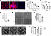
Autosomal dominant polycystic kidney disease (ADPKD) is a leading genetic cause of kidney failure, characterized by progressive cyst growth, inflammation, and interstitial fibrosis. Renal fibrosis, driven by myofibroblast activation and excessive extracellular matrix (ECM) deposition, is increasingly recognized as a key contributor to disease progression, yet targeted antifibrotic therapies remain limited. Here, we evaluated the therapeutic potential of pirfenidone to suppress fibrosis and disease progression in ADPKD. Single-nucleus RNA sequencing of human ADPKD kidneys identified fibroblasts as the predominant source of fibrous and adhesive ECM, with higher ECM-associated gene expression compared with that in normal kidney fibroblasts. In vitro, primary human ADPKD renal myofibroblasts displayed a similar profibrotic gene expression profile, and pirfenidone treatment suppressed ECM gene expression, cell proliferation, migration, and contractility. In the Pkd1RC/RC mouse model of ADPKD, pirfenidone reduced renal fibrosis, myofibroblast accumulation, ECM deposition, profibrotic gene expression, and associated signaling pathways and improved kidney function. Pirfenidone also reduced kidney enlargement but reduced cyst burden only in female mice. Collectively, these findings demonstrate that pirfenidone attenuates renal fibrosis and improves kidney function in ADPKD by suppressing myofibroblast activation and ECM production, supporting fibrosis as a therapeutic target and highlighting pirfenidone as a potential adjunct to cyst-directed therapies.
Authors
Viji Remadevi, Abeda Jamadar, Meekha M. Varghese, Haichun Yang, Sumedha Gunewardena, Darren P. Wallace, Reena Rao
Abstract
The adult mammalian heart has a limited ability to regenerate lost myocardium following myocardial infarction (MI), largely due to the poor proliferative capacity of cardiomyocytes (CMs). Dual-specificity tyrosine phosphorylation-regulated kinase 1A (DYRK1A) is a known regulator of cell quiescence, though the mechanisms underlying its function remain unclear. Previous studies have shown that pharmacological inhibition of DYRK1A using harmine induces CM cell cycle re-entry after ischemia/reperfusion (I/R) MI. Here, we developed a computational network model of DYRK1A-mediated regulation of the cell cycle, which predicts how DYRK1A inhibition promotes CM re-entry. To validate these predictions, we tested selective DYRK1A inhibitors and observed robust induction of cell cycle activity in neonatal rat cardiomyocytes (NRCMs). Integrating our network model with bulk RNA-sequencing data from DYRK1A inhibitor-treated NRCMs, we identified E2F1 as a key transcriptional driver of cell cycle gene expression. Finally, we demonstrate that both pharmacological and post-developmental inhibition of DYRK1A enhances heart function and increases CM cycling following I/R MI. Our findings suggest that functional recovery induced by small molecule inhibitor of DYRK1A is mediated by the induction of cycling CMs.
Authors
Bryce C. Murillo, Alexander Young, Kaitlyn L. Wintruba, Alexander J. Eichert, Klara Siejda, Dennon Hoernig, Leigh A. Bradley, Bryana N. Harris, Catherine Zhao, MIchelle Wu, Emmanuel Deau, Mattias F. Lindberg, Laurent Meijer, Jeffrey J. Saucerman, Matthew J. Wolf
Abstract
Cell metabolic rewiring is associated with resistance to venetoclax-azacitidine (Ven-Aza) combination therapy and relapse in acute myeloid leukemia (AML) patients. Drug-resistant cells exhibit an enhanced reliance on oxidative phosphorylation (OXPHOS) for energy production. Therefore, impairing mitochondrial metabolism represents an exciting strategy to face this unmet clinical need. We recently demonstrated that the specific activation of the phosphatase PP2A-B56α enhances the pro-apoptotic efficacy of venetoclax in AML. Here, through leveraging unbiased multi-omics-based approaches and using both genetic and pharmacological tools, we define key roles for the tumor suppressor PP2A-B56α complex in OXPHOS regulation and treatment response in disease-relevant AML models. From a translational perspective, the specific stabilization of PP2A-B56α heterocomplex with the novel PP2A molecular glue activator, RPT04402, reduces OXPHOS levels in treatment-resistant AML cells and improves treatment response in both Ven-Aza-sensitive and -resistant AML cell lines, primary cells, and in vivo models. Together, our work supports further research on targeted combination therapy approaches based on PP2A-B56α stabilization to counteract OXPHOS-related treatment resistance and improve AML responses in a patient population with historically poor outcomes.
Authors
Silvia Romero-Murillo, Irene Peris, Anna Maria Lucianò, Nerea Marcotegui, Carmen Vicente, Brian Tran, Kelsey Barrie, Caitlin M. O'Connor, Andrea Torres-López, Maria C. Mateos, Maria L. Cayuela, Victoriano Mulero, Joaquín Fernández-Irigoyen, Enrique Santamaría, Maria D. Odero, Goutham Narla
Abstract
Wnt signaling drives tumorigenesis in multiple cancers, in part through complex interactions with other oncogenic pathways including the MAPK cascade. In Wnt-addicted cancers, pharmacologic and genetic inhibition of Wnt signaling activates multiple receptor tyrosine kinases (RTKs), increases ERK phosphorylation and induces MAPK target gene expression, but the specific RTKs responsible for this MAPK hyperactivation are not known. Here we performed phosphotyrosine-targeted mass spectrometry, which revealed robust phosphorylation of EPHA2 and EGFR upon Wnt inhibition. Unexpectedly, we find that in xenografts, EPHA2 suppresses EGFR and ERK activation. Most notably, the increased ERK phosphorylation observed in EPHA2 KO tumors is transcriptionally inert, as there is no concomitant increase in MAPK target gene expression until concomitant Wnt inhibition. This suggests a Wnt-activated transcriptional repressor such as GATA3 that gates MAPK signaling in Wnt-high cancers. While Wnt-high KRAS-mutant cancers are resistant to erlotinib alone, adding Wnt inhibitor mitigates this resistance. Additionally, loss of EPHA2 enhances their sensitivity to both erlotinib and Wnt inhibitors. These studies therefore identify therapeutic vulnerabilities in Wnt-high tumors, even within traditionally EGFR inhibitor-resistant, RAS-mutant contexts.
Authors
Shawn R. Wadia, Changyuan Hu, Siddhi Patnaik, Shreya Sridharan, Roger J. Daly, David M. Virshup, Babita Madan

Abstract
Wound healing is a highly dynamic and metabolically demanding process. However, the primary drivers of metabolic alterations involved in this process remain incompletely understood. Here, we employed multiomics profiling of clinical samples to investigate metabolic alterations during wound healing. Our analyses revealed significant activation of the TCA cycle and identified α-ketoglutarate (αKG) as a central regulator orchestrating the reparative phase. Systemic administration of αKG promoted wound closure and re-epithelialization, characterized by enhanced neo-tissue formation with an extended epithelial tongue. Mechanistically, αKG promoted cell proliferation via the cell cycle pathway and enhanced fibroblast-derived TGF-β signaling to induce epithelial-mesenchymal transition–like programs in epithelial cells. To address the spatial metabolic heterogeneity, we developed a transdermal MN platform based on gelatin methacryloyl for localized αKG delivery, further accelerating tissue repair. Collectively, these findings identify αKG as a metabolic driver of wound repair, reveal its dual role in modulating the epithelial-fibroblast microenvironment, and introduce a targeted bioengineering strategy with translational potential for both acute and chronic wound management.
Authors
Yuhan Li, Weimin Lin, Denghao Huang, Yueying Wang, Yimeng Cai, Jie Xiang, Linfeng Liu, Xinxing Shuai, Qi Yin, Shuang Jiang, Malcolm Xing, Yuan Wang, Leixiao Yu, Quan Yuan
Abstract
Fractures heal by rapid formation of mineralized callus, a process requiring periosteal cell proliferation and differentiation. Our objective was to dissect the contribution of proliferating osteoblast lineage cells to fracture callus formation. First, mice expressing thymidine kinase (TK) in 3.6Col1a1-lineage cells were treated with ganciclovir (GCV) to ablate proliferating osteolineage cells for 5 or 10 days. Immunostaining demonstrated that this approach specifically depleted TK+ proliferating cells in the bony regions of the callus, while sparing other proliferating cells. Single-cell RNA-seq of callus cells revealed that GCV-treated Col1-TK mice had fewer osteoblasts and chondrocytes than controls, with more myofibroblasts and immune cells, consistent with fibrous nonunion. In controls, 15-30% of callus cells expressing the early osteoblast markers osterix (Sp7) and the late marker osteocalcin (Bglap) were in the cell cycle. Next, we targeted proliferating osteolineage cells at different stages of differentiation by crossing Osx-CreERT2, Ocn-Cre and Dmp1-CreERT2 mice with ROSA-TK mice. Following fracture, each Cre;ROSA-TK mouse line exhibited decreased callus bone volume and a shift from callus bone to fibrous tissue. Therefore, during fracture repair, proliferation of callus cells at early and mature stages of osteoblast differentiation is critical to the formation of a mineralized callus that is essential for healing.
Authors
Nicole R. Gould, Andre F. Coello, Jennifer A. McKenzie, Mariam Obaji, Tiandao Li, Katherine R. Hixon, Leyi Chen, Kristen Barwick, Tiffany Lee, Bo A. Zhang, David Ornitz, Matthew J. Silva
Abstract
Menin is a scaffolding protein that interacts with context-specific partners to regulate gene expression. In MLL-rearranged leukemias, Menin:MLL interactions drive leukemogenesis and Menin inhibitors have been FDA approved for these cancers. We previously reported that Menin promotes oncogenic phenotypes in Ewing sarcoma (EwS). Here, we sought to define EwS-specific functions of Menin and determine if Menin inhibitors could be therapeutically leveraged for these tumors. Genetic knockout of Menin had no impact on EwS cell proliferation in vitro, but metastatic potential of Menin-depleted cells in vivo was impaired. Transcriptional profiling of Menin knockout cells in vitro showed reproducible downregulation of MYC signature genes and upregulation of developmental programs. Conversely, transcriptional rewiring of developmental genes and restoration of MYC target gene expression were evident in tumors that arose from Menin knockout cells. Exposing EwS cells to the Menin inhibitor VTP50469 (revumenib) inhibited expression of MYC targets and co-immunoprecipitation studies detected Menin:MYC interactions that were partially disrupted by the drug. Metastatic colonization of disseminated EwS cells in vivo was significantly inhibited in mice fed VTP50469 chow. Together these findings implicate Menin as a mediator of EwS metastasis and suggest that Menin inhibitors warrant investigation as novel therapeutics for patients with high-risk disease.
Authors
Katherine A. Braun, Nicolas M. Garcia, Mohamed A. Ahmed, Darleen S. Tu, Stephanie I. Walter, Emma D. Wrenn, Megan E. B. Dean, Neerja Katiyar, Elizabeth R. Lawlor
Abstract
The pathogenesis of Bartter syndrome (BS) has long been attributed to decreased salt reabsorption in the thick ascending limb of Henle’s loop (TAL). By studying Clc-k2 (mouse ortholog of ClC-Kb)-knockout (Clc-k2-/-) mice, we recently uncovered an additional mechanism in which loss of Clc-k2 induces TAL hypoplasia in neonatal kidneys, exacerbating BS severity. Here, we further investigated this mechanism. TALs and distal convoluted tubules (DCTs) isolated from Clc-k2-/- and wild-type mice were used for transcriptome, proteomics, cell cycle, and proliferation assays. Mitochondrial morphology and function were studied using electron microscopy and mitochondrial respiration assays. Our results revealed impairments in cell proliferation, S-to-G2/M cell cycle transition, mitochondrial biogenesis, oxidative phosphorylation, glycolysis, and fatty acid oxidation in Clc-k2-/- TALs and DCTs. Increasing transport function by introducing a gain-of-function with-no-lysine kinase 4 mutation in Clc-k2-/- mice restored these metabolic and proliferative impairments and improved phenotype. Transgenic expression of peroxisome proliferator-activated receptor gamma coactivator-1α, a master regulator of mitochondrial biogenesis, in Clc-k2-/- mice also alleviated mitochondrial dysfunction and phenotype. These findings support the hypothesis that mitochondrial hypofunction, resulting from decreased transport function, contributes to cell cycle arrest and tubular hypoplasia in BS. Targeting mitochondria early in life could be a potential therapeutic approach for BS.
Authors
Chiao-Hui Hsieh, Yu-Jen Chen, Chih-Chien Sung, Emily Morrison, Chou-Long Huang, Chih-Jen Cheng
No posts were found with this tag.


