
Research ArticleCell biologyHematology
Open Access |  10.1172/jci.insight.205218
Stabilization of the PP2A-B56α complex overcomes venetoclax-azacitidine resistance by impairing OXPHOS in AML
Silvia Romero-Murillo,1,2,3,4 Irene Peris,5,6 Anna Maria Lucianò,7,8,9 Nerea Marcotegui,3 Carmen Vicente,3 Brian Tran,10 Kelsey Barrie,5 Caitlin M. O’Connor,5,6 Andrea Torres-López,11 Maria C. Mateos,2,11 Maria L. Cayuela,12 Victoriano Mulero,7,8,9 Joaquín Fernández-Irigoyen,1,2 Enrique Santamaría,1,2 Maria D. Odero,2,3,4 and Goutham Narla5,6,13
1Clinical Neuroproteomics Unit, Navarrabiomed, Hospital Universitario de Navarra (HUN), UPNA, Navarra, Spain.
2Instituto de Investigación Sanitaria de Navarra, Pamplona, Spain.
3Centro de Investigación Médica Aplicada and
4Department of Biochemistry and Genetics, University of Navarra, Pamplona, Spain.
5Department of Internal Medicine, Division of Genetic Medicine, The University of Michigan Medical School, Ann Arbor, Michigan, USA.
6Roger Cancer Center, The University of Michigan Health, Ann Arbor, Michigan, USA.
7Departamento de Biología Celular e Histología, Facultad de Biología, Universidad de Murcia, Murcia, Spain.
8Instituto Murciano de Investigación Biosanitaria (IMIB) Pascual Parrilla, Murcia, Spain.
9Centro de Investigación Biomédica en Red de Enfermedades Raras (CIBERER), Instituto de Salud Carlos III, Madrid, Spain.
10Department of Pharmacology, Rogel Cancer Center, University of Michigan, Ann Arbor, Michigan, USA.
11Hematology Service, HUN, Pamplona, Spain.
12Cancer and Aging Group, Hospital Universitario Virgen de la Arrixaca, and IMIB, Murcia, Spain.
13Department of Cancer Biology, The University of Michigan, Ann Arbor, Michigan, USA.
Address correspondence to: Goutham Narla, University of Michigan, 1500 East Medical Center Drive, Rogel Cancer Center Room 3215, Ann Arbor, Michigan 48109, USA. Email: gnarla@med.umich.edu.
Authorship note: SRM and IP contributed equally to this work and are co–first authors.
Find articles by Romero-Murillo, S. in: PubMed | Google Scholar
1Clinical Neuroproteomics Unit, Navarrabiomed, Hospital Universitario de Navarra (HUN), UPNA, Navarra, Spain.
2Instituto de Investigación Sanitaria de Navarra, Pamplona, Spain.
3Centro de Investigación Médica Aplicada and
4Department of Biochemistry and Genetics, University of Navarra, Pamplona, Spain.
5Department of Internal Medicine, Division of Genetic Medicine, The University of Michigan Medical School, Ann Arbor, Michigan, USA.
6Roger Cancer Center, The University of Michigan Health, Ann Arbor, Michigan, USA.
7Departamento de Biología Celular e Histología, Facultad de Biología, Universidad de Murcia, Murcia, Spain.
8Instituto Murciano de Investigación Biosanitaria (IMIB) Pascual Parrilla, Murcia, Spain.
9Centro de Investigación Biomédica en Red de Enfermedades Raras (CIBERER), Instituto de Salud Carlos III, Madrid, Spain.
10Department of Pharmacology, Rogel Cancer Center, University of Michigan, Ann Arbor, Michigan, USA.
11Hematology Service, HUN, Pamplona, Spain.
12Cancer and Aging Group, Hospital Universitario Virgen de la Arrixaca, and IMIB, Murcia, Spain.
13Department of Cancer Biology, The University of Michigan, Ann Arbor, Michigan, USA.
Address correspondence to: Goutham Narla, University of Michigan, 1500 East Medical Center Drive, Rogel Cancer Center Room 3215, Ann Arbor, Michigan 48109, USA. Email: gnarla@med.umich.edu.
Authorship note: SRM and IP contributed equally to this work and are co–first authors.
Find articles by Peris, I. in: PubMed | Google Scholar
1Clinical Neuroproteomics Unit, Navarrabiomed, Hospital Universitario de Navarra (HUN), UPNA, Navarra, Spain.
2Instituto de Investigación Sanitaria de Navarra, Pamplona, Spain.
3Centro de Investigación Médica Aplicada and
4Department of Biochemistry and Genetics, University of Navarra, Pamplona, Spain.
5Department of Internal Medicine, Division of Genetic Medicine, The University of Michigan Medical School, Ann Arbor, Michigan, USA.
6Roger Cancer Center, The University of Michigan Health, Ann Arbor, Michigan, USA.
7Departamento de Biología Celular e Histología, Facultad de Biología, Universidad de Murcia, Murcia, Spain.
8Instituto Murciano de Investigación Biosanitaria (IMIB) Pascual Parrilla, Murcia, Spain.
9Centro de Investigación Biomédica en Red de Enfermedades Raras (CIBERER), Instituto de Salud Carlos III, Madrid, Spain.
10Department of Pharmacology, Rogel Cancer Center, University of Michigan, Ann Arbor, Michigan, USA.
11Hematology Service, HUN, Pamplona, Spain.
12Cancer and Aging Group, Hospital Universitario Virgen de la Arrixaca, and IMIB, Murcia, Spain.
13Department of Cancer Biology, The University of Michigan, Ann Arbor, Michigan, USA.
Address correspondence to: Goutham Narla, University of Michigan, 1500 East Medical Center Drive, Rogel Cancer Center Room 3215, Ann Arbor, Michigan 48109, USA. Email: gnarla@med.umich.edu.
Authorship note: SRM and IP contributed equally to this work and are co–first authors.
Find articles by Lucianò, A. in: PubMed | Google Scholar
1Clinical Neuroproteomics Unit, Navarrabiomed, Hospital Universitario de Navarra (HUN), UPNA, Navarra, Spain.
2Instituto de Investigación Sanitaria de Navarra, Pamplona, Spain.
3Centro de Investigación Médica Aplicada and
4Department of Biochemistry and Genetics, University of Navarra, Pamplona, Spain.
5Department of Internal Medicine, Division of Genetic Medicine, The University of Michigan Medical School, Ann Arbor, Michigan, USA.
6Roger Cancer Center, The University of Michigan Health, Ann Arbor, Michigan, USA.
7Departamento de Biología Celular e Histología, Facultad de Biología, Universidad de Murcia, Murcia, Spain.
8Instituto Murciano de Investigación Biosanitaria (IMIB) Pascual Parrilla, Murcia, Spain.
9Centro de Investigación Biomédica en Red de Enfermedades Raras (CIBERER), Instituto de Salud Carlos III, Madrid, Spain.
10Department of Pharmacology, Rogel Cancer Center, University of Michigan, Ann Arbor, Michigan, USA.
11Hematology Service, HUN, Pamplona, Spain.
12Cancer and Aging Group, Hospital Universitario Virgen de la Arrixaca, and IMIB, Murcia, Spain.
13Department of Cancer Biology, The University of Michigan, Ann Arbor, Michigan, USA.
Address correspondence to: Goutham Narla, University of Michigan, 1500 East Medical Center Drive, Rogel Cancer Center Room 3215, Ann Arbor, Michigan 48109, USA. Email: gnarla@med.umich.edu.
Authorship note: SRM and IP contributed equally to this work and are co–first authors.
Find articles by Marcotegui, N. in: PubMed | Google Scholar
1Clinical Neuroproteomics Unit, Navarrabiomed, Hospital Universitario de Navarra (HUN), UPNA, Navarra, Spain.
2Instituto de Investigación Sanitaria de Navarra, Pamplona, Spain.
3Centro de Investigación Médica Aplicada and
4Department of Biochemistry and Genetics, University of Navarra, Pamplona, Spain.
5Department of Internal Medicine, Division of Genetic Medicine, The University of Michigan Medical School, Ann Arbor, Michigan, USA.
6Roger Cancer Center, The University of Michigan Health, Ann Arbor, Michigan, USA.
7Departamento de Biología Celular e Histología, Facultad de Biología, Universidad de Murcia, Murcia, Spain.
8Instituto Murciano de Investigación Biosanitaria (IMIB) Pascual Parrilla, Murcia, Spain.
9Centro de Investigación Biomédica en Red de Enfermedades Raras (CIBERER), Instituto de Salud Carlos III, Madrid, Spain.
10Department of Pharmacology, Rogel Cancer Center, University of Michigan, Ann Arbor, Michigan, USA.
11Hematology Service, HUN, Pamplona, Spain.
12Cancer and Aging Group, Hospital Universitario Virgen de la Arrixaca, and IMIB, Murcia, Spain.
13Department of Cancer Biology, The University of Michigan, Ann Arbor, Michigan, USA.
Address correspondence to: Goutham Narla, University of Michigan, 1500 East Medical Center Drive, Rogel Cancer Center Room 3215, Ann Arbor, Michigan 48109, USA. Email: gnarla@med.umich.edu.
Authorship note: SRM and IP contributed equally to this work and are co–first authors.
Find articles by Vicente, C. in: PubMed | Google Scholar
1Clinical Neuroproteomics Unit, Navarrabiomed, Hospital Universitario de Navarra (HUN), UPNA, Navarra, Spain.
2Instituto de Investigación Sanitaria de Navarra, Pamplona, Spain.
3Centro de Investigación Médica Aplicada and
4Department of Biochemistry and Genetics, University of Navarra, Pamplona, Spain.
5Department of Internal Medicine, Division of Genetic Medicine, The University of Michigan Medical School, Ann Arbor, Michigan, USA.
6Roger Cancer Center, The University of Michigan Health, Ann Arbor, Michigan, USA.
7Departamento de Biología Celular e Histología, Facultad de Biología, Universidad de Murcia, Murcia, Spain.
8Instituto Murciano de Investigación Biosanitaria (IMIB) Pascual Parrilla, Murcia, Spain.
9Centro de Investigación Biomédica en Red de Enfermedades Raras (CIBERER), Instituto de Salud Carlos III, Madrid, Spain.
10Department of Pharmacology, Rogel Cancer Center, University of Michigan, Ann Arbor, Michigan, USA.
11Hematology Service, HUN, Pamplona, Spain.
12Cancer and Aging Group, Hospital Universitario Virgen de la Arrixaca, and IMIB, Murcia, Spain.
13Department of Cancer Biology, The University of Michigan, Ann Arbor, Michigan, USA.
Address correspondence to: Goutham Narla, University of Michigan, 1500 East Medical Center Drive, Rogel Cancer Center Room 3215, Ann Arbor, Michigan 48109, USA. Email: gnarla@med.umich.edu.
Authorship note: SRM and IP contributed equally to this work and are co–first authors.
Find articles by
Tran, B.
in:
PubMed
|
Google Scholar
|

1Clinical Neuroproteomics Unit, Navarrabiomed, Hospital Universitario de Navarra (HUN), UPNA, Navarra, Spain.
2Instituto de Investigación Sanitaria de Navarra, Pamplona, Spain.
3Centro de Investigación Médica Aplicada and
4Department of Biochemistry and Genetics, University of Navarra, Pamplona, Spain.
5Department of Internal Medicine, Division of Genetic Medicine, The University of Michigan Medical School, Ann Arbor, Michigan, USA.
6Roger Cancer Center, The University of Michigan Health, Ann Arbor, Michigan, USA.
7Departamento de Biología Celular e Histología, Facultad de Biología, Universidad de Murcia, Murcia, Spain.
8Instituto Murciano de Investigación Biosanitaria (IMIB) Pascual Parrilla, Murcia, Spain.
9Centro de Investigación Biomédica en Red de Enfermedades Raras (CIBERER), Instituto de Salud Carlos III, Madrid, Spain.
10Department of Pharmacology, Rogel Cancer Center, University of Michigan, Ann Arbor, Michigan, USA.
11Hematology Service, HUN, Pamplona, Spain.
12Cancer and Aging Group, Hospital Universitario Virgen de la Arrixaca, and IMIB, Murcia, Spain.
13Department of Cancer Biology, The University of Michigan, Ann Arbor, Michigan, USA.
Address correspondence to: Goutham Narla, University of Michigan, 1500 East Medical Center Drive, Rogel Cancer Center Room 3215, Ann Arbor, Michigan 48109, USA. Email: gnarla@med.umich.edu.
Authorship note: SRM and IP contributed equally to this work and are co–first authors.
Find articles by Barrie, K. in: PubMed | Google Scholar
1Clinical Neuroproteomics Unit, Navarrabiomed, Hospital Universitario de Navarra (HUN), UPNA, Navarra, Spain.
2Instituto de Investigación Sanitaria de Navarra, Pamplona, Spain.
3Centro de Investigación Médica Aplicada and
4Department of Biochemistry and Genetics, University of Navarra, Pamplona, Spain.
5Department of Internal Medicine, Division of Genetic Medicine, The University of Michigan Medical School, Ann Arbor, Michigan, USA.
6Roger Cancer Center, The University of Michigan Health, Ann Arbor, Michigan, USA.
7Departamento de Biología Celular e Histología, Facultad de Biología, Universidad de Murcia, Murcia, Spain.
8Instituto Murciano de Investigación Biosanitaria (IMIB) Pascual Parrilla, Murcia, Spain.
9Centro de Investigación Biomédica en Red de Enfermedades Raras (CIBERER), Instituto de Salud Carlos III, Madrid, Spain.
10Department of Pharmacology, Rogel Cancer Center, University of Michigan, Ann Arbor, Michigan, USA.
11Hematology Service, HUN, Pamplona, Spain.
12Cancer and Aging Group, Hospital Universitario Virgen de la Arrixaca, and IMIB, Murcia, Spain.
13Department of Cancer Biology, The University of Michigan, Ann Arbor, Michigan, USA.
Address correspondence to: Goutham Narla, University of Michigan, 1500 East Medical Center Drive, Rogel Cancer Center Room 3215, Ann Arbor, Michigan 48109, USA. Email: gnarla@med.umich.edu.
Authorship note: SRM and IP contributed equally to this work and are co–first authors.
Find articles by
O’Connor, C.
in:
PubMed
|
Google Scholar
|
1Clinical Neuroproteomics Unit, Navarrabiomed, Hospital Universitario de Navarra (HUN), UPNA, Navarra, Spain.
2Instituto de Investigación Sanitaria de Navarra, Pamplona, Spain.
3Centro de Investigación Médica Aplicada and
4Department of Biochemistry and Genetics, University of Navarra, Pamplona, Spain.
5Department of Internal Medicine, Division of Genetic Medicine, The University of Michigan Medical School, Ann Arbor, Michigan, USA.
6Roger Cancer Center, The University of Michigan Health, Ann Arbor, Michigan, USA.
7Departamento de Biología Celular e Histología, Facultad de Biología, Universidad de Murcia, Murcia, Spain.
8Instituto Murciano de Investigación Biosanitaria (IMIB) Pascual Parrilla, Murcia, Spain.
9Centro de Investigación Biomédica en Red de Enfermedades Raras (CIBERER), Instituto de Salud Carlos III, Madrid, Spain.
10Department of Pharmacology, Rogel Cancer Center, University of Michigan, Ann Arbor, Michigan, USA.
11Hematology Service, HUN, Pamplona, Spain.
12Cancer and Aging Group, Hospital Universitario Virgen de la Arrixaca, and IMIB, Murcia, Spain.
13Department of Cancer Biology, The University of Michigan, Ann Arbor, Michigan, USA.
Address correspondence to: Goutham Narla, University of Michigan, 1500 East Medical Center Drive, Rogel Cancer Center Room 3215, Ann Arbor, Michigan 48109, USA. Email: gnarla@med.umich.edu.
Authorship note: SRM and IP contributed equally to this work and are co–first authors.
Find articles by Torres-López, A. in: PubMed | Google Scholar
1Clinical Neuroproteomics Unit, Navarrabiomed, Hospital Universitario de Navarra (HUN), UPNA, Navarra, Spain.
2Instituto de Investigación Sanitaria de Navarra, Pamplona, Spain.
3Centro de Investigación Médica Aplicada and
4Department of Biochemistry and Genetics, University of Navarra, Pamplona, Spain.
5Department of Internal Medicine, Division of Genetic Medicine, The University of Michigan Medical School, Ann Arbor, Michigan, USA.
6Roger Cancer Center, The University of Michigan Health, Ann Arbor, Michigan, USA.
7Departamento de Biología Celular e Histología, Facultad de Biología, Universidad de Murcia, Murcia, Spain.
8Instituto Murciano de Investigación Biosanitaria (IMIB) Pascual Parrilla, Murcia, Spain.
9Centro de Investigación Biomédica en Red de Enfermedades Raras (CIBERER), Instituto de Salud Carlos III, Madrid, Spain.
10Department of Pharmacology, Rogel Cancer Center, University of Michigan, Ann Arbor, Michigan, USA.
11Hematology Service, HUN, Pamplona, Spain.
12Cancer and Aging Group, Hospital Universitario Virgen de la Arrixaca, and IMIB, Murcia, Spain.
13Department of Cancer Biology, The University of Michigan, Ann Arbor, Michigan, USA.
Address correspondence to: Goutham Narla, University of Michigan, 1500 East Medical Center Drive, Rogel Cancer Center Room 3215, Ann Arbor, Michigan 48109, USA. Email: gnarla@med.umich.edu.
Authorship note: SRM and IP contributed equally to this work and are co–first authors.
Find articles by Mateos, M. in: PubMed | Google Scholar
1Clinical Neuroproteomics Unit, Navarrabiomed, Hospital Universitario de Navarra (HUN), UPNA, Navarra, Spain.
2Instituto de Investigación Sanitaria de Navarra, Pamplona, Spain.
3Centro de Investigación Médica Aplicada and
4Department of Biochemistry and Genetics, University of Navarra, Pamplona, Spain.
5Department of Internal Medicine, Division of Genetic Medicine, The University of Michigan Medical School, Ann Arbor, Michigan, USA.
6Roger Cancer Center, The University of Michigan Health, Ann Arbor, Michigan, USA.
7Departamento de Biología Celular e Histología, Facultad de Biología, Universidad de Murcia, Murcia, Spain.
8Instituto Murciano de Investigación Biosanitaria (IMIB) Pascual Parrilla, Murcia, Spain.
9Centro de Investigación Biomédica en Red de Enfermedades Raras (CIBERER), Instituto de Salud Carlos III, Madrid, Spain.
10Department of Pharmacology, Rogel Cancer Center, University of Michigan, Ann Arbor, Michigan, USA.
11Hematology Service, HUN, Pamplona, Spain.
12Cancer and Aging Group, Hospital Universitario Virgen de la Arrixaca, and IMIB, Murcia, Spain.
13Department of Cancer Biology, The University of Michigan, Ann Arbor, Michigan, USA.
Address correspondence to: Goutham Narla, University of Michigan, 1500 East Medical Center Drive, Rogel Cancer Center Room 3215, Ann Arbor, Michigan 48109, USA. Email: gnarla@med.umich.edu.
Authorship note: SRM and IP contributed equally to this work and are co–first authors.
Find articles by Cayuela, M. in: PubMed | Google Scholar
1Clinical Neuroproteomics Unit, Navarrabiomed, Hospital Universitario de Navarra (HUN), UPNA, Navarra, Spain.
2Instituto de Investigación Sanitaria de Navarra, Pamplona, Spain.
3Centro de Investigación Médica Aplicada and
4Department of Biochemistry and Genetics, University of Navarra, Pamplona, Spain.
5Department of Internal Medicine, Division of Genetic Medicine, The University of Michigan Medical School, Ann Arbor, Michigan, USA.
6Roger Cancer Center, The University of Michigan Health, Ann Arbor, Michigan, USA.
7Departamento de Biología Celular e Histología, Facultad de Biología, Universidad de Murcia, Murcia, Spain.
8Instituto Murciano de Investigación Biosanitaria (IMIB) Pascual Parrilla, Murcia, Spain.
9Centro de Investigación Biomédica en Red de Enfermedades Raras (CIBERER), Instituto de Salud Carlos III, Madrid, Spain.
10Department of Pharmacology, Rogel Cancer Center, University of Michigan, Ann Arbor, Michigan, USA.
11Hematology Service, HUN, Pamplona, Spain.
12Cancer and Aging Group, Hospital Universitario Virgen de la Arrixaca, and IMIB, Murcia, Spain.
13Department of Cancer Biology, The University of Michigan, Ann Arbor, Michigan, USA.
Address correspondence to: Goutham Narla, University of Michigan, 1500 East Medical Center Drive, Rogel Cancer Center Room 3215, Ann Arbor, Michigan 48109, USA. Email: gnarla@med.umich.edu.
Authorship note: SRM and IP contributed equally to this work and are co–first authors.
Find articles by
Mulero, V.
in:
PubMed
|
Google Scholar
|
1Clinical Neuroproteomics Unit, Navarrabiomed, Hospital Universitario de Navarra (HUN), UPNA, Navarra, Spain.
2Instituto de Investigación Sanitaria de Navarra, Pamplona, Spain.
3Centro de Investigación Médica Aplicada and
4Department of Biochemistry and Genetics, University of Navarra, Pamplona, Spain.
5Department of Internal Medicine, Division of Genetic Medicine, The University of Michigan Medical School, Ann Arbor, Michigan, USA.
6Roger Cancer Center, The University of Michigan Health, Ann Arbor, Michigan, USA.
7Departamento de Biología Celular e Histología, Facultad de Biología, Universidad de Murcia, Murcia, Spain.
8Instituto Murciano de Investigación Biosanitaria (IMIB) Pascual Parrilla, Murcia, Spain.
9Centro de Investigación Biomédica en Red de Enfermedades Raras (CIBERER), Instituto de Salud Carlos III, Madrid, Spain.
10Department of Pharmacology, Rogel Cancer Center, University of Michigan, Ann Arbor, Michigan, USA.
11Hematology Service, HUN, Pamplona, Spain.
12Cancer and Aging Group, Hospital Universitario Virgen de la Arrixaca, and IMIB, Murcia, Spain.
13Department of Cancer Biology, The University of Michigan, Ann Arbor, Michigan, USA.
Address correspondence to: Goutham Narla, University of Michigan, 1500 East Medical Center Drive, Rogel Cancer Center Room 3215, Ann Arbor, Michigan 48109, USA. Email: gnarla@med.umich.edu.
Authorship note: SRM and IP contributed equally to this work and are co–first authors.
Find articles by Fernández-Irigoyen, J. in: PubMed | Google Scholar
1Clinical Neuroproteomics Unit, Navarrabiomed, Hospital Universitario de Navarra (HUN), UPNA, Navarra, Spain.
2Instituto de Investigación Sanitaria de Navarra, Pamplona, Spain.
3Centro de Investigación Médica Aplicada and
4Department of Biochemistry and Genetics, University of Navarra, Pamplona, Spain.
5Department of Internal Medicine, Division of Genetic Medicine, The University of Michigan Medical School, Ann Arbor, Michigan, USA.
6Roger Cancer Center, The University of Michigan Health, Ann Arbor, Michigan, USA.
7Departamento de Biología Celular e Histología, Facultad de Biología, Universidad de Murcia, Murcia, Spain.
8Instituto Murciano de Investigación Biosanitaria (IMIB) Pascual Parrilla, Murcia, Spain.
9Centro de Investigación Biomédica en Red de Enfermedades Raras (CIBERER), Instituto de Salud Carlos III, Madrid, Spain.
10Department of Pharmacology, Rogel Cancer Center, University of Michigan, Ann Arbor, Michigan, USA.
11Hematology Service, HUN, Pamplona, Spain.
12Cancer and Aging Group, Hospital Universitario Virgen de la Arrixaca, and IMIB, Murcia, Spain.
13Department of Cancer Biology, The University of Michigan, Ann Arbor, Michigan, USA.
Address correspondence to: Goutham Narla, University of Michigan, 1500 East Medical Center Drive, Rogel Cancer Center Room 3215, Ann Arbor, Michigan 48109, USA. Email: gnarla@med.umich.edu.
Authorship note: SRM and IP contributed equally to this work and are co–first authors.
Find articles by Santamaría, E. in: PubMed | Google Scholar
1Clinical Neuroproteomics Unit, Navarrabiomed, Hospital Universitario de Navarra (HUN), UPNA, Navarra, Spain.
2Instituto de Investigación Sanitaria de Navarra, Pamplona, Spain.
3Centro de Investigación Médica Aplicada and
4Department of Biochemistry and Genetics, University of Navarra, Pamplona, Spain.
5Department of Internal Medicine, Division of Genetic Medicine, The University of Michigan Medical School, Ann Arbor, Michigan, USA.
6Roger Cancer Center, The University of Michigan Health, Ann Arbor, Michigan, USA.
7Departamento de Biología Celular e Histología, Facultad de Biología, Universidad de Murcia, Murcia, Spain.
8Instituto Murciano de Investigación Biosanitaria (IMIB) Pascual Parrilla, Murcia, Spain.
9Centro de Investigación Biomédica en Red de Enfermedades Raras (CIBERER), Instituto de Salud Carlos III, Madrid, Spain.
10Department of Pharmacology, Rogel Cancer Center, University of Michigan, Ann Arbor, Michigan, USA.
11Hematology Service, HUN, Pamplona, Spain.
12Cancer and Aging Group, Hospital Universitario Virgen de la Arrixaca, and IMIB, Murcia, Spain.
13Department of Cancer Biology, The University of Michigan, Ann Arbor, Michigan, USA.
Address correspondence to: Goutham Narla, University of Michigan, 1500 East Medical Center Drive, Rogel Cancer Center Room 3215, Ann Arbor, Michigan 48109, USA. Email: gnarla@med.umich.edu.
Authorship note: SRM and IP contributed equally to this work and are co–first authors.
Find articles by Odero, M. in: PubMed | Google Scholar
1Clinical Neuroproteomics Unit, Navarrabiomed, Hospital Universitario de Navarra (HUN), UPNA, Navarra, Spain.
2Instituto de Investigación Sanitaria de Navarra, Pamplona, Spain.
3Centro de Investigación Médica Aplicada and
4Department of Biochemistry and Genetics, University of Navarra, Pamplona, Spain.
5Department of Internal Medicine, Division of Genetic Medicine, The University of Michigan Medical School, Ann Arbor, Michigan, USA.
6Roger Cancer Center, The University of Michigan Health, Ann Arbor, Michigan, USA.
7Departamento de Biología Celular e Histología, Facultad de Biología, Universidad de Murcia, Murcia, Spain.
8Instituto Murciano de Investigación Biosanitaria (IMIB) Pascual Parrilla, Murcia, Spain.
9Centro de Investigación Biomédica en Red de Enfermedades Raras (CIBERER), Instituto de Salud Carlos III, Madrid, Spain.
10Department of Pharmacology, Rogel Cancer Center, University of Michigan, Ann Arbor, Michigan, USA.
11Hematology Service, HUN, Pamplona, Spain.
12Cancer and Aging Group, Hospital Universitario Virgen de la Arrixaca, and IMIB, Murcia, Spain.
13Department of Cancer Biology, The University of Michigan, Ann Arbor, Michigan, USA.
Address correspondence to: Goutham Narla, University of Michigan, 1500 East Medical Center Drive, Rogel Cancer Center Room 3215, Ann Arbor, Michigan 48109, USA. Email: gnarla@med.umich.edu.
Authorship note: SRM and IP contributed equally to this work and are co–first authors.
Find articles by
Narla, G.
in:
PubMed
|
Google Scholar
|
Published September 15, 2026 - More info
JCI Insight. 2026;11(20):e205218. https://doi.org/10.1172/jci.insight.205218.
© 2026 Romero-Murillo et al. This work is licensed under the Creative Commons Attribution 4.0 International License. To view a copy of this license, visit http://creativecommons.org/licenses/by/4.0/.
Received: January 30, 2026; Accepted: September 10, 2026


-
Results
Reduced B56α expression impairs Ven-Aza treatment response. Our group previously reported that PPP2R5A mRNA levels (encoding B56α protein) might act as a potential predictive biomarker for treatment response to venetoclax-based therapies combined with PP2A pharmacological activation in AML (16). These results led us to wonder whether B56α subunit expression could affect treatment response to the clinically approved Ven-Aza combination therapy. To evaluate this hypothesis, we first explored the OS of a subset of patients from the OHSU BeatAML2 cohort treated with the Ven-Aza regimen, for whom PPP2R5A expression data were available (n = 15) (18). Based on PPP2R5A z-score median values, these patients were grouped into groups expressing low PPP2R5A (n = 8, normalized [norm.] expression = 5.99–6.90) and high PPP2R5A (n = 7, norm. expression = 6.91–7.25). Interestingly, patients with AML with reduced PPP2R5A expression demonstrated a significantly shorter median OS as compared with patients with high PPP2R5A expression levels (9.15 versus 17.13 months, respectively; P = 0.0129) (Figure 1A). Conversely, low mRNA expression of the other tumor suppressor PP2A regulatory subunits from the B56 family or B55α (PPP2R2A) did not show reduced OS (Supplemental Figure 1; supplemental material available online with this article; https://doi.org/10.1172/jci.insight.205218DS1). Next, PPP2R5A expression and synergy scores for Ven-Aza treatment were calculated in a series of 11 AML primary samples (Supplemental Table 1). A positive correlation was observed (r = 0.62, P = 0.044); samples with lower PPP2R5A expression showed lower synergistic scores upon Ven-Aza treatment (Figure 1B), suggesting that B56α might play a role in regulating combination therapy response in patients with AML.
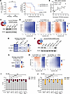 Figure 1
Low B56α expression levels impair the response to venetoclax-azacitidine treatment. (A) mRNA PPP2R5A expression levels in Ven-Aza–treated patients from the OHSU Beat AML cohort grouped into low (n = 8) or high PPP2R5A expressing groups (n = 7). Statistical significance was determined by unpaired t test. On the right, survival curves comparison of low versus high PPP2R5A expressing groups analyzed by the log-rank (Mantel-Cox) test. (B) Correlation scatter plot between PPP2R5A mRNA expression and highest single agent (HSA) synergy scores of 11 AML primary cells. (C) Graphical representation and Western blot analysis of B56α protein expression in HL-60 parental cell line, PPP2R5A CRISPRCas9 WT clone (B56αWT), and sublines with B56α genetic inactivation (B56α–/– g1 and B56α–/– g2). (D) HSA synergy scores of each drug combination in HL-60 B56αWT and B56α–/– g1 and B56α–/– g2 clones. (E) Heatmap representation of HSA synergy score means of Ven-Aza combination in HL-60, OCI-AML3, and MV4-11 B56αWT and B56α–/– g1 and B56α–/– g2 clones (48 hours after treatment). (F) Graphical representation and Western blot analysis of B56α protein expression in HL-60 parental cell line, B56α–/– clones, and B56α–/– g1 after viral transduction with EGFP (B56α–/– g1 + EGFP) or PPP2R5A (B56α–/– g1 + B56α) vectors. (G and H) Heatmap representation of HSA synergy score means of Ven-Aza combination and HSA synergy scores of each drug combination in HL-60 B56α–/– g1 + EGFP and B56α–/– g1 + B56α cells. (I and J) Average percentages ± SEM of viable and annexin V/propidium iodide positive (AnV/PI+) cells and with mitochondrial membrane potential loss in HL-60 B56αWT, B56α–/–, B56α–/– g1 +EGFP, and B56α–/– g1 + B56α cells after 48 and 24 hours upon Ven-Aza treatment (125 nM and 625 nM, respectively). Data were analyzed by 2-way ANOVA and Tukey post hoc test. In vitro results were obtained from 3 biological replicates. *P < 0.05, **P < 0.01, ***P < 0.001, ****P < 0.0001.
To determine if reduced PP2A-B56α expression drives combination therapy resistance, B56α–/–sublines were generated by CRISPR-Cas9 in HL-60, OCI-AML3, and MV4-11 AML cell lines (Figure 1C and Supplemental Figure 2, A and B). Loss of B56α subunit expression did not generally affect baseline cell growth or GI50 values upon single venetoclax and azacitidine treatments (Supplemental Figure 2, C and D). Interestingly, the Ven-Aza combination did not show synergy in B56α–/– cells as compared with B56αWT cells in all 3 lines tested (Figure 1, D and E, and Supplemental Figure 2, E and F). Remarkably, the reconstitution of B56α protein expression in HL-60 B56α–/– g1 clone recovered Ven-Aza treatment sensitivity (Figure 1, F–H, and Supplemental Figure 3, A and B). Indeed, Ven-Aza treatment was significantly less effective in inducing apoptosis in the HL-60 B56α–/– clones (approximately 20% of cell death in B56α–/– clones versus 38.9% in B56αWT cells). This effect was reversed upon B56α reconstitution (Figure 1I). Furthermore, the loss of mitochondrial membrane potential (MMP) after Ven-Aza treatment was also significantly reversed in HL-60 B56α–/– cells (approximately 30% of cells with loss of MMP in B56α–/– versus 51.7% in B56αWT cells) and recovered up to 49.6% of TMRM– cells after B56α reconstitution (Figure 1J). Cell death and MMP were also measured in OCI-AML3 and MV4-11 B56α–/– CRISPR sublines upon Ven-Aza regimen, showing similar results (Supplemental Figure 3, C and D). Importantly, B56α–/– cells did not present an intrinsic resistance to apoptosis (Supplemental Figure 3E).
To further corroborate whether the observed effects were B56α specific and not a result of general tumor suppressive activity of PP2A, we generated HL-60 sublines with genetic inactivation of B55α regulatory subunit, where no differences in Ven-Aza treatment response, apoptosis induction, or loss of MMP were noted between B55αWT and B55α–/– cells (Supplemental Figure 4).
Collectively, these results highlight the relevance of the B56α regulatory subunit in modulating the response to Ven-Aza combination therapy in AML and suggest that a resistance mechanism to this drug combination could be regulated by PP2A-B56α signaling.
Transcriptomic and proteomic analyses indicate that OXPHOS is upregulated in HL-60 B56α–/– cells. To better define the pathways regulated by PP2A-B56α in AML cells, we compared the transcriptomic profile of the HL-60 B56αWT and B56α–/– clones using RNA-seq (Figure 2A and Supplemental Table 2). Gene set enrichment analysis (GSEA) was performed using the hallmark gene set collection (19). HL-60 B56α–/– cells presented “Oxidative_phosphorylation” and “MYC_targets_V1” hallmarks positively regulated, and 13 other gene sets downregulated, such as “Apoptosis” and several immune response-related pathways (P < 0.05) (Figure 2, B and C, and Supplemental Table 3). Notably, transcriptomic AML patient data from the low PPP2R5A expression group (OHSU BeatAML2 database; ref. 18) showed that “Oxidative_phosphorylation” and “MYC_targets_V1” hallmarks were also upregulated, with the “TNFA_signaling_via_NFKB” pathway being among the most downregulated in both analyses (Figure 2, B and C; Supplemental Figure 5, A–C; and Supplemental Tables 4–7). Eighty-four genes from the OXPHOS hallmark gene set (48.6% of genes) were commonly deregulated in HL-60 B56α–/– clones and patients with low PPP2R5A expression (Supplemental Figure 5D and Supplemental Table 8). These data point out similar phenotypes between our HL-60 clones lacking the B56α subunit and patients with AML with low PPP2R5A expression levels. Some of these pathways, such as MYC and NF-κB, have already been described to be regulated by PP2A-B56α (20, 21), validating our experimental approach. To our knowledge, no previous studies have identified a role for B56α in OXPHOS in AML.
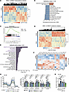 Figure 2
Transcriptomics and proteomics analyses indicate that OXPHOS is upregulated in HL-60 B56α–/– models. (A) Heatmap representation of genes differentially upregulated (red) and downregulated (blue) at transcriptional level in HL-60 B56α–/– clones compared with B56αWT cells (n = ~3 per subline). (B) Hallmark gene-set enrichment analysis of differential gene expression in B56α–/– clones. Positive normalized enrichment score (NES) represents upregulated hallmark gene sets, while negative NES indicates the gene sets downregulated with P < 0.05. (C) Enrichment plot of upregulated OXPHOS hallmark gene set with its NES and false discovery rate (FDR) in B56α–/– clones compared with B56αWT cells. (D) Heatmap representation of proteomics analyses in HL-60 B56α–/– clones compared with B56αWT cells (n = 4 per subline). (E) Hallmark gene set pathways altered in B56α–/– clones compared with B56αWT cells. Hallmarks in purple represent metabolism-related pathways. Statistical significance is expressed as –Log10(P value). (F) Heatmap representation of the differential expression of the mitochondrial proteins from the ETC complex in HL-60 B56αWT and B56α–/– clones (n = 4 per subline). (G) Dynamic representation of oxygen consumption rate (OCR) in HL-60 B56αWT, B56α–/–, and reconstituted B56α clones at basal conditions obtained from 3 biological replicates. (Right) Comparison of the basal respiration, ATP production, maximal respiratory capacity, proton leak, and spare respiratory capacity of the clones expressed as fold change of the normalized HL-60 B56αWT clone values. Data represent mean ± SD and were analyzed by 2-way ANOVA and Tukey post hoc test. (H) Bar graph show average ± SD mitochondrial complex I activity of HL-60 clones measured as the change in absorbance per minute per amount of protein loaded and expressed as the fold change of HL-60 B56αWT clone values. These results were obtained from 3 biological replicates. Statistical significance was determined by 1-way ANOVA and Tukey post hoc test analyses.
To better define the biologically significant pathways regulated by the PP2A-B56α complex in AML, proteomic analyses were also performed in our CRISPR-Cas9 HL-60 B56α sublines (Figure 2D and Supplemental Table 9). Expression of 16% (638 proteins) of the total proteins detected was significantly altered (P < 0.05) in HL-60 B56α–/– sublines compared with HL-60 B56αWT cells (Supplemental Table 9). Pathway analysis of the proteomic data was conducted using Hallmark Gene Sets enrichment analysis of the Metascape database (22). Twenty-seven pathways were significantly deregulated at proteomic level (P < 0.01), including those which PP2A has been shown to regulate, including E2F and MYC targets, apoptosis, p53, and PI3K/AKT/mTOR (17). Interestingly, these results also reveal important differences in cell metabolism, encompassing adipogenesis, glycolysis, fatty acid metabolism, and OXPHOS, which was the most significantly altered pathway in B56α-deficient cells (P < 0.0001) (Figure 2E and Supplemental Table 10). Within OXPHOS pathway, upregulated proteins were mostly mitochondrial proteins related to complex I of the electron-transporting chain (ETC) (NDUF family proteins) (Figure 2F and Supplemental Table 10). Moreover, among the differentially expressed proteins detected in HL-60 B56α–/– cells, 38% were also deregulated transcriptionally, with OXPHOS pathway components significantly upregulated at both the transcriptional and proteomic levels (Supplemental Figure 5, E–G). Measurement of OCR confirmed the activation of the OXPHOS pathway in HL-60 B56α–/– cells when compared with HL-60 B56αWT and B56α-reconstituted cells in terms of basal and maximal mitochondrial respiration rates and spare respiratory capacity (SRC) (Figure 2G). Moreover, HL-60 B56α–/– clones showed higher mitochondrial complex I activity as compared with the B56αWT and B56α-reconstituted clones (Figure 2H). Genetic inactivation of B56α regulatory subunit consistently increased basal and maximal mitochondrial respiration across different B56–/–AML sublines, indicating a conserved enhancement of mitochondrial respiratory capacity (Supplemental Figure 6, A–D). However, the downstream bioenergetic adaptations differed between cell lines: HL-60 and MV4-11 B56α–/– cells enhanced complex I–dependent respiration, whereas OCI-AML3 maintained increased mitochondrial respiration through Complex I–independent electron entry into the ETC, pointing to alternative metabolic pathway engagement (Supplemental Figure 6, A–D). Only MV4-11 translated the increased respiratory activity into higher ATP production and SRC, suggesting improved coupling efficiency. HL-60 instead showed an enhanced SRC (Figure 2G), consistent with expanded respiratory reserve rather than ATP output. No differences were found in the OCR between HL-60 B55α–/– and B55αWT cells (Supplemental Figure 6E).
Collectively, these observations reveal a key role for the PP2A-B56α holoenzyme in regulating OXPHOS in AML.
Ven-Aza–resistant AML models demonstrate upregulated OXPHOS activity. Ven-Aza treatment disrupts OXPHOS in de novo AML blasts (23, 24). However, drug-resistant leukemia cells rewire their metabolism to bypass this inhibition and still rely on mitochondrial respiration for survival, upregulating OXPHOS to acquire resistance to Ven-Aza treatment (7–9, 12, 25). To further explore the mechanisms of treatment resistance, we generated Ven-Aza–resistant AML cell models. HL-60, OCI-AML3, and MV4-11 cell lines were exposed to increasing doses of both drugs until they developed resistance to single and combination therapy (HL-60-R, OCI-AML3-R, and MV4-11-R) (Figure 3, A and B, and Supplemental Figure 7, A and B). Resistant cell lines showed no induction of apoptosis or loss of MMP after treatment with over 8 times higher doses than the parental cell lines (Supplemental Figure 7, C and D). In a s.c. mouse model, HL-60-R cells were completely resistant to Ven-Aza therapy as compared with the parental cells (P < 0.0001), reproducing in vitro results (Figure 3C). Proteomic analyses of the HL-60-R compared with HL-60 parental cells revealed altered metabolic processes in HL-60-R cells, with OXPHOS being the most deregulated pathway, followed by fatty acid metabolism and adipogenesis (P < 0.0001) (Figure 3D, Supplemental Figure 8, and Supplemental Tables 11–13). Seahorse assays confirmed the significantly augmented OXPHOS activity in Ven-Aza–resistant cells. Although the resistant metabolic phenotype across AML cell lines was not identical, all resistant models exhibited increased maximal respiratory capacity and complex I activity (Figure 3, E–G), indicating that enhanced mitochondrial respiratory potential is a common adaptation to Ven-Aza resistance in AML. Importantly, it strengthens our observations on the connection between OXPHOS upregulation and Ven-Aza resistance in patients with AML with low PPP2R5A expression and in cell lines harboring loss of B56α.
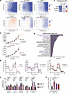 Figure 3
AML Venetoclax-azacitidine resistant models upregulate OXPHOS. (A and B) Two-dimensional contour plots represent highest single agent (HSA) synergy scores of each drug combination (y axis: venetoclax; x axis: azacitidine) and heatmap representation of HSA synergy score means of Ven-Aza combination in HL-60, OCI-AML3, and MV4-11 parental and Ven-Aza–resistant cell lines (48-hour treatment). (C) Time-dependent tumor growth upon vehicle or Ven-Aza treatment of injected NSG mice with HL-60 and HL-60-R cell lines. Doses: 100 and 3 mg/kg of venetoclax and azacitidine, respectively (n = ~15 mice per group). Statistical significance was determined by 2-way ANOVA and Tukey post hoc test. Data represent mean ± SEM. (D) Hallmark gene set pathways altered in HL-60 Ven-Aza resistant cell line compared with HL-60 parental. Hallmarks in purple represent metabolism-related pathways. Statistical significance is expressed as –Log10(P value). (E) Dynamic representation of oxygen consumption rate (OCR) in HL-60, OCI-AML3, and MV4-11 parental and Ven-Aza–resistant cell lines under basal conditions. (F) Bar graphs show comparison of the basal respiration, ATP production, maximal respiratory capacity, proton leak, and spare respiratory capacity of parental and Ven-Aza–resistant cell lines expressed as fold change of the normalized parental cell values for each cell line, respectively. Data were analyzed by 2-way ANOVA and Tukey post hoc test. (G) Bar graph represents mitochondrial complex I activity in parental and Ven-Aza–resistant cell lines expressed as the fold change of their respective parental cell line. Statistical significance was determined by 1-way ANOVA and Tukey post hoc test analyses. In vitro results were obtained from at least 3 biological replicates and represent mean ± SD.
PP2A-B56α heterocomplex stabilization decreases OXPHOS in Ven-Aza AML-resistant models. Given the role of B56α in OXPHOS regulation and the importance of mitochondrial metabolism in driving AML treatment resistance, we tested whether RPT04402, a potentially novel PMG that specifically stabilizes active PP2A-B56α complexes (26), was able to reverse the OXPHOS-related Ven-Aza–resistant phenotype. RPT04402 treatment caused a broad-spectrum effect on mitochondrial respiration in our 3 AML parental cell lines, as evidenced by dose-dependent decreases in the levels of basal and maximal respiration and ATP production (Figure 4A and Supplemental Figure 9, A and B). These effects were significantly attenuated in B56α–/– clones, especially the maximal respiration levels, highlighting the specificity of the compound to PP2A-B56α complexes and defining a key role for the PP2A-B56α heterotrimer in regulating mitochondrial maximal respiration (Figure 4B and Supplemental Figure 9, C and D). To further corroborate the specificity of RPT04402, we ectopically expressed a V5-tagged Aα protein in HL-60 cells (Supplemental Figure 10A). Then, Aα-V5 interactions with the catalytic subunit and the 5 members of the B56 regulatory family (α, β, γ, δ, and ε) were analyzed using a Proximity Ligation Assay (PLA). PMG treatment resulted in a significant increase in only PP2A-B56α complexes, enhancing B56α heterotrimerization over the rest of the B56 subunits and confirming the high specificity of RPT04402 to this heterotrimer (Figure 4, C and D, and Supplemental Figure 10, B and C). Western blots of all these subunits were performed to verify that the increase in Aα-V5/B56α binding was not a result of expression variations upon drug treatment (Supplemental Figure 10, D and E). Additionally, RPT04402 treatment of the OCI-AML3 and MV4-11 B56αWT sublines drove a broad-spectrum decrease in OCR, effects that were generally reversed when B56α was knocked down (Supplemental Figure 11).
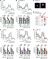 Figure 4
PP2A-B56α heterocomplex stabilization decreases OXPHOS in venetoclax-azacitidine AML-resistant models. (A, B, E–G)Seahorse extracellular flux analysis shows dynamic representation of oxygen consumption rate (OCR) in HL-60 (A HL-60 B56α–/– g1 (B), HL-60-R (E), OCI-AML3-R (F), MV4-11-R cell lines (G) under basal conditions and treated with increasing concentrations of RPT04402 (12-hour treatment). Bar graphs show that RPT04402 treatment decreases basal respiration, ATP production, and maximal respiratory capacity of parental and Ven-Aza–resistant AML cell lines in a dose-dependent manner, expressed as a fold change of the control. (C) Representative images of the proximity ligation analysis (PLA) targeting the interaction between Aα-V5 tagged and B56α subunit (Texas Red) in the absence or presence of RPT04402 (20 μM). Experiment performed on HL-60 cells transfected with Aα-V5. Nuclei stained with DAPI. All images 40x magnification. Scale bar: 12.5 μm. (D) Relative quantification of PLA fluorescence signal of either B56α, B56β, B56γ, B56δ, B56ε, or Cα/β measured by calculating the corrected total cell fluorescence (CTCF) of multiple images (7–10 images) per biological replicate (n = 3). Data were analyzed by 2- (A, B, and E–G) or 1-way (D) ANOVA and Tukey post hoc test. All experiments were performed in 3 independent biological replicates. Data represent mean ± SD.
B56α subunit expression was verified at mRNA and protein levels in our resistant models before testing the effects of RPT04402 treatment (Supplemental Figure 12, A–C). The Ven-Aza–resistant phenotype did not affect RPT04402 sensitivity in these cells (Supplemental Figure 12D). Importantly, OCR was significantly reduced in resistant models upon RPT04402 treatment, similarly to the parental baseline levels (Figure 4, E–G). Collectively, these results demonstrate that the stabilization of PP2A-B56α complex specifically decreases OXPHOS-dependent energy production in both Ven-Aza–sensitive and –resistant AML models.
Combined treatment with RPT04402, venetoclax, and azacitidine disrupts OXPHOS and overcomes Ven-Aza resistance in AML. Previous results from our group demonstrated that PP2A activators enhance responses to venetoclax-based combinations (16). Therefore, we evaluated the efficacy of the triple combination Ven-Aza–RPT04402 in our AML models. RPT04402 synergistically enhanced the antileukemic effect of the standard-of-care Ven-Aza combination in both parental and treatment-resistant AML cells (Figure 5A and Supplemental Figure 13A). Indeed, only the triple combination therapy markedly reduced basal and maximal respiration and ATP production, disrupting mitochondrial respiration in Ven-Aza–resistant models (Figure 5, B–D). In the parental cell lines (Ven-Aza–sensitive cells), both Ven-Aza and triple treatments decreased OXPHOS (Supplemental Figure 13B). Since these viability assays were performed using an MTS activity assay, which measures changes in metabolic activity, these cell viability changes were also confirmed using an independent DNA-based viability assay (Supplemental Figure 13, C and D). The efficacy of Ven-Aza–RPT04402 therapy relied specifically on the B56α PP2A regulatory subunit, since synergy score values were not significantly increased by the triple therapy in HL-60 B56α–/– clones as observed in the HL-60 B56αWT and in HL-60 B55α CRISPR-Cas9 sublines (Supplemental Figure 14).
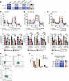 Figure 5
RPT04402 combined with venetoclax and azacitidine disrupts OXPHOS and overcomes venetoclax-azacitidine resistance in AML. (A) Heatmap representation of HSA synergy score means for Ven-Aza treatment and the triple therapy (Ven-Aza–RPT04402) in parental and Ven-Aza–resistant HL60, OCI-AML3, and MV4-11 cell lines (48-hour treatment) (synergistic when HSA > 10). (B–D) Dynamic representation of oxygen consumption rate (OCR) in HL-60-R (B), OCI-AML3-R (C), and MV4-11-R cell lines (D) at baseline (control) and after 12 hours of RPT04402, Ven-Aza, and Ven-Aza–RPT04402 (Triple combo) treatment. Bar graph represents basal respiration, ATP production, and maximal respiratory capacity levels expressed as fold change of control. RPT04402 concentrations for Triple combo treatment in HL-60-R, OCI-AML3-R, and MV4-11-R cell lines were 30, 15, and 10 μM, respectively. Asterisks on the top of each bar indicate the statistical significance of each condition compared with the control. Results were obtained from 3 biological replicates. Statistical significance was determined by 2-way ANOVA and Tukey post hoc test. Data represent mean ± SD. (E and F) Flow cytometry characterization of AML-1, AML-2, and AML-3-R primary samples of CD34/CD38 markers and quantification of the percentage of cells expressing CD45RA, CD123, and IL1RAP markers. (G) Heatmap representation of HSA synergy score means of Ven-Aza and Ven-Aza–RPT04402 treatment in 3 AML primary samples (60-hour treatment) obtained from ~ 2 biological replicates. *P < 0.05, **P < 0.01, ***P < 0.001, ****P < 0.0001.
Although RPT04402 alone did not significantly induce apoptosis or mitochondrial depolarization at tested doses, its addition to the Ven-Aza therapy significantly enhanced both apoptotic cell death and mitochondrial membrane depolarization in parental and resistant AML cells (Supplemental Figure 15). This observation demonstrates that the observed metabolic alterations are accompanied by functional activation of mitochondrial apoptosis, thereby reinforcing the therapeutic potential of the triple combination and contributing to overcome resistance to the Ven-Aza regimen.
After checking that RPT04402 alone or combined with venetoclax and azacitidine did not present a significant reduction in the number or type of normal human CD34+ hematopoietic stem/progenitor colonies (Supplemental Figure 16, A–C), we next tested the triple combination in 3 AML patient–derived primary cells. Samples AML-1 and AML-2 were obtained at diagnosis, while sample AML-3-relapsed AML-3-R was obtained at relapse after 6 cycles of Ven-Aza treatment (Supplemental Figure 16D and Supplemental Table 1). Interestingly, the AML-3-R sample was enriched in CD34+CD38–CD45RA+ cells (Figure 5, E and F, and Supplemental Figure 16E), an immunophenotype related to leukemia stem cells (LSCs) (8, 27, 28). In both the newly diagnosed and relapsed samples (Ven-Aza–sensitive or –resistant) carrying either ELN-favorable or adverse-risk mutations (Supplemental Table 1), the addition of RPT04402 doubled the synergy score of Ven-Aza therapy (Figure 5G and Supplemental Figure 16F).
Collectively, the stabilization of the PP2A-B56α complex by RPT04402 combined with Ven-Aza treatment counteracts dysregulated OXPHOS signaling, one of the key mechanisms driving treatment resistance, and improves the efficacy of Ven-Aza treatment in de novo and relapsed AML samples.
RPT04402 improves the response to Ven-Aza in treatment-resistant in vivo models. To evaluate the triple treatment efficacy in vivo, we took advantage of zebrafish xenograft models, as they allow noninvasive imaging of tumor growth, high experimental throughput, and short-term results; they also conserve similar hematopoietic regulation to humans (29–31). HL-60-R and OCI-AML3-R cells were xenotransplanted into zebrafish larvae orthotopically, and RPT04402, Ven-Aza, and the triple combination were added to water for 72 hours (Figure 6A). Consistent with our results described above, Ven-Aza treatment was not effective in reducing HL-60-R or OCI-AML3-R cell infiltration (P = 0.06 and P = 0.35, respectively) (Figure 6, B–E). Remarkably, RPT04402 single-agent treatment and RPT04402 in combination with Ven-Aza significantly reduced cell infiltration in both Ven-Aza–resistant cells (RPT04402: 36.6% and 41.6%; triple combo: 66.8% and 53.2%; in HL-60-R and OCI-AML3-R, respectively) and Ven-Aza–sensitive cells (RPT04402: 27.3% and 44%; triple combo: 57.8% and 75.7%; in HL-60 and OCI-AML3, respectively) (Figure 6, B–E). S.c. mouse xenograft experiments in Ven-Aza–resistant cell line models (Figure 6F) corroborated the in vivo activity of RPT04402 (23% and 15% of tumor growth inhibition [TGI] in HL-60-R and OCI-AML3-R, respectively). Combination treatment of RPT04402 with Ven-Aza improved the efficacy of the therapy in these fast-growing Ven-Aza–resistant tumors (29% TGI in HL-60-R and 27.3% in OCI-AML3-R) without any sign of toxicity (Figure 6, G and H).
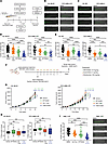 Figure 6
RPT04402 improves the response to venetoclax-azacitidine therapy in resistant models in vivo. (A) Schematic representation of zebrafish xenotransplantation and treatment workflow. Drug doses: 500 nM Ven, 1 μM Aza, and/or 20 μM RPT04402. (B and C) Representative images of HL-60-R and OCI-AML3-R (B) as well as HL-60 and OCI-AML3 cell lines (C) in xenograft larvae tail at 72 hours postinjection (hpi). AML cells were tracked as green-emitting fluorescent cells. (D and E) Cell invasion capacity of parental and Ven-Aza resistant cell lines were measured as the number of cells present in the caudal hematopoietic tissue after 70 hours of treatment. Each point represents one zebrafish larvae. Statistical significance was determined by 1-way ANOVA and Tukey post hoc test in HL-60 and HL-60-R cells, and by Kruskal-Wallis and Dunn post hoc test analyses for OCI-AML3 and OCI-AML3-R cell lines. (F) Schematic representation of in vivo subcutaneous efficacy study (detailed explanation in methods section). (G) Time-dependent tumor growth upon treatment of injected NSG mice with HL-60-R and OCI-AML3-R cell lines (n = ~15 mice per group). TGI, tumor growth inhibition. Statistical significance was determined by 2-way ANOVA and Tukey post hoc test. Data represent mean ± SEM. (H) Percentages of body weight change of NSG mice injected with HL-60-R and OCI-AML3-R cell lines after 15 and 22 days of treatment, respectively. Statistical significance was determined by 1-way ANOVA and Tukey post hoc test. Treatment doses: 10, 100, and 3 mg/kg for RPT04402, venetoclax, and azacitidine, respectively. (I) Cell invasion capacity of AML-3-R cells (72-hour treatment). Statistical significance was determined by Kruskal-Wallis and Dunn post hoc test analyses. On the right, representative images of AML-3-R cells of zebrafish PDX tail at 72 hours post-injection. Scale bar: 0.25 mm.
Finally, we generated a zebrafish patient-derived xenograft (PDX) using the AML-3-R sample at relapse after Ven-Aza treatment, following the same workflow as previously described (Figure 6A). RPT04402 treatment alone effectively reduced cell infiltration (30.8% of cell infiltration inhibition) and synergized with Ven-Aza therapy (62.9% of cell infiltration inhibition) in this AML relapsed model in vivo (Figure 6I).
Altogether, RPT04402 shows in vivo activity in both zebrafish and mice, and Ven-Aza–RPT04402 combination is effective on distinct Ven-Aza–resistant AML models, setting the basis for a promising therapeutic approach to overcome treatment resistance in AML.


