
Research ArticleCell biologyInflammation
Open Access |  10.1172/jci.insight.199507
Versican regulating viscoelasticity drives pleural fibrosis via mechanotransductive signaling
Zi-Heng Jia,1 Xin-Liang He,1,2 Xiao-Lin Cui,3 Qian Li,3 Pei-Pei Cheng,3 Li-Qin Zhao,1 Shu-Yi Ye,1 Shi-He Hu,3 Chen-Yue Lian,1 He-De Zhang,3 Li-Mei Liang,1,2 Lin-Jie Song,1,2 Fan Yu,1,2 Liang Xiong,1,2 Fei Xiang,1,2 Xiaorong Wang,1,2 Meng Wang,2 Xiyong Dai,4 Hong Ye,2,3 and Wan-Li Ma1,2
1Department of Respiratory and Critical Care Medicine, Union Hospital, Tongji Medical College, Huazhong University of Science and Technology, Wuhan, China.
2Key Laboratory of Respiratory Diseases of National Health Commission of China, Wuhan, China.
3Department of Pathophysiology, School of Basic Medicine, Tongji Medical College, Huazhong University of Science and Technology, Wuhan, China.
4Department of Thoracic Surgery, Wuhan Pulmonary Hospital, Wuhan, China.
Address correspondence to: Wan-Li Ma, Department of Respiratory and Critical Care Medicine, Union Hospital, Tongji Medical College, Huazhong University of Science and Technology, Wuhan 430022, China, Email: whmawl@hust.edu.cn. Or to: Hong Ye, Department of Pathophysiology, School of Basic Medicine, Tongji Medical College, Huazhong University of Science and Technology, Wuhan 430030, China. Email: yehmwl@hust.edu.cn. Or to: Xiyong Dai, Department of Thoracic Surgery, Wuhan Pulmonary Hospital, 430030 Wuhan, China. Email: daixiyong71@126.com.
Authorship note: ZHJ and XLH are co-first authors and contributed equally to this work.
Find articles by Jia, Z. in: PubMed | Google Scholar
1Department of Respiratory and Critical Care Medicine, Union Hospital, Tongji Medical College, Huazhong University of Science and Technology, Wuhan, China.
2Key Laboratory of Respiratory Diseases of National Health Commission of China, Wuhan, China.
3Department of Pathophysiology, School of Basic Medicine, Tongji Medical College, Huazhong University of Science and Technology, Wuhan, China.
4Department of Thoracic Surgery, Wuhan Pulmonary Hospital, Wuhan, China.
Address correspondence to: Wan-Li Ma, Department of Respiratory and Critical Care Medicine, Union Hospital, Tongji Medical College, Huazhong University of Science and Technology, Wuhan 430022, China, Email: whmawl@hust.edu.cn. Or to: Hong Ye, Department of Pathophysiology, School of Basic Medicine, Tongji Medical College, Huazhong University of Science and Technology, Wuhan 430030, China. Email: yehmwl@hust.edu.cn. Or to: Xiyong Dai, Department of Thoracic Surgery, Wuhan Pulmonary Hospital, 430030 Wuhan, China. Email: daixiyong71@126.com.
Authorship note: ZHJ and XLH are co-first authors and contributed equally to this work.
Find articles by He, X. in: PubMed | Google Scholar
1Department of Respiratory and Critical Care Medicine, Union Hospital, Tongji Medical College, Huazhong University of Science and Technology, Wuhan, China.
2Key Laboratory of Respiratory Diseases of National Health Commission of China, Wuhan, China.
3Department of Pathophysiology, School of Basic Medicine, Tongji Medical College, Huazhong University of Science and Technology, Wuhan, China.
4Department of Thoracic Surgery, Wuhan Pulmonary Hospital, Wuhan, China.
Address correspondence to: Wan-Li Ma, Department of Respiratory and Critical Care Medicine, Union Hospital, Tongji Medical College, Huazhong University of Science and Technology, Wuhan 430022, China, Email: whmawl@hust.edu.cn. Or to: Hong Ye, Department of Pathophysiology, School of Basic Medicine, Tongji Medical College, Huazhong University of Science and Technology, Wuhan 430030, China. Email: yehmwl@hust.edu.cn. Or to: Xiyong Dai, Department of Thoracic Surgery, Wuhan Pulmonary Hospital, 430030 Wuhan, China. Email: daixiyong71@126.com.
Authorship note: ZHJ and XLH are co-first authors and contributed equally to this work.
Find articles by Cui, X. in: PubMed | Google Scholar
1Department of Respiratory and Critical Care Medicine, Union Hospital, Tongji Medical College, Huazhong University of Science and Technology, Wuhan, China.
2Key Laboratory of Respiratory Diseases of National Health Commission of China, Wuhan, China.
3Department of Pathophysiology, School of Basic Medicine, Tongji Medical College, Huazhong University of Science and Technology, Wuhan, China.
4Department of Thoracic Surgery, Wuhan Pulmonary Hospital, Wuhan, China.
Address correspondence to: Wan-Li Ma, Department of Respiratory and Critical Care Medicine, Union Hospital, Tongji Medical College, Huazhong University of Science and Technology, Wuhan 430022, China, Email: whmawl@hust.edu.cn. Or to: Hong Ye, Department of Pathophysiology, School of Basic Medicine, Tongji Medical College, Huazhong University of Science and Technology, Wuhan 430030, China. Email: yehmwl@hust.edu.cn. Or to: Xiyong Dai, Department of Thoracic Surgery, Wuhan Pulmonary Hospital, 430030 Wuhan, China. Email: daixiyong71@126.com.
Authorship note: ZHJ and XLH are co-first authors and contributed equally to this work.
Find articles by Li, Q. in: PubMed | Google Scholar
1Department of Respiratory and Critical Care Medicine, Union Hospital, Tongji Medical College, Huazhong University of Science and Technology, Wuhan, China.
2Key Laboratory of Respiratory Diseases of National Health Commission of China, Wuhan, China.
3Department of Pathophysiology, School of Basic Medicine, Tongji Medical College, Huazhong University of Science and Technology, Wuhan, China.
4Department of Thoracic Surgery, Wuhan Pulmonary Hospital, Wuhan, China.
Address correspondence to: Wan-Li Ma, Department of Respiratory and Critical Care Medicine, Union Hospital, Tongji Medical College, Huazhong University of Science and Technology, Wuhan 430022, China, Email: whmawl@hust.edu.cn. Or to: Hong Ye, Department of Pathophysiology, School of Basic Medicine, Tongji Medical College, Huazhong University of Science and Technology, Wuhan 430030, China. Email: yehmwl@hust.edu.cn. Or to: Xiyong Dai, Department of Thoracic Surgery, Wuhan Pulmonary Hospital, 430030 Wuhan, China. Email: daixiyong71@126.com.
Authorship note: ZHJ and XLH are co-first authors and contributed equally to this work.
Find articles by Cheng, P. in: PubMed | Google Scholar
1Department of Respiratory and Critical Care Medicine, Union Hospital, Tongji Medical College, Huazhong University of Science and Technology, Wuhan, China.
2Key Laboratory of Respiratory Diseases of National Health Commission of China, Wuhan, China.
3Department of Pathophysiology, School of Basic Medicine, Tongji Medical College, Huazhong University of Science and Technology, Wuhan, China.
4Department of Thoracic Surgery, Wuhan Pulmonary Hospital, Wuhan, China.
Address correspondence to: Wan-Li Ma, Department of Respiratory and Critical Care Medicine, Union Hospital, Tongji Medical College, Huazhong University of Science and Technology, Wuhan 430022, China, Email: whmawl@hust.edu.cn. Or to: Hong Ye, Department of Pathophysiology, School of Basic Medicine, Tongji Medical College, Huazhong University of Science and Technology, Wuhan 430030, China. Email: yehmwl@hust.edu.cn. Or to: Xiyong Dai, Department of Thoracic Surgery, Wuhan Pulmonary Hospital, 430030 Wuhan, China. Email: daixiyong71@126.com.
Authorship note: ZHJ and XLH are co-first authors and contributed equally to this work.
Find articles by Zhao, L. in: PubMed | Google Scholar
1Department of Respiratory and Critical Care Medicine, Union Hospital, Tongji Medical College, Huazhong University of Science and Technology, Wuhan, China.
2Key Laboratory of Respiratory Diseases of National Health Commission of China, Wuhan, China.
3Department of Pathophysiology, School of Basic Medicine, Tongji Medical College, Huazhong University of Science and Technology, Wuhan, China.
4Department of Thoracic Surgery, Wuhan Pulmonary Hospital, Wuhan, China.
Address correspondence to: Wan-Li Ma, Department of Respiratory and Critical Care Medicine, Union Hospital, Tongji Medical College, Huazhong University of Science and Technology, Wuhan 430022, China, Email: whmawl@hust.edu.cn. Or to: Hong Ye, Department of Pathophysiology, School of Basic Medicine, Tongji Medical College, Huazhong University of Science and Technology, Wuhan 430030, China. Email: yehmwl@hust.edu.cn. Or to: Xiyong Dai, Department of Thoracic Surgery, Wuhan Pulmonary Hospital, 430030 Wuhan, China. Email: daixiyong71@126.com.
Authorship note: ZHJ and XLH are co-first authors and contributed equally to this work.
Find articles by Ye, S. in: PubMed | Google Scholar
1Department of Respiratory and Critical Care Medicine, Union Hospital, Tongji Medical College, Huazhong University of Science and Technology, Wuhan, China.
2Key Laboratory of Respiratory Diseases of National Health Commission of China, Wuhan, China.
3Department of Pathophysiology, School of Basic Medicine, Tongji Medical College, Huazhong University of Science and Technology, Wuhan, China.
4Department of Thoracic Surgery, Wuhan Pulmonary Hospital, Wuhan, China.
Address correspondence to: Wan-Li Ma, Department of Respiratory and Critical Care Medicine, Union Hospital, Tongji Medical College, Huazhong University of Science and Technology, Wuhan 430022, China, Email: whmawl@hust.edu.cn. Or to: Hong Ye, Department of Pathophysiology, School of Basic Medicine, Tongji Medical College, Huazhong University of Science and Technology, Wuhan 430030, China. Email: yehmwl@hust.edu.cn. Or to: Xiyong Dai, Department of Thoracic Surgery, Wuhan Pulmonary Hospital, 430030 Wuhan, China. Email: daixiyong71@126.com.
Authorship note: ZHJ and XLH are co-first authors and contributed equally to this work.
Find articles by Hu, S. in: PubMed | Google Scholar
1Department of Respiratory and Critical Care Medicine, Union Hospital, Tongji Medical College, Huazhong University of Science and Technology, Wuhan, China.
2Key Laboratory of Respiratory Diseases of National Health Commission of China, Wuhan, China.
3Department of Pathophysiology, School of Basic Medicine, Tongji Medical College, Huazhong University of Science and Technology, Wuhan, China.
4Department of Thoracic Surgery, Wuhan Pulmonary Hospital, Wuhan, China.
Address correspondence to: Wan-Li Ma, Department of Respiratory and Critical Care Medicine, Union Hospital, Tongji Medical College, Huazhong University of Science and Technology, Wuhan 430022, China, Email: whmawl@hust.edu.cn. Or to: Hong Ye, Department of Pathophysiology, School of Basic Medicine, Tongji Medical College, Huazhong University of Science and Technology, Wuhan 430030, China. Email: yehmwl@hust.edu.cn. Or to: Xiyong Dai, Department of Thoracic Surgery, Wuhan Pulmonary Hospital, 430030 Wuhan, China. Email: daixiyong71@126.com.
Authorship note: ZHJ and XLH are co-first authors and contributed equally to this work.
Find articles by Lian, C. in: PubMed | Google Scholar
1Department of Respiratory and Critical Care Medicine, Union Hospital, Tongji Medical College, Huazhong University of Science and Technology, Wuhan, China.
2Key Laboratory of Respiratory Diseases of National Health Commission of China, Wuhan, China.
3Department of Pathophysiology, School of Basic Medicine, Tongji Medical College, Huazhong University of Science and Technology, Wuhan, China.
4Department of Thoracic Surgery, Wuhan Pulmonary Hospital, Wuhan, China.
Address correspondence to: Wan-Li Ma, Department of Respiratory and Critical Care Medicine, Union Hospital, Tongji Medical College, Huazhong University of Science and Technology, Wuhan 430022, China, Email: whmawl@hust.edu.cn. Or to: Hong Ye, Department of Pathophysiology, School of Basic Medicine, Tongji Medical College, Huazhong University of Science and Technology, Wuhan 430030, China. Email: yehmwl@hust.edu.cn. Or to: Xiyong Dai, Department of Thoracic Surgery, Wuhan Pulmonary Hospital, 430030 Wuhan, China. Email: daixiyong71@126.com.
Authorship note: ZHJ and XLH are co-first authors and contributed equally to this work.
Find articles by Zhang, H. in: PubMed | Google Scholar
1Department of Respiratory and Critical Care Medicine, Union Hospital, Tongji Medical College, Huazhong University of Science and Technology, Wuhan, China.
2Key Laboratory of Respiratory Diseases of National Health Commission of China, Wuhan, China.
3Department of Pathophysiology, School of Basic Medicine, Tongji Medical College, Huazhong University of Science and Technology, Wuhan, China.
4Department of Thoracic Surgery, Wuhan Pulmonary Hospital, Wuhan, China.
Address correspondence to: Wan-Li Ma, Department of Respiratory and Critical Care Medicine, Union Hospital, Tongji Medical College, Huazhong University of Science and Technology, Wuhan 430022, China, Email: whmawl@hust.edu.cn. Or to: Hong Ye, Department of Pathophysiology, School of Basic Medicine, Tongji Medical College, Huazhong University of Science and Technology, Wuhan 430030, China. Email: yehmwl@hust.edu.cn. Or to: Xiyong Dai, Department of Thoracic Surgery, Wuhan Pulmonary Hospital, 430030 Wuhan, China. Email: daixiyong71@126.com.
Authorship note: ZHJ and XLH are co-first authors and contributed equally to this work.
Find articles by
Liang, L.
in:
PubMed
|
Google Scholar
|

1Department of Respiratory and Critical Care Medicine, Union Hospital, Tongji Medical College, Huazhong University of Science and Technology, Wuhan, China.
2Key Laboratory of Respiratory Diseases of National Health Commission of China, Wuhan, China.
3Department of Pathophysiology, School of Basic Medicine, Tongji Medical College, Huazhong University of Science and Technology, Wuhan, China.
4Department of Thoracic Surgery, Wuhan Pulmonary Hospital, Wuhan, China.
Address correspondence to: Wan-Li Ma, Department of Respiratory and Critical Care Medicine, Union Hospital, Tongji Medical College, Huazhong University of Science and Technology, Wuhan 430022, China, Email: whmawl@hust.edu.cn. Or to: Hong Ye, Department of Pathophysiology, School of Basic Medicine, Tongji Medical College, Huazhong University of Science and Technology, Wuhan 430030, China. Email: yehmwl@hust.edu.cn. Or to: Xiyong Dai, Department of Thoracic Surgery, Wuhan Pulmonary Hospital, 430030 Wuhan, China. Email: daixiyong71@126.com.
Authorship note: ZHJ and XLH are co-first authors and contributed equally to this work.
Find articles by Song, L. in: PubMed | Google Scholar
1Department of Respiratory and Critical Care Medicine, Union Hospital, Tongji Medical College, Huazhong University of Science and Technology, Wuhan, China.
2Key Laboratory of Respiratory Diseases of National Health Commission of China, Wuhan, China.
3Department of Pathophysiology, School of Basic Medicine, Tongji Medical College, Huazhong University of Science and Technology, Wuhan, China.
4Department of Thoracic Surgery, Wuhan Pulmonary Hospital, Wuhan, China.
Address correspondence to: Wan-Li Ma, Department of Respiratory and Critical Care Medicine, Union Hospital, Tongji Medical College, Huazhong University of Science and Technology, Wuhan 430022, China, Email: whmawl@hust.edu.cn. Or to: Hong Ye, Department of Pathophysiology, School of Basic Medicine, Tongji Medical College, Huazhong University of Science and Technology, Wuhan 430030, China. Email: yehmwl@hust.edu.cn. Or to: Xiyong Dai, Department of Thoracic Surgery, Wuhan Pulmonary Hospital, 430030 Wuhan, China. Email: daixiyong71@126.com.
Authorship note: ZHJ and XLH are co-first authors and contributed equally to this work.
Find articles by
Yu, F.
in:
PubMed
|
Google Scholar
|
1Department of Respiratory and Critical Care Medicine, Union Hospital, Tongji Medical College, Huazhong University of Science and Technology, Wuhan, China.
2Key Laboratory of Respiratory Diseases of National Health Commission of China, Wuhan, China.
3Department of Pathophysiology, School of Basic Medicine, Tongji Medical College, Huazhong University of Science and Technology, Wuhan, China.
4Department of Thoracic Surgery, Wuhan Pulmonary Hospital, Wuhan, China.
Address correspondence to: Wan-Li Ma, Department of Respiratory and Critical Care Medicine, Union Hospital, Tongji Medical College, Huazhong University of Science and Technology, Wuhan 430022, China, Email: whmawl@hust.edu.cn. Or to: Hong Ye, Department of Pathophysiology, School of Basic Medicine, Tongji Medical College, Huazhong University of Science and Technology, Wuhan 430030, China. Email: yehmwl@hust.edu.cn. Or to: Xiyong Dai, Department of Thoracic Surgery, Wuhan Pulmonary Hospital, 430030 Wuhan, China. Email: daixiyong71@126.com.
Authorship note: ZHJ and XLH are co-first authors and contributed equally to this work.
Find articles by Xiong, L. in: PubMed | Google Scholar
1Department of Respiratory and Critical Care Medicine, Union Hospital, Tongji Medical College, Huazhong University of Science and Technology, Wuhan, China.
2Key Laboratory of Respiratory Diseases of National Health Commission of China, Wuhan, China.
3Department of Pathophysiology, School of Basic Medicine, Tongji Medical College, Huazhong University of Science and Technology, Wuhan, China.
4Department of Thoracic Surgery, Wuhan Pulmonary Hospital, Wuhan, China.
Address correspondence to: Wan-Li Ma, Department of Respiratory and Critical Care Medicine, Union Hospital, Tongji Medical College, Huazhong University of Science and Technology, Wuhan 430022, China, Email: whmawl@hust.edu.cn. Or to: Hong Ye, Department of Pathophysiology, School of Basic Medicine, Tongji Medical College, Huazhong University of Science and Technology, Wuhan 430030, China. Email: yehmwl@hust.edu.cn. Or to: Xiyong Dai, Department of Thoracic Surgery, Wuhan Pulmonary Hospital, 430030 Wuhan, China. Email: daixiyong71@126.com.
Authorship note: ZHJ and XLH are co-first authors and contributed equally to this work.
Find articles by Xiang, F. in: PubMed | Google Scholar
1Department of Respiratory and Critical Care Medicine, Union Hospital, Tongji Medical College, Huazhong University of Science and Technology, Wuhan, China.
2Key Laboratory of Respiratory Diseases of National Health Commission of China, Wuhan, China.
3Department of Pathophysiology, School of Basic Medicine, Tongji Medical College, Huazhong University of Science and Technology, Wuhan, China.
4Department of Thoracic Surgery, Wuhan Pulmonary Hospital, Wuhan, China.
Address correspondence to: Wan-Li Ma, Department of Respiratory and Critical Care Medicine, Union Hospital, Tongji Medical College, Huazhong University of Science and Technology, Wuhan 430022, China, Email: whmawl@hust.edu.cn. Or to: Hong Ye, Department of Pathophysiology, School of Basic Medicine, Tongji Medical College, Huazhong University of Science and Technology, Wuhan 430030, China. Email: yehmwl@hust.edu.cn. Or to: Xiyong Dai, Department of Thoracic Surgery, Wuhan Pulmonary Hospital, 430030 Wuhan, China. Email: daixiyong71@126.com.
Authorship note: ZHJ and XLH are co-first authors and contributed equally to this work.
Find articles by Wang, X. in: PubMed | Google Scholar
1Department of Respiratory and Critical Care Medicine, Union Hospital, Tongji Medical College, Huazhong University of Science and Technology, Wuhan, China.
2Key Laboratory of Respiratory Diseases of National Health Commission of China, Wuhan, China.
3Department of Pathophysiology, School of Basic Medicine, Tongji Medical College, Huazhong University of Science and Technology, Wuhan, China.
4Department of Thoracic Surgery, Wuhan Pulmonary Hospital, Wuhan, China.
Address correspondence to: Wan-Li Ma, Department of Respiratory and Critical Care Medicine, Union Hospital, Tongji Medical College, Huazhong University of Science and Technology, Wuhan 430022, China, Email: whmawl@hust.edu.cn. Or to: Hong Ye, Department of Pathophysiology, School of Basic Medicine, Tongji Medical College, Huazhong University of Science and Technology, Wuhan 430030, China. Email: yehmwl@hust.edu.cn. Or to: Xiyong Dai, Department of Thoracic Surgery, Wuhan Pulmonary Hospital, 430030 Wuhan, China. Email: daixiyong71@126.com.
Authorship note: ZHJ and XLH are co-first authors and contributed equally to this work.
Find articles by Wang, M. in: PubMed | Google Scholar
1Department of Respiratory and Critical Care Medicine, Union Hospital, Tongji Medical College, Huazhong University of Science and Technology, Wuhan, China.
2Key Laboratory of Respiratory Diseases of National Health Commission of China, Wuhan, China.
3Department of Pathophysiology, School of Basic Medicine, Tongji Medical College, Huazhong University of Science and Technology, Wuhan, China.
4Department of Thoracic Surgery, Wuhan Pulmonary Hospital, Wuhan, China.
Address correspondence to: Wan-Li Ma, Department of Respiratory and Critical Care Medicine, Union Hospital, Tongji Medical College, Huazhong University of Science and Technology, Wuhan 430022, China, Email: whmawl@hust.edu.cn. Or to: Hong Ye, Department of Pathophysiology, School of Basic Medicine, Tongji Medical College, Huazhong University of Science and Technology, Wuhan 430030, China. Email: yehmwl@hust.edu.cn. Or to: Xiyong Dai, Department of Thoracic Surgery, Wuhan Pulmonary Hospital, 430030 Wuhan, China. Email: daixiyong71@126.com.
Authorship note: ZHJ and XLH are co-first authors and contributed equally to this work.
Find articles by Dai, X. in: PubMed | Google Scholar
1Department of Respiratory and Critical Care Medicine, Union Hospital, Tongji Medical College, Huazhong University of Science and Technology, Wuhan, China.
2Key Laboratory of Respiratory Diseases of National Health Commission of China, Wuhan, China.
3Department of Pathophysiology, School of Basic Medicine, Tongji Medical College, Huazhong University of Science and Technology, Wuhan, China.
4Department of Thoracic Surgery, Wuhan Pulmonary Hospital, Wuhan, China.
Address correspondence to: Wan-Li Ma, Department of Respiratory and Critical Care Medicine, Union Hospital, Tongji Medical College, Huazhong University of Science and Technology, Wuhan 430022, China, Email: whmawl@hust.edu.cn. Or to: Hong Ye, Department of Pathophysiology, School of Basic Medicine, Tongji Medical College, Huazhong University of Science and Technology, Wuhan 430030, China. Email: yehmwl@hust.edu.cn. Or to: Xiyong Dai, Department of Thoracic Surgery, Wuhan Pulmonary Hospital, 430030 Wuhan, China. Email: daixiyong71@126.com.
Authorship note: ZHJ and XLH are co-first authors and contributed equally to this work.
Find articles by
Ye, H.
in:
PubMed
|
Google Scholar
|
1Department of Respiratory and Critical Care Medicine, Union Hospital, Tongji Medical College, Huazhong University of Science and Technology, Wuhan, China.
2Key Laboratory of Respiratory Diseases of National Health Commission of China, Wuhan, China.
3Department of Pathophysiology, School of Basic Medicine, Tongji Medical College, Huazhong University of Science and Technology, Wuhan, China.
4Department of Thoracic Surgery, Wuhan Pulmonary Hospital, Wuhan, China.
Address correspondence to: Wan-Li Ma, Department of Respiratory and Critical Care Medicine, Union Hospital, Tongji Medical College, Huazhong University of Science and Technology, Wuhan 430022, China, Email: whmawl@hust.edu.cn. Or to: Hong Ye, Department of Pathophysiology, School of Basic Medicine, Tongji Medical College, Huazhong University of Science and Technology, Wuhan 430030, China. Email: yehmwl@hust.edu.cn. Or to: Xiyong Dai, Department of Thoracic Surgery, Wuhan Pulmonary Hospital, 430030 Wuhan, China. Email: daixiyong71@126.com.
Authorship note: ZHJ and XLH are co-first authors and contributed equally to this work.
Find articles by
Ma, W.
in:
PubMed
|
Google Scholar
|
Published April 23, 2026 - More info
JCI Insight. 2026;11(12):e199507. https://doi.org/10.1172/jci.insight.199507.
© 2026 Jia et al. This work is licensed under the Creative Commons Attribution 4.0 International License. To view a copy of this license, visit http://creativecommons.org/licenses/by/4.0/.
Received: September 2, 2025; Accepted: April 20, 2026


-
Results
Tuberculous pleural ECM-mediated profibrogenic phenotype of PMCs. To explore whether tuberculous pleural ECM contributes to pleural fibrosis, acellular pleural ECM was prepared by decellularizing tuberculous pleura and normal control pleura. Primary human PMCs were identified (Supplemental Figure 1; supplemental material available online with this article; https://doi.org/10.1172/jci.insight.199507DS1) and seeded into the acellular pleural ECM (Figure 1A). As shown in Figure 1, B and C, and Supplemental Figure 2, the acellular pleural ECM from tuberculous pleura increased protein expression of fibronectin, collagen I, and α–smooth muscle actin (α-SMA) in PMCs, which indicated PMCs were undergoing mesothelial-mesenchymal transition (MesoMT). Consistently, qRT-PCR analysis demonstrated increasing mRNA levels of fibronectin, collagen I, and α-SMA in the cells exposed to the acellular pleural ECM from tuberculous pleura (Figure 1D). To confirm the above results, primary rat PMCs were isolated and cultured without or with TGF-β1 for 24 hours. Then, rat PMC ECM was prepared by decellularization, and TGF-β1–induced ECM and control ECM were obtained and used to culture new primary rat PMCs (Figure 1E). As shown in Figure 1, F–H, TGF-β1–induced ECM treatment increased protein and mRNA levels of fibronectin, collagen I, and α-SMA, indicating MesoMT in PMCs. Collectively, these data indicated that tuberculous pleural ECM and fibrotic ECM promoted PMC MesoMT.
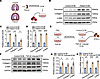 Figure 1
Fibrotic pleural ECM mediated profibrogenic phenotypes of PMCs. (A) Diagram shows the preparation of acellular pleural ECM from patients with tuberculous pleural fibrosis and control group. (B–D) Primary human PMCs were isolated and cultured with the acellular pleural ECM. After 24 hours, the cells were harvested. Relative protein and mRNA levels of fibronectin, collagen I, and α-SMA were detected by Western blotting (B and C) and quantitative real-time PCR (qRT-PCR) (D). Control group, n = 3; patients, n = 3. (E) Primary rat PMCs were isolated and cultured without or with TGF-β1 for 24 hours. Then, rat PMC ECM was prepared by decellularization. (F–H) New primary rat PMCs were treated with TGF-β1–induced ECM or control ECM for 24 hours. Relative protein and mRNA levels of fibronectin, collagen I, and α-SMA were detected by Western blotting (F and G) and qRT-PCR (H). Data are presented as mean ± SEM. Statistical analysis was performed by unpaired Student’s t tests. n = 3, *P < 0.05, **P < 0.01, ***P < 0.001, ****P < 0.0001.
Versican expression is upregulated in pleural fibrosis. Using tandem mass tag–based proteomic analysis, the protein profile of pleura was investigated based on samples from 4 patients with tuberculous pleural fibrosis (TBPF) and 4 control individuals. Versican, an ECM protein, was increased in TBPF (Figure 2A). For visualization in a volcano plot, the raw P values were transformed using –log10(P value) (Figure 2B). Then, Masson’s trichrome staining was performed to show collagen in pleura samples from patients with TBPF and control participants (Figure 2C). Versican expression was further examined by tissue staining in the pleura samples. As shown in Figure 2, D–F, IHC and immunofluorescence staining showed that versican was almost undetectable in control pleura, whereas it was substantially increased in the pleura from patients with TBPF with high-level expression of α-SMA. Furthermore, versican protein was colocalized with the protein Wilms tumor 1, a marker of PMCs. More samples were examined that confirmed a marked increase of versican expression in the pleura of patients with TBPF (Supplemental Figure 3). The level of versican was higher in tuberculous pleural effusion than in transudative pleural effusion (Figure 2G). Moreover, the concentration of versican in the pleural effusion was positively correlated with pleural thickness (Figure 2H). At the same time, in vitro immunofluorescence, Western blotting, and qRT-PCR analysis showed that TGF-β1 and bleomycin substantially increased the expression of versican with increased protein collagen I and α-SMA in PMCs (Figure 2, I–L, and Supplemental Figure 4, A–C). Then, acellular fibrotic ECM with or without knockdown of versican was prepared by treating PMCs transfected with versican siRNA or control siRNA (Supplemental Figure 4, D–F) with TGF-β. After decellularization, new PMCs were seeded in versican-deficient ECM or control ECM. The control ECM induced upregulation of fibronectin, collagen I, and α-SMA; versican- deficient ECM attenuated these increases (Figure 2, M–O). Moreover, administration of recombinant versican restored the changes (Figure 2, M–O). These data indicated that versican mediated fibronectin, collagen I, and α-SMA synthesis in PMCs in vitro.
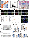 Figure 2
Versican expression was upregulated in pleural fibrosis. (A) Protein profile of human fibrotic pleura was performed by tandem mass tag–based proteomics. Ctrl, pleura from control group; TBPF, pleura from patients with tuberculous pleural fibrosis. (n = 4). (B) Volcano plot of differentially expressed genes. (C) Representative images of Masson’s trichrome staining in pleura from control group and patients with TBPF. Scale bars: 100 μm. (D) Representative images of IHC staining of versican in pleura of controls or patients. Scale bar: 100 μm. (E) Representative images of immunofluorescence staining of versican (red) and Wilms tumor 1 (green). Scale bars: 100 μm. (F) Representative images of immunofluorescence images of α-SMA (red) and Wilms tumor 1 (green). Scale bars: 100 μm. (G) Pleural effusion versican content by ELISA. (H) Correlation analysis between versican concentration in effusion and pleural thickness in patients with TBPF, r = 0.6614, P < 0.001. (I) Representative images of immunofluorescence staining of versican in PMCs incubated with TGF-β (5 ng/mL) or bleomycin (0.2 μg/mL) for 24 hours. (J–L) PMCs were incubated with TGF-β (5 ng/mL) for 24 hours, after which intracellular protein levels of versican, collagen I, and α-SMA were measured by Western blotting; mRNA levels of versican, collagen I, and α-SMA were detected by qRT-PCR at 12 hours. Data are presented as mean ± SEM. Statistical analyses were performed with unpaired Student’s t tests. n = 3, **P < 0.01, ***P < 0.001, ****P < 0.0001. (M–O) Primary rat PMCs were isolated and cultured with VCAN siRNA or control siRNA with TGF-β1 for 36 hours. Then, rat PMC ECM was prepared by decellularization. PMCs were treated by control ECM or versican-deficient ECM with or without recombinant versican (1 μg/mL) for 24 hours, after which fibronectin, collagen I, and α-SMA were detected by Western blotting (M and N) or qRT-PCR (O). Statistical analysis was performed by 1-way ANOVA. n = 3, **P < 0.01, ***P < 0.001, ****P < 0.0001.
The pleura is mainly composed of PMCs and a few fibroblasts. To confirm whether the main source of versican is PMCs, primary human and rat PMCs and fibroblasts were examined. As shown in Supplemental Figure 4, G and H, the basic expression level of versican in primary human PMCs was higher than that in primary human lung fibroblasts. Bleomycin induced versican expression in primary human PMCs, primary human lung fibroblasts, rat PMCs, and rat lung fibroblasts. Moreover, the upregulation of versican in primary human PMCs in response to bleomycin was much greater than that in primary human lung fibroblasts (Supplemental Figure 4G). Consistently, similar results were found in primary rat PMCs and rat lung fibroblasts (Supplemental Figure 4H). These data indicated that versican was upregulated in pleural fibrosis, and it was mainly from activated PMCs.
Loss of versican reduced viscoelasticity and attenuated pleural fibrosis. To verify whether versican mediates pleural fibrosis, mouse pleural fibrosis models were made by intrapleural injection of bleomycin plus carbon particles (Figure 3A). At the same time, versican shRNAs loaded into lentivirus were used to knock down versican (Figure 3B and Supplemental Figure 5). Bleomycin plus carbon induced mouse pleural thickening and fibrosis in visceral, parietal, and diaphragmatic pleura, and versican shRNA attenuated pleural fibrosis (Figure 3, C–E). The versican shRNA also restored declines in mouse lung function, including chord compliance, inspiratory capacity, mean mid-expiratory flow, forced vital capacity, and forced expiratory flow (Figure 3, F–J).
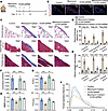 Figure 3
Versican mediated pleural fibrosis in vivo. (A) Mouse pleural fibrosis model was induced by intrapleural injection of bleomycin (0.48 μg/mouse) plus carbon particles (0.1 mg/mouse). Lentivirus expressing shRNA directed against versican (VCAN) shRNA or scrambled sequence shRNA was administrated by intrapleural injection at a dose of 2 × 10⁶ TU on days 4, 7, and 10. After a lung function test, all mice were euthanized at day 21, and then tissues were taken for analysis. (B) Immunofluorescence staining of versican in visceral pleura of mouse pleural fibrosis model. Scale bars: 50 μm. (C) Representative Masson’s trichrome staining images of visceral pleura from lung sections, parietal pleura from chest wall, and diaphragm sections. Scale bar: 100 μm. (D) Changes in pleural thickness. (E) Changes in collagen percentages of pleura. (F–J) Changes in lung function. Cchord, chord compliance.
To confirm whether versican mediates fibrotic changes in PMCs in vitro, bleomycin and TGF-β1 were used to treat PMCs. We found that bleomycin and TGF-β1 induced upregulation of fibronectin, collagen I, and α-SMA mRNA as well as their proteins, which were attenuated by versican siRNA (Supplemental Figure 6, A–F). At the same time, recombinant versican proteins were used to treat PMCs. As shown in Supplemental Figure 6, G–I, versican increased mRNA and protein levels of fibronectin, collagen I, and α-SMA.
The above results suggested that increased versican in fibrotic ECM in turn mediated fibrotic changes in pleura. The underlying mechanisms were further explored. It has been reported that versican increases tissue viscoelasticity (25, 26). Here, rheometry was used to evaluate the viscoelasticity of pleural ECM. As shown in Figure 4, A–G, we confirmed that the pleura of patients with TBPF had a higher loss tangent and faster stress relaxation under a constant deformation. The pleura of patients with pleural fibrosis had higher viscoelasticity than that of control pleura at an identical shear rate. The increased viscoelasticity was also found in visceral pleura of a mouse pleural fibrosis model induced by intrapleural injections of bleomycin plus carbon particles. Knockdown of versican with shRNA lentivirus attenuated the changes in viscoelasticity (Figure 4, H–M). Together, these data indicated that versican in fibrotic ECM increased the viscoelasticity of pleura, which might be related to pleural fibrosis.
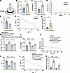 Figure 4
Loss of versican reduced viscoelasticity of fibrotic pleura. (A) Schematic of rheometry analysis of fresh pleural tissues. (B) Rheometry analysis of the storage modulus in control and fibrotic pleura. (C) The loss tangent (viscoelasticity) in control and fibrotic pleura. (D) Stress relaxation curves in pleura samples from control and patient groups. Norm., normalized. (E) Stress was normalized to the initial stress and depicted as τ1/2 (the timescale at which the stress is relaxed to half its original value). (F) Pleural tissues were taken from patients with pleural fibrosis and controls. Shear viscosity was detected and corresponding curves were made. (G) Bar graphs showing relative fold-changes in viscosity with shear rate at 10 (s–1). (B, C, E, and G) Data are presented as mean ± SEM. Statistical analyses were performed with unpaired Student’s t tests. n = 5, *P < 0.05. (H–M) Mouse pleural fibrosis model was induced by intrapleural injection of bleomycin plus carbon particles. Lentivirus expressing shRNA directed against versican (VCAN) shRNA or scrambled sequence shRNA was administrated by intrapleural injection at a dose of 2 × 10⁶ TU on days 4, 7, and 10. After a lung function test, all mice were euthanized at day 21, and then tissues were taken for analysis. (H) Rheometry analysis of the storage modulus. (I) The loss tangent (viscoelasticity) in mouse pleura. (J) Stress relaxation curves in mouse pleura. (K) Stress was normalized to the initial stress and depicted as τ1/2 (the timescale at which the stress is relaxed to half its original value). (L) Shear viscosity was detected and corresponding curves were made. (M) Bar graphs showing relative fold-changes in viscosity with shear rate at 10 (s–1). (H, I, K, and M) Data are presented as mean ± SEM. Statistical analyses were performed with 1-way ANOVA. n = 4, **P < 0.01, ***P < 0.001.
Increased viscoelasticity of pleural ECM promoted pleural fibrosis through CD44. Versican binds to cell surface proteins (receptors) to activate downstream signal pathways. CD44, integrin β1, and EGFR are reported to interact with versican (27–30). To investigate whether these cell surface receptors contribute to versican mediating matrix viscoelasticity and pleural fibrosis, primary rat PMCs were cultured in hydrogels with different degrees of viscoelasticity for 24 hours. As shown in Figure 5, A and B, CD44 increased and no obvious changes in integrin β1 or EGFR were observed in PMCs cultured in high viscoelastic hydrogels. Next, expression of CD44, integrin β1, and EGFR in response to recombinant versican was measured in primary human PMCs. Recombinant versican increased CD44 and did not change integrin β1or EGFR (Figure 5, C–E). Furthermore, inhibition of integrin β1 and EGFR by siRNA did not change the fibrotic process in versican-treated primary rat PMCs (Supplemental Figure 7).
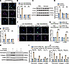 Figure 5
CD44 upregulated and mediated fibrotic phenotype in PMCs. (A and B) PMCs were cultured in low- or high-viscoelasticity hydrogels for 24 hours. Then PMCs were harvested for immunostaining (A) and qRT-PCR (B) to detect protein and mRNA expression of CD44, integrin β1 (ITGB1), and EGFR. (C and D) PMCs were treated by recombinant versican (1 μg/mL) for 24 hours, after which CD44, ITGB1, and EGFR were detected by Western blotting. (E) qRT-PCR analysis of CD44, ITGB1, and EGFR expression in PMCs treated with recombinant versican (1 μg/mL) for 12 hours. (B, D, and E) Data are presented as mean ± SEM. Statistical analyses were performed with unpaired Student’s t tests. n = 3, **P < 0.01, ***P < 0.001. (F and G) After transfection with CD44 siRNA (siRNA3 was selected and used) for 36 hours, PMCs were cultured in low- or high-viscoelasticity hydrogels for 48 hours. Then PMCs were harvested for immunostaining (F) and qRT-PCR (G) to detect protein and mRNA expression of collagen I and α-SMA. Scale bar: 20 μm. (H–J) After transfection with CD44 siRNA for 36 hours, PMCs were cultured with or without recombinant versican (1 μg/mL) for 24 hours. Then PMCs were harvested for Western blotting (H and I) and qRT-PCR (J) to detect protein and mRNA expression of fibronectin, collagen I, and α-SMA. (G, I, and J) Data are presented as mean ± SEM. Statistical analyses were performed with 1-way ANOVA. n = 3, *P < 0.05, **P < 0.01, ***P < 0.001, ****P < 0.0001.
To confirm the role of CD44 in versican-mediated pleural fibrosis, knocking down CD44 was done with CD44 siRNA in primary rat PMCs (Supplemental Figure 8). To directly study the effect of viscoelasticity in fibrosis, primary rat PMCs were cultured in hydrogels with different levels of viscoelasticity. In high-viscoelasticity gels, the expression of MesoMT markers collagen I and α-SMA increased, and both were attenuated by CD44 siRNA (Figure 5, F and G). Moreover, qRT-PCR and Western blotting showed that versican induced mRNA and protein expression of fibronectin, collagen I, and α-SMA. The expression of fibronectin, collagen I, and α-SMA were prevented by CD44 siRNA (Figure 5, H–J).
To further confirm the role of CD44 in versican-mediated pleural fibrosis, in vivo experiments were performed. In clinical samples, CD44hi was expressed with versican in patients with pleural fibrotic tissue compared with control tissue (Figure 6A). Recombinant versican proteins plus carbon were intrapleurally injected in mice to develop models. As shown in Figure 6, B–I, versican proteins plus carbon increased pleural thickness in visceral, parietal, and diaphragmatic pleura and decreased lung function metrics including chord compliance, inspiratory capacity, inspiratory capacity, forced vital capacity, and forced expiratory flow. Anti-mouse CD44 neutralizing antibody (IM7) attenuated these changes, which indicated that IM7 prevented pleural fibrosis.
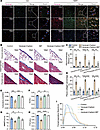 Figure 6
Anti-mouse CD44 neutralizing antibody (IM7) alleviated versican-mediated pleural fibrosis in vivo. (A) Representative results for immunostaining of versican and CD44 in pleura from patients and controls. Scale bars: 100 μm. (B–J) The mouse pleural fibrosis model was induced by intrapleural injections of recombinant versican proteins (150 μg/kg) plus carbon particles (0.1 mg/mouse). IM7 was administrated by intrapleural injection at a dose of 100 μg at days 4, 7, and 10. After lung function testing, all mice were euthanized at day 21, and then pleura were taken for analysis. (B) Representative images of Masson’s trichrome staining of visceral pleura from lung sections, parietal pleura from chest wall, and diaphragm sections. Scale bar: 100 μm. (C) Changes in pleural thickness. (D) Changes in collagen percentages of pleura. (E–I) Changes in lung function test. Data are presented as mean ± SEM. Statistical analyses were performed with 1-way ANOVA. n = 4, **P < 0.01, ***P < 0.001, ****P < 0.0001.
To define whether versican and CD44 had a common effect in pleural fibrosis that resulted from different etiological factors, a mouse pleural fibrosis model was induced by intrapleural injections of Bacillus Calmette-Guérin (BCG) or tuberculous pleural effusion (TBPE). Immunofluorescence analysis demonstrated that versican expression was upregulated in TBPE- and BCG-induced models (Supplemental Figure 9). BCG and TBPE increased pleural thickness in visceral, parietal, and diaphragmatic pleura (Supplemental Figure 10, A–F) and decreased lung function metrics including chord compliance, inspiratory capacity, mean mid-expiratory flow, forced vital capacity, and forced expiratory flow (Supplemental Figure 10, G–P). Versican shRNA or IM7 attenuated these changes, which indicated that versican and CD44 had a common effect in pleural fibrosis. These data provide solid evidence that versican-induced high viscoelasticity mediated pleural fibrosis through CD44.
Versican/CD44 increased Smad4 through deubiquitination by ubiquitin-specific peptidase 10 in PMCs. To further explore the downstream signal pathway of versican and CD44 in the pathogenesis of pleural fibrosis, the downstream pathway of TGF-β signal Smad2/3/4 expression was assessed in primary rat PMCs. Interestingly, versican increased Smad4 protein rather than Smad2/3 (Supplemental Figure 11, A and B) without a change in the Smad4 mRNA level (Supplemental Figure 11C). However, Western blotting showed that versican increased phosphorylation of Smad2/3 in the nucleus (Supplemental Figure 11, D–F). These results suggest that versican facilitated translocation of phosphorylated Smad2/3 into the nucleus through Smad4 rather than increasing the total protein level of Smad2/3 in the cells. Ubiquitination of Smad4 has been found to regulate fibrosis; therefore, we investigated the effect of versican on ubiquitination of Smad4 in primary rat PMCs. As shown in Figure 7A, recombinant rat versican protein suppressed ubiquitination and degradation of Smad4, which led to the aggregation of Smad4 in primary rat PMCs. Next, ubiquitin-specific peptidase 10 (USP10) was detected. The confocal images demonstrated that USP10 and Smad4 were colocalized in primary rat PMCs (Supplemental Figure 12A), and USP10 directly interacted with Smad4 in primary rat PMCs (Supplemental Figure 12B). Immunostaining analysis showed that high viscoelasticity increased the protein expression of USP10 and Smad4 in primary rat PMCs (Figure 7B). High viscoelasticity increasing USP10 was attenuated by CD44 siRNA without a change in Smad4 mRNA (Figure 7C).
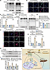 Figure 7
Versican reinforced deubiquitination of Smad4 by recruiting USP10 in PMCs. (A) Ubiquitination of Smad4 was detected by IP in PMCs. (B and C) After transfection with CD44 siRNA for 36 hours, PMCs were cultured in low- or high-viscoelasticity hydrogels for 48 hours. Then PMCs were harvested for immunostaining (B) and qRT-PCR (C) to detect protein and mRNA levels of collagen I and α-SMA. (D and E) After transfection with USP10 siRNA2 for 36 hours, PMCs were cultured with or without recombinant versican (1 μg/mL) for 24 hours; ubiquitination of Smad4 was detected by IP. (F and G) After transfection with USP10 siRNA for 36 hours, PMCs were cultured in low- or high-viscoelasticity hydrogels for 48 hours. Then PMCs were harvested for immunostaining (F) and qRT-PCR (G) to detect protein and mRNA expression of Smad4, collagen I, and α-SMA. (H and I) After transfection with control and USP10 siRNA for 36 hours, PMCs were cultured with or without recombinant versican (1 μg/mL) for 24 hours. Then PMCs were harvested for Western blotting to detect protein expression of fibronectin, collagen I, and α-SMA. (J) Schematic illustration of the mechanisms of versican in PMCs. Data are presented as mean ± SEM. Statistical analyses were performed with 1-way ANOVA. n = 3, *P < 0.05, **P < 0.01, ***P < 0.001, ****P < 0.0001.
To further confirm that USP10 mediated posttranslational modification of Smad4, USP10 siRNA was used in PMCs (Supplemental Figure 13). Versican inhibited deubiquitination of Smad4 and increased the Smad4 level, and these changes were prevented by USP10 siRNA (Figure 7, D and E). More importantly, recombinant versican proteins and high viscoelasticity induced upregulation of collagen I and α-SMA mRNAs and proteins, and USP10 siRNA attenuated their mRNA and protein levels (Figure 7, F–I, and Supplemental Figure 14).
To demonstrate traction-independent CD44 signaling as the mechanistic basis of the observed effects, we conducted key experiments in the versican-hydrogels system in the presence of the myosin II inhibitor blebbistatin to suppress actomyosin contractility. Under this condition, the specific activation of CD44 and the associated downstream cellular effects induced by the high-viscoelastic matrix persisted (Supplemental Figure 15). This finding demonstrated that the observed CD44 signaling is independent of the integrin/actomyosin traction force pathway. To directly distinguish between the “HA-CD44 ligation” and “altered matrix physical properties” hypotheses, we constructed versican and an HA binding–deficient versican mutant (Supplemental Figure 16A). Co-IP assays confirmed that this deficient mutant cannot bind to CD44 (Supplemental Figure 16B). However, it still promoted cellular fibrosis (Supplemental Figure 16, C–E), indicating that fibrosis is driven not by HA-CD44 ligation but by altered matrix viscoelasticity.
Collectively, as shown in Figure 7J, this study demonstrated that versican/CD44 increased Smad4 through USP10 deubiquitination in PMCs, which contributed to versican-mediated fibrogenesis in pleura.


