
Research ArticleDevelopmentPulmonology
Open Access |  10.1172/jci.insight.200486
Genetic background influences developmental airway smooth muscle program and susceptibility to airway hyperresponsiveness in mice
Takehiro Otoshi,1 Benjamin D. Kotton,1 Ayyappa K.S. Kameshwar,2 Yoshinori Seki,2 Zachary Cardell,3 Xiangyi Ke,1 Yuta Matsuno,2 Pooja Rajaram,2 Youn-Kyung Kim,4 Sarah M. Sharpton,5 Loredana Quadro,4 Wellington V. Cardoso,1 and Masako Suzuki2,3
1Columbia Center for Human Development, Department of Medicine, Division of Pulmonary Allergy and Critical Care Medicine, and Department of Genetics and Development, Columbia University, New York, New York, USA.
2Department of Nutrition, Texas A&M University, College Station, Texas, USA.
3Department of Genetics, Albert Einstein College of Medicine, Bronx, New York, USA.
4Department of Food Science and Rutgers Center for Lipid Research, and New Jersey Institute for Food, Nutrition, and Health, Rutgers University, New Brunswick, New Jersey, USA.
5Molecular Genomics Core, Institute for Genome Sciences and Society, Texas A&M University, College Station, Texas, USA.
Address correspondence to: Wellington V. Cardoso, Columbia Center for Human Development, Columbia University Irving Medical Center, Department of Medicine. 650 West 168th Street, BB 8-812, New York, New York, 10032, USA. Phone: 781.690.7550; Email: wvc2104@cumc.columbia.edu. Or to: Masako Suzuki, Texas A&M University, 498 Olson Blvd., College Station, Texas, 77840, USA. Phone: 979.321.7076; Email: masako.suzuki@ag.tamu.edu.
Authorship note: WVC and MS contributed equally to this work
Find articles by Otoshi, T. in: PubMed | Google Scholar
1Columbia Center for Human Development, Department of Medicine, Division of Pulmonary Allergy and Critical Care Medicine, and Department of Genetics and Development, Columbia University, New York, New York, USA.
2Department of Nutrition, Texas A&M University, College Station, Texas, USA.
3Department of Genetics, Albert Einstein College of Medicine, Bronx, New York, USA.
4Department of Food Science and Rutgers Center for Lipid Research, and New Jersey Institute for Food, Nutrition, and Health, Rutgers University, New Brunswick, New Jersey, USA.
5Molecular Genomics Core, Institute for Genome Sciences and Society, Texas A&M University, College Station, Texas, USA.
Address correspondence to: Wellington V. Cardoso, Columbia Center for Human Development, Columbia University Irving Medical Center, Department of Medicine. 650 West 168th Street, BB 8-812, New York, New York, 10032, USA. Phone: 781.690.7550; Email: wvc2104@cumc.columbia.edu. Or to: Masako Suzuki, Texas A&M University, 498 Olson Blvd., College Station, Texas, 77840, USA. Phone: 979.321.7076; Email: masako.suzuki@ag.tamu.edu.
Authorship note: WVC and MS contributed equally to this work
Find articles by Kotton, B. in: PubMed | Google Scholar
1Columbia Center for Human Development, Department of Medicine, Division of Pulmonary Allergy and Critical Care Medicine, and Department of Genetics and Development, Columbia University, New York, New York, USA.
2Department of Nutrition, Texas A&M University, College Station, Texas, USA.
3Department of Genetics, Albert Einstein College of Medicine, Bronx, New York, USA.
4Department of Food Science and Rutgers Center for Lipid Research, and New Jersey Institute for Food, Nutrition, and Health, Rutgers University, New Brunswick, New Jersey, USA.
5Molecular Genomics Core, Institute for Genome Sciences and Society, Texas A&M University, College Station, Texas, USA.
Address correspondence to: Wellington V. Cardoso, Columbia Center for Human Development, Columbia University Irving Medical Center, Department of Medicine. 650 West 168th Street, BB 8-812, New York, New York, 10032, USA. Phone: 781.690.7550; Email: wvc2104@cumc.columbia.edu. Or to: Masako Suzuki, Texas A&M University, 498 Olson Blvd., College Station, Texas, 77840, USA. Phone: 979.321.7076; Email: masako.suzuki@ag.tamu.edu.
Authorship note: WVC and MS contributed equally to this work
Find articles by Kameshwar, A. in: PubMed | Google Scholar
1Columbia Center for Human Development, Department of Medicine, Division of Pulmonary Allergy and Critical Care Medicine, and Department of Genetics and Development, Columbia University, New York, New York, USA.
2Department of Nutrition, Texas A&M University, College Station, Texas, USA.
3Department of Genetics, Albert Einstein College of Medicine, Bronx, New York, USA.
4Department of Food Science and Rutgers Center for Lipid Research, and New Jersey Institute for Food, Nutrition, and Health, Rutgers University, New Brunswick, New Jersey, USA.
5Molecular Genomics Core, Institute for Genome Sciences and Society, Texas A&M University, College Station, Texas, USA.
Address correspondence to: Wellington V. Cardoso, Columbia Center for Human Development, Columbia University Irving Medical Center, Department of Medicine. 650 West 168th Street, BB 8-812, New York, New York, 10032, USA. Phone: 781.690.7550; Email: wvc2104@cumc.columbia.edu. Or to: Masako Suzuki, Texas A&M University, 498 Olson Blvd., College Station, Texas, 77840, USA. Phone: 979.321.7076; Email: masako.suzuki@ag.tamu.edu.
Authorship note: WVC and MS contributed equally to this work
Find articles by Seki, Y. in: PubMed | Google Scholar
1Columbia Center for Human Development, Department of Medicine, Division of Pulmonary Allergy and Critical Care Medicine, and Department of Genetics and Development, Columbia University, New York, New York, USA.
2Department of Nutrition, Texas A&M University, College Station, Texas, USA.
3Department of Genetics, Albert Einstein College of Medicine, Bronx, New York, USA.
4Department of Food Science and Rutgers Center for Lipid Research, and New Jersey Institute for Food, Nutrition, and Health, Rutgers University, New Brunswick, New Jersey, USA.
5Molecular Genomics Core, Institute for Genome Sciences and Society, Texas A&M University, College Station, Texas, USA.
Address correspondence to: Wellington V. Cardoso, Columbia Center for Human Development, Columbia University Irving Medical Center, Department of Medicine. 650 West 168th Street, BB 8-812, New York, New York, 10032, USA. Phone: 781.690.7550; Email: wvc2104@cumc.columbia.edu. Or to: Masako Suzuki, Texas A&M University, 498 Olson Blvd., College Station, Texas, 77840, USA. Phone: 979.321.7076; Email: masako.suzuki@ag.tamu.edu.
Authorship note: WVC and MS contributed equally to this work
Find articles by Cardell, Z. in: PubMed | Google Scholar
1Columbia Center for Human Development, Department of Medicine, Division of Pulmonary Allergy and Critical Care Medicine, and Department of Genetics and Development, Columbia University, New York, New York, USA.
2Department of Nutrition, Texas A&M University, College Station, Texas, USA.
3Department of Genetics, Albert Einstein College of Medicine, Bronx, New York, USA.
4Department of Food Science and Rutgers Center for Lipid Research, and New Jersey Institute for Food, Nutrition, and Health, Rutgers University, New Brunswick, New Jersey, USA.
5Molecular Genomics Core, Institute for Genome Sciences and Society, Texas A&M University, College Station, Texas, USA.
Address correspondence to: Wellington V. Cardoso, Columbia Center for Human Development, Columbia University Irving Medical Center, Department of Medicine. 650 West 168th Street, BB 8-812, New York, New York, 10032, USA. Phone: 781.690.7550; Email: wvc2104@cumc.columbia.edu. Or to: Masako Suzuki, Texas A&M University, 498 Olson Blvd., College Station, Texas, 77840, USA. Phone: 979.321.7076; Email: masako.suzuki@ag.tamu.edu.
Authorship note: WVC and MS contributed equally to this work
Find articles by Ke, X. in: PubMed | Google Scholar
1Columbia Center for Human Development, Department of Medicine, Division of Pulmonary Allergy and Critical Care Medicine, and Department of Genetics and Development, Columbia University, New York, New York, USA.
2Department of Nutrition, Texas A&M University, College Station, Texas, USA.
3Department of Genetics, Albert Einstein College of Medicine, Bronx, New York, USA.
4Department of Food Science and Rutgers Center for Lipid Research, and New Jersey Institute for Food, Nutrition, and Health, Rutgers University, New Brunswick, New Jersey, USA.
5Molecular Genomics Core, Institute for Genome Sciences and Society, Texas A&M University, College Station, Texas, USA.
Address correspondence to: Wellington V. Cardoso, Columbia Center for Human Development, Columbia University Irving Medical Center, Department of Medicine. 650 West 168th Street, BB 8-812, New York, New York, 10032, USA. Phone: 781.690.7550; Email: wvc2104@cumc.columbia.edu. Or to: Masako Suzuki, Texas A&M University, 498 Olson Blvd., College Station, Texas, 77840, USA. Phone: 979.321.7076; Email: masako.suzuki@ag.tamu.edu.
Authorship note: WVC and MS contributed equally to this work
Find articles by Matsuno, Y. in: PubMed | Google Scholar
1Columbia Center for Human Development, Department of Medicine, Division of Pulmonary Allergy and Critical Care Medicine, and Department of Genetics and Development, Columbia University, New York, New York, USA.
2Department of Nutrition, Texas A&M University, College Station, Texas, USA.
3Department of Genetics, Albert Einstein College of Medicine, Bronx, New York, USA.
4Department of Food Science and Rutgers Center for Lipid Research, and New Jersey Institute for Food, Nutrition, and Health, Rutgers University, New Brunswick, New Jersey, USA.
5Molecular Genomics Core, Institute for Genome Sciences and Society, Texas A&M University, College Station, Texas, USA.
Address correspondence to: Wellington V. Cardoso, Columbia Center for Human Development, Columbia University Irving Medical Center, Department of Medicine. 650 West 168th Street, BB 8-812, New York, New York, 10032, USA. Phone: 781.690.7550; Email: wvc2104@cumc.columbia.edu. Or to: Masako Suzuki, Texas A&M University, 498 Olson Blvd., College Station, Texas, 77840, USA. Phone: 979.321.7076; Email: masako.suzuki@ag.tamu.edu.
Authorship note: WVC and MS contributed equally to this work
Find articles by Rajaram, P. in: PubMed | Google Scholar
1Columbia Center for Human Development, Department of Medicine, Division of Pulmonary Allergy and Critical Care Medicine, and Department of Genetics and Development, Columbia University, New York, New York, USA.
2Department of Nutrition, Texas A&M University, College Station, Texas, USA.
3Department of Genetics, Albert Einstein College of Medicine, Bronx, New York, USA.
4Department of Food Science and Rutgers Center for Lipid Research, and New Jersey Institute for Food, Nutrition, and Health, Rutgers University, New Brunswick, New Jersey, USA.
5Molecular Genomics Core, Institute for Genome Sciences and Society, Texas A&M University, College Station, Texas, USA.
Address correspondence to: Wellington V. Cardoso, Columbia Center for Human Development, Columbia University Irving Medical Center, Department of Medicine. 650 West 168th Street, BB 8-812, New York, New York, 10032, USA. Phone: 781.690.7550; Email: wvc2104@cumc.columbia.edu. Or to: Masako Suzuki, Texas A&M University, 498 Olson Blvd., College Station, Texas, 77840, USA. Phone: 979.321.7076; Email: masako.suzuki@ag.tamu.edu.
Authorship note: WVC and MS contributed equally to this work
Find articles by Kim, Y. in: PubMed | Google Scholar
1Columbia Center for Human Development, Department of Medicine, Division of Pulmonary Allergy and Critical Care Medicine, and Department of Genetics and Development, Columbia University, New York, New York, USA.
2Department of Nutrition, Texas A&M University, College Station, Texas, USA.
3Department of Genetics, Albert Einstein College of Medicine, Bronx, New York, USA.
4Department of Food Science and Rutgers Center for Lipid Research, and New Jersey Institute for Food, Nutrition, and Health, Rutgers University, New Brunswick, New Jersey, USA.
5Molecular Genomics Core, Institute for Genome Sciences and Society, Texas A&M University, College Station, Texas, USA.
Address correspondence to: Wellington V. Cardoso, Columbia Center for Human Development, Columbia University Irving Medical Center, Department of Medicine. 650 West 168th Street, BB 8-812, New York, New York, 10032, USA. Phone: 781.690.7550; Email: wvc2104@cumc.columbia.edu. Or to: Masako Suzuki, Texas A&M University, 498 Olson Blvd., College Station, Texas, 77840, USA. Phone: 979.321.7076; Email: masako.suzuki@ag.tamu.edu.
Authorship note: WVC and MS contributed equally to this work
Find articles by Sharpton, S. in: PubMed | Google Scholar
1Columbia Center for Human Development, Department of Medicine, Division of Pulmonary Allergy and Critical Care Medicine, and Department of Genetics and Development, Columbia University, New York, New York, USA.
2Department of Nutrition, Texas A&M University, College Station, Texas, USA.
3Department of Genetics, Albert Einstein College of Medicine, Bronx, New York, USA.
4Department of Food Science and Rutgers Center for Lipid Research, and New Jersey Institute for Food, Nutrition, and Health, Rutgers University, New Brunswick, New Jersey, USA.
5Molecular Genomics Core, Institute for Genome Sciences and Society, Texas A&M University, College Station, Texas, USA.
Address correspondence to: Wellington V. Cardoso, Columbia Center for Human Development, Columbia University Irving Medical Center, Department of Medicine. 650 West 168th Street, BB 8-812, New York, New York, 10032, USA. Phone: 781.690.7550; Email: wvc2104@cumc.columbia.edu. Or to: Masako Suzuki, Texas A&M University, 498 Olson Blvd., College Station, Texas, 77840, USA. Phone: 979.321.7076; Email: masako.suzuki@ag.tamu.edu.
Authorship note: WVC and MS contributed equally to this work
Find articles by Quadro, L. in: PubMed | Google Scholar
1Columbia Center for Human Development, Department of Medicine, Division of Pulmonary Allergy and Critical Care Medicine, and Department of Genetics and Development, Columbia University, New York, New York, USA.
2Department of Nutrition, Texas A&M University, College Station, Texas, USA.
3Department of Genetics, Albert Einstein College of Medicine, Bronx, New York, USA.
4Department of Food Science and Rutgers Center for Lipid Research, and New Jersey Institute for Food, Nutrition, and Health, Rutgers University, New Brunswick, New Jersey, USA.
5Molecular Genomics Core, Institute for Genome Sciences and Society, Texas A&M University, College Station, Texas, USA.
Address correspondence to: Wellington V. Cardoso, Columbia Center for Human Development, Columbia University Irving Medical Center, Department of Medicine. 650 West 168th Street, BB 8-812, New York, New York, 10032, USA. Phone: 781.690.7550; Email: wvc2104@cumc.columbia.edu. Or to: Masako Suzuki, Texas A&M University, 498 Olson Blvd., College Station, Texas, 77840, USA. Phone: 979.321.7076; Email: masako.suzuki@ag.tamu.edu.
Authorship note: WVC and MS contributed equally to this work
Find articles by Cardoso, W. in: PubMed | Google Scholar
1Columbia Center for Human Development, Department of Medicine, Division of Pulmonary Allergy and Critical Care Medicine, and Department of Genetics and Development, Columbia University, New York, New York, USA.
2Department of Nutrition, Texas A&M University, College Station, Texas, USA.
3Department of Genetics, Albert Einstein College of Medicine, Bronx, New York, USA.
4Department of Food Science and Rutgers Center for Lipid Research, and New Jersey Institute for Food, Nutrition, and Health, Rutgers University, New Brunswick, New Jersey, USA.
5Molecular Genomics Core, Institute for Genome Sciences and Society, Texas A&M University, College Station, Texas, USA.
Address correspondence to: Wellington V. Cardoso, Columbia Center for Human Development, Columbia University Irving Medical Center, Department of Medicine. 650 West 168th Street, BB 8-812, New York, New York, 10032, USA. Phone: 781.690.7550; Email: wvc2104@cumc.columbia.edu. Or to: Masako Suzuki, Texas A&M University, 498 Olson Blvd., College Station, Texas, 77840, USA. Phone: 979.321.7076; Email: masako.suzuki@ag.tamu.edu.
Authorship note: WVC and MS contributed equally to this work
Find articles by Suzuki, M. in: PubMed | Google Scholar
Published July 21, 2026 - More info
JCI Insight. 2026;11(17):e200486. https://doi.org/10.1172/jci.insight.200486.
© 2026 Otoshi et al. This work is licensed under the Creative Commons Attribution 4.0 International License. To view a copy of this license, visit http://creativecommons.org/licenses/by/4.0/.
Received: September 22, 2025; Accepted: July 14, 2026


-
Results
Genetic background is a determinant of the aberrant response of the embryonic lung to an intrauterine RA-deficient environment and the susceptibility to AHR in adulthood. A comprehensive analysis of the impact of genetic background on pulmonary function across 36 distinct inbred mouse strains showed remarkable differences in AHR in response to MCh (3). AHR can result from multiple factors, including abnormal inflammatory, structural, and neural responses. There is compelling evidence of an association between AHR and airway remodeling with prenatal disruption of vitamin A signaling in mice (10). We reasoned that further examining this association in mice with distinct genetic backgrounds could provide relevant insights into the developmental origins and prenatal events influencing AHR susceptibility. However, we had no evidence that the observations above, which were reported in an outbred (CD-1) line, could be reproduced in mice with defined genetic backgrounds. Even less certain was the relationship between vitamin A/RA status and AHR in mice of different backgrounds. Thus, to investigate these issues we selected AJ and B6, 2 widely used inbred mouse strains known for their markedly distinct responses to cholinergic agonist challenge (3). To confirm the suitability of these strains for our subsequent studies, adult 16-week-old male AJ and B6 mice were subjected to whole-body plethysmography (FinePoint), and specific airway resistance (sRaw) was measured under the nonchallenged state (baseline) or increasing concentrations of MCh (6.25, 12.5, 25, and 50 mg/mL). Airway resistance increased in both strains in response to increasing MCh concentration (Figure 1A). A 2-way ANOVA showed a significant main effect of MCh concentration (P = 0.016) and a significant main effect of strain, with AJ mice exhibiting significantly higher airway resistance than B6 mice (P = 0.008). Our findings of sRaw and AHR were consistent with airway resistance reported in the Mouse Phenome Database (AJ, 0.424 ± 0.096; B6, 0.321 ± 0.080; P < 0.05).
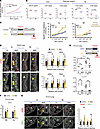 Figure 1
Genetic background determines airway smooth muscle (SM) program and airway hyperresponsiveness after transient prenatal RA deficiency. (A) Specific airway resistance (sRaw) in adult mice under methacholine (MCh) challenge. Significant differences in sRaw between AJ and B6 (P = 0.008, 2-way ANOVA). Mean ± SEM (n = 3 per group). (B) Retinoid concentrations (retinol [ROH], retinyl ester [RE]) in adult mice. n = 5 per group. Two-tailed Student’s t test. (C) Graphs: sRaw (mean ± SEM): AJ exhibited significantly higher BMS-induced airway resistance differences over Control than B6 as shown by 2-way ANOVA. AJ Control (n = 4), AJ BMS (n = 3), B6 Control (n = 9), B6 BMS (n = 6). (D) Immunofluorescence (IF) and quantitative analysis in adult mouse lungs exposed prenatally to BMS or control conditions. Two-tailed Student’s t test. AJ Control (n = 39), AJ BMS (n = 37), B6 Control (n = 45), B6 BMS (n = 43). Scale bars: 20 μm. (E) Expression of RA pathway components in E14.5 lungs (qPCR). Significant Rarb and Cyp26b1 downregulation by BMS in both strains. One-way ANOVA followed by Tukey’s post hoc test. *P < 0.05, **P < 0.01. AJ Control (n = 3), AJ BMS (n = 3), B6 Control (n = 15), B6 BMS (n = 15). (F) qPCR of Sftpc in E14.5 lungs. Mean ± SEM. Two-tailed Student’s t test. AJ Control (n = 3), AJ BMS (n = 3), B6 Control (n = 6), B6 BMS (n = 5). (G) Analysis of E14.5 lungs. aSMA and SM22 IF and morphometric assessment of airway SM. Arrows: normal distribution (white) and ectopic accumulation (yellow) distribution. Graph: Mean ± SEM for each marker. *P < 0.05. Two-tailed Student’s t test. AJ Control (n = 9), AJ BMS (n = 6), B6 Control (n = 9), B6 BMS (n = 9). Scale bars: 20 μm.
Given that differences in retinol (ROH) and retinyl ester (RE) levels have been reported in tissues from different adult inbred mouse strains, and that inadequate adult vitamin A/RA status has been linked to AHR, we asked whether the distinct sRaw/AHR of AJ and B6 reflected strain-dependent differences in retinoid bioavailability (11). The concentrations of ROH and RE in serum and in homogenates from lung and liver were measured in 8-week-old adult mice under a standard vitamin A–sufficient diet (Lab Diet, 5LG4). ROH levels in lung, liver, and serum were similar between strains (Figure 1B). Although AJ showed significantly higher RE levels in the liver (P < 0.05) and a trend toward higher RE levels in lung (P = 0.0565), overall, the differences in retinoid content between adult AJ and B6 mice were unlikely to explain the distinct AHR behavior between strains.
We then tested whether AJ and B6 embryos were equally susceptible to developing AHR in adulthood when briefly exposed to a vitamin A/RA deficient intrauterine environment during early lung development (10). To ensure effective and transient disruption of RA signaling in both strains, we used a more direct approach to prevent RAR activation in the embryo by orally administering mothers with a well-established pan-RAR reverse agonist, BMS493 (BMS) (10, 12). BMS administration could also minimize the variability in vitamin A uptake and retinoid bioavailability seen across strains. We conducted pilot studies to identify a BMS concentration that could simulate the effects of a mild-moderate vitamin A deficiency without the known teratogenic effects in embryonic growth or organogenesis, allowing offspring to survive to adulthood. Thus, AJ and B6 dams were daily gavaged with BMS at different concentrations (3.75–15 μg per body weight per day; μg/BW/day) or vehicle, corn oil (control group). Embryos were exposed to BMS in vivo from E9.5–E14.5, a developmental window encompassing the initial stages of branching morphogenesis and airway differentiation (10) (Supplemental Figure 1A; supplemental material available online with this article; https://doi.org/10.1172/jci.insight.200486DS1). Morphological analysis of E14.5 embryos exposed to the highest BMS concentration (15 μg/BW/day) showed severe effects in the body size and multiple developmental abnormalities, including the lung, most evident in AJ mice (Supplemental Figure 1, A and B). AJ lungs were severely hypoplastic compared with respective controls and B6 lungs. These abnormalities were largely undetected at 3.75 μg/BW/day. Lungs from both AJ and B6 embryos exposed to BMS at this concentration appeared macroscopically comparable to respective controls (Supplemental Figure 1, A and B).
qPCR analysis of these embryonic lungs confirmed that intrauterine exposure to BMS 3.75 μg/BW/day effectively disrupted RA signaling, as seen by the significant decrease in Rarb and Cyp26b1 expression compared with controls (n ≥ 3, P < 0.05). Importantly, Sftpc, which marks the epithelium of growing distal buds, showed no significant differences in expression between control and BMS lungs in both strains (Figure 1, E and F). This ensured that BMS was effective in disrupting RA signaling but not lung epithelial growth and branching. To further demonstrate that the prenatal perturbation in RA signaling did not result in the teratogenic effects that prevent viability, we monitored these mice until adulthood. Embryos from both AJ and B6 mothers exposed to BMS (3.75 μg/BW/day; E9.5–E14.5) were viable, comparable with controls, and reached adulthood. No difference in BW or liver weight relative to BW was found in mice prenatally exposed to BMS or control in both strains. Although we detected a small difference in lung weight relative to BW between adult mice prenatally exposed to BMS compared with control conditions, this difference was similarly found in both AJ and B6 (n > 3, from at least 2 dams) and, importantly, did not prevent animals to undergo postnatal development and reach adulthood as in controls (Supplemental Figure 1, C and D).
Next, we asked whether the brief disruption of RA signaling in embryos exposed to BMS differentially influenced the susceptibility of AJ or B6 mice to develop AHR in adult life. sRaw was assessed at baseline and after MCh challenge in adult 16-week-old male mice from all groups as before. Brief intrauterine exposure to an RA-deficient environment consistently resulted in aberrant higher sRaw at baseline and across all MCh concentrations in adult AJ, but not B6, which responded to the challenge only at the highest MCh concentration (Figure 1C). Two-way ANOVA revealed that both strain and BMS treatment contributed to explaining the variance in AHR at the increasing MCh concentrations tested. Crucially, it also showed that AJ-specific BMS-induced airway resistance over control was significantly higher than that found in B6 (strain-treatment interaction P value: 0.018).
We reasoned that the hyperresponsiveness of BMS-AJ was associated with some structural changes in the airway SM, as suspected by similar observations previously reported in CD-1 mice. Indeed, immunofluorescence (IF) and quantitative analysis confirmed the increased thickness in airway SM labelled by α-smooth muscle actin (aSMA) and SM22 in the adult AJ lungs compared with B6 (Figure 1D). To confirm the developmental origin of this phenotype, we examined embryonic lungs from both AJ and B6 mice immediately after the last day of exposure to maternal BMS administration. E14.5 BMS-AJ lungs showed no obvious abnormalities in epithelial patterning or growth; however, aSMA and SM22 IF revealed increased ectopic accumulation of airway SM, consistent with the pattern previously reported in vitamin A–deficient embryos in CD-1 mice (10). By contrast, disruption of RA signaling had no effect on SM markers in BMS lungs from B6 embryos (Figure 1G).
These findings were consistent with the idea that differences in genetic background are major determinants of how the lung responds to prenatal perturbations in RA signaling and the potential link to the susceptibility to AHR in adult life.
AJ and B6 lungs differ broadly in gene expression signatures, with minimal differences in cellular composition in response to prenatal RA deficiency. The distinct response of AJ and B6 embryos to prenatal disruption of RA signaling led us to examine differences in lung gene expression signatures associated with these distinct backgrounds. First, we performed bulk RNA-seq in E14.5 lungs from AJ and B6 embryos intrauterine exposed to BMS or control conditions (n = 4 per group). Interestingly, principal component analysis (PCA) showed that the strain-related differences in gene expression between control AJ and B6 lungs were greater than those resulting from the prenatal exposure to BMS in each strain (87% variance vs. 4% variance) (Figure 2, A and B, and Supplemental Table 1). Indeed, control AJ and B6 lungs differed greatly in their signatures, as revealed by 1258 differentially expressed genes (DEGs) (Figure 2C and Supplemental Figure 2B).
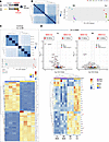 Figure 2
AJ and B6 developing lungs have distinct gene expression signatures under RA deficiency. (A) Experimental design. (B) Heatmap of sample-to-sample distances: segregation by strain. PCA plot shows PC1 (strain effect) and PC2 (BMS effect). (C) Differential gene expression between controls. Unsupervised heatmap depicting the top genes enriched in each strain (*Lrat enriched in AJ). (D) Volcano plots: DEGs in BMS versus control embryonic lungs. Number of DEGs depicted in boxes (Padj < 0.05 and |log2[fold change]| > 0.58). Supervised heatmap, highlighting BMS-downregulated markers of RA activity (*).
We searched for putative direct RA targets among these genes, initially by looking for RA-responsive element–containing (RARE-containing) open chromatin regions (RARE-OCRs) in publicly available ENCODE ATAC-seq mouse datasets from E14.5–E16.5 lungs, representative of early stages of lung differentiation (5) (Supplemental Figure 2A). This revealed 28,355 RARE-OCRs with an irreproducible discovery rate of less than 0.05; nearly half of these (44.2%) were identified within the promoter region of genes, potential RA targets (RARE-OCR genes, Supplemental Table 2). The data suggested a robust RA-responsive gene network at these developmental stages. The proportion of RARE-OCRs that overlapped with genes differentially expressed between control AJ and B6 lungs was not markedly different (AJ: 39.7%; B6: 54.4%). However, gene set enrichment analysis (GSEA) suggested that they differed considerably in downstream pathways and presumably function. For example, analysis of RARE-OCR–containing genes enriched in AJ included Gene Ontology (GO) terms, such as growth factor activity, lyase activity, and transferase activity, while those enriched in B6 included ECM-related pathways and growth factor binding (Supplemental Figure 2B). Moreover, confirming our initial findings, Stra6 and Lrat, key genes associated with vitamin A uptake and retinoid bioprocessing/storage, were among the DEGs upregulated in control AJ compared with B6 (Supplemental Figure 2D and Supplemental Table 3) (13, 14).
We then compared the impact of maternal BMS exposure on gene expression signatures of AJ and B6 embryonic lungs. Efficient disruption of RA signaling was confirmed in both strains by downregulation of RA pathway target genes (Rarb, Cyp26b1, and Dhrs3) as well as other known RARE-OCR–containing genes such as Hox family members (Figure 2D, Supplemental Figure 2, C and E, and Supplemental Table 4). GSEA of DEGs in BMS versus control lungs from AJ embryos revealed significant enrichment for terms associated with the suppression of RA-related molecular function (e.g., RA binding), as well as enrichment for terms associated with activation of muscle contraction and microfilament activity, none of which were found in B6 lungs (Supplemental Figure 2F and Supplemental Figure 3A). Consistent with this, analysis of transcription factor activity using the R package decoupleR with the univariate linear model (ULM) approach showed decreased Rarg, Rarb, and Rora activity and increased activity of transcription factors, such as Egr1, Id2, Med1, Tbx2, and Tbx3 associated with muscle development (GO: 0060537) in AJ BMS (Supplemental Figure 3B). This was not observed in BMS versus control B6 lungs, suggesting a less robust RA-related functional activity in these events. Together, the data pointed to broad differences in the gene expression signatures of the developing AJ and B6 lungs, including regulators of retinoid bioavailability and RA targets. However, these differences could hardly justify their role as determinants of the altered response of AJ mice to an RA-deficient environment.
To further investigate whether the distinct behavior of AJ and B6 resulted from predominant changes in gene expression in a specific cell population in response to BMS, we performed single-nucleus multiome analysis (snRNA-seq and snATAC-seq) of these lungs exposed to BMS under the same conditions. After filtering low-quality entries, 21,293 nuclei were examined across all conditions (AJ: control = 4750 and BMS = 6363; B6: control = 5321 and BMS = 4859). Using weighted nearest neighbors (WNN) to integrate snRNA-seq and snATAC-seq data, 4 populations were identified broadly as epithelial, mesenchymal, endothelial, and immune cell based on reported markers and chromatin structure profiles (Supplemental Figure 4A and Supplemental Figure 5A) (15). No significant strain-specific differences in cell proportion were found in any of these 4 cell populations in response to prenatal disruption of RA signaling (Supplemental Figure 4B). This was somewhat unexpected since the genetic background can account for subtle but often important variations in cellular composition, resulting in strain-dependent differences in organ size and patterns of growth (16). By contrast, we found major changes in gene expression and to a lesser extent open chromatin accessibility in response to BMS in both strains. Consistent with our bulk RNA-seq findings, BMS exposure resulted in a higher number of DEGs in AJ lungs (n = 548) compared with B6 (n = 214). Moreover, BMS-AJ also had a higher number of differentially accessible regions (DARs) (n = 117) than BMS-B6 (n = 71); of these, 61 out of 117 (52%) and 37 out of 71 (52%), respectively, harbored RARE-OCRs (Supplemental Figure 5B and Supplemental Tables 5 and 6). The lower number of DARs compared with DEGs suggested that differences in transcription factor expression were more likely to be the primary driver of the RA-mediated events rather than shifts in the epigenetic state. An initial analysis of the top DEGs in each of these populations was unremarkable. The collectively higher number of DEGs and DARs in the mesenchymal cell population of AJ compared with B6 and the AJ-distinctive SM differentiation program in response to BMS, led us to further focus our analysis on the lung mesenchyme.
Distinct subpopulations emerge from the mesenchymal cell compartment during airway morphogenesis in both AJ and B6 lungs. Given the higher number of DEGs and DARs in the mesenchymal compartment of BMS-AJ compared with BMS-B6 (Supplemental Figure 5B), the known diversity of lung mesenchymal cell types, and the dynamic morphogenetic changes taking place in the E14.5 lung, we performed a comprehensive analysis of this compartment to understand how BMS influenced its gene expression and behavior. First, we computationally filtered for the mesenchymal cells from our multiome datasets, combining all groups. Leiden clustering identified 8 subpopulations, annotated based on established markers (Seurat FindMarkers with the Wilcoxon rank-sum test) and 2 other small indistinct subpopulations with mixed signatures, not further analyzed (Figure 3, A–D, and Supplemental Table 7). Cluster 0 was identified as undifferentiated proliferative cells for their enrichment in cell cycle–related genes (Top2a, Kif15, and Mki67) (Supplemental Figure 6A). Clusters 1 and 6 consisted of 2 populations of fibroblast-like cells based on their expression of ECM-related genes (Fibroblast 1: Col25a1, Mfap4 and Fibroblast 2: Bnc2, Fap) (Supplemental Figure 6C). Cluster 4 was annotated as “mitochondria-enriched mesenchyme” (mt-Co3, mt-Atp6, and mt-Nd1) (Supplemental Figure 6B). Cluster 3 was characterized by enrichment in genes associated with biological processes, such as cell migration, morphogenetic movements, and chemoattraction reported mostly in neuronal studies (Sema3d, Epha3, and Ptn), but also featuring key regulators of lung pattern formation and mesenchymal differentiation (Wnt2, Fgf10, Rspo2, and Ctnna2) (Supplemental Figure 7A). Two populations (Clusters 5 and 7) have common expression statuses of genes associated with the SM program (Supplemental Figure 6E). However, Cluster 5 was annotated as “vascular smooth muscle” (pericytes) based on the differential enrichment in Pdgfrb, Heyl, and Notch3, compared with Cluster 7 “airway smooth muscle” identified by markers such as Lgr6, Actg2, Cdh4, Acta2, Myom1, Chrm2, and others (Figure 4B and Supplemental Figure 6, D and E). Lastly, Cluster 2 was enigmatic as it encompassed a population of undifferentiated mesenchymal progenitors marked by Zfp536, Tgfbi, and Igf1, but also expressing early stages of the SM cell program (Myocd and Myh11), overall distinct from Clusters 5 and 7 (Figure 4A).
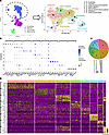 Figure 3
Single-nucleus multiomics identifies multiple subpopulations of lung mesenchymal cells. (A) UMAP projection of reclustered mesenchymal cells (all conditions and strains). Eight subpopulations identified by established markers. (B) Dot plot of top differentially enriched genes for each of the mesenchymal cluster. (C) Pie chart depicting the relative proportion of each mesenchymal cell subpopulation. (D) Heatmap of top differentially enriched genes for each of the mesenchymal cluster.
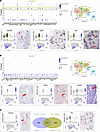 Figure 4
Multiomics analysis identifies 2 distinct stages of airway SM cell program during lung morphogenesis. (A and B) Clusters 2 and 7: Dot plots displaying top DEGs (Padj < 0.05). Representative genes from each cluster are depicted in violin and feature plots. In situ hybridization (GenePaint; http://www.genepaint.org/): Distinct spatial distribution of these populations in distal mesenchyme where the SM emerges (Cluster 2) or in already established airway SM at E14.5 (Cluster 7). (C) Violin plots and Venn diagram depicting unique and shared genes between Clusters 2 and 7. Red arrows and dashed box depict gene expression in airway smooth muscle.
A systematic survey of the spatial localization of top markers from each cluster showed partially overlapping but also distinct expression patterns in the E14.5 developing lung. Transcripts from Cluster 0 markers were found throughout the lung mesenchymal compartment, consistent with the broad distribution of proliferative cells at this stage (Supplemental Figure 6A). This contrasted with the more spatially restricted distribution of Cluster 2, 3, 5, and 7 genes (Figure 4, A and B, Supplemental Figure 6, D and E, and Supplemental Figure 7, A and C). Notably, analysis of Cluster 2 markers revealed a remarkable pattern of transcript distribution in the mesenchyme at the stalk region of distal buds from where the SM program emerges (Figure 4A and Supplemental Figure 8A). Based on its signature, gene ontology, and spatial distribution, Cluster 2 was annotated as “airway smooth muscle progenitor,” reflecting its enrichment in genes associated with initiation of the cell fate program toward this lineage. Indeed, evidence from multiple studies, as well as our GSEA/GO results, showed Cluster 2 top differentially enriched genes, components of the TGF-β, IGF, and PDGF pathways (Tgfbi, Pdgfra, and Igf1) reported as early regulators of the SM program (17–19). Also enriched in Cluster 2 we found Ptch1 and Hhip, components of the Shh pathway reported as crucial for initiation of the airway SM program (20). By contrast, Cluster 7 cells were most distinctly identified by the expression of more definitive markers of SM differentiation and for their localization to proximal airways (Figure 4, B and C, and Supplemental Figure 8B). The analysis also underscored the closer relationship in the signature of Clusters 7 and 2 compared with the others. For example, among the 714 and the 514 genes identified as markers of Clusters 7 and 2, respectively, 30% or more genes were common to both clusters (221 genes; Padj < 0.05, absolute log2[fold change] > 0.585) (Figure 4C and Supplemental Table 14). These overall (e.g., Myocd and Myh11) differed in levels or percentages of cells expressed in Clusters 7 or 2 and likely include cells undergoing a transition from commitment to initiation of the SM program in the highly dynamic niche of the E14.5 distal lung (dot plots in Figure 4, A and B). By contrast, Clusters 7 and 5, in spite of being identified as SM (airway and vascular, respectively), shared only approximately 10% of their DEG signatures (Supplemental Figure 6E). Notably, DEGs in Cluster 3 overall showed a characteristic spatial transcript distribution in the distal mesenchyme, often in a subpleural location and found collectively by GO to be regulators of cell migration, morphogenetic movements, and chemoattraction, widely reported in neurons and the lung. Additional Cluster 3 enriched genes, such as Wnt2 and Fgf10, are known for their prominent role in lung pattern formation and shown by lineage tracing analysis to label early progenitors of the airway SM program (Supplemental Figure 7, A and B) (21). Moreover, Wnt2 has been shown to regulate the airway SM program through MRTFB/myocadin and FGF10 (22).
Altogether, from these annotations we propose a developmental model that aligns well with that proposed by Kumar et al. (21), which describes a progression from undifferentiated tip bud mesenchymal progenitors (Cluster 3) to recruitment to the airway SM lineage around the stalk region of distal lung buds (Cluster 2) to ultimately differentiate into mature SM cells (Cluster 7) (Supplemental Figure 7D).
Endogenous RA signaling is differentially required in a specific TGF-β–activating mesenchymal progenitor population to prevent aberrant SM program of AJ lungs during morphogenesis. Having identified the signature characteristic of these mesenchymal clusters and their spatial distribution in the E14.5 lung, we then asked what differences in cellular composition and gene expression could be attributed solely to the genetic background of each strain, selectively in the mesenchymal cell compartment. For this, we compared the gene expression signature of E14.5 AJ and B6 under unperturbed (control) conditions. Transcriptome analysis showed that AJ and B6 control lungs differed significantly in the number of DEGs in Cluster 0 (478 DEGs), Cluster 2 (273 DEGs), and Cluster 3 (161 DEGs), with fewer differences in the remaining mesenchymal clusters (Supplemental Figure 9A and Supplemental Table 10). Although interesting, we could not rule out that this could be in part due to an overall higher number of cells in these clusters. Notably, we had no evidence that endogenous RA signaling was preferentially more active in the mesenchymal populations of control AJ compared with B6 lungs. Rarb, a surrogate marker of RA activation, was similarly expressed in all clusters of controls from both strains (Supplemental Figure 9B). A comparison of the gene expression signature of Clusters 0, 2, and 3 from control AJ and B6 lungs revealed Mgp, Negr1, Col1a1, and Opcml among the top DEGs significantly downregulated in AJ in these 3 clusters. This signature was also found in AJ controls when all mesenchymal clusters were examined collectively (Clusters 0–9) and when AJ and B6 were analyzed by bulk RNA-seq of whole-lung homogenates. These results strongly suggest that these genes represent key components of the signatures that distinguish AJ from B6 in E14.5 lungs, ultimately associated with the strain differences in genetic background (Supplemental Figure 9, C–F and Supplemental Table 12).
Next, we assessed the impact of intrauterine exposure to BMS on the gene expression signature of AJ and B6 developing lungs. Single-nucleus multiomics showed a significantly higher impact of BMS on the transcriptomics of AJ lungs. Altogether, the number of DEGs between control and BMS in all clusters was almost twice as high in AJ compared with B6 lungs (n = 142 and n = 78, respectively), with the majority of these found in Clusters 0, 2, and 3, regardless of differences in cell proportions (Figure 5, A and B, and Supplemental Tables 8 and 9). We leveraged the statistical power of a global analysis collectively of all mesenchymal clusters from our AJ and B6 single-nucleus transcriptome dataset to identify DEGs between control and BMS-exposed lungs. Although mesenchymal disruption of RA signaling was confirmed by downregulation of Rarb in both strains, strain-specific differences in gene expression in response to BMS were clearly detected between AJ and B6. These included downregulation of additional components of the RA pathway (Lrat and Dhrs3), accompanied by upregulation of TGF-β targets and known regulators and markers of SM program in the mesenchymal population of BMS-AJ lungs (Supplemental Figure 10, A and B, and Supplemental Table 11).
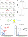 Figure 5
Cluster 2 cells are enriched in regulators of SM program and respond differentially to an RA-deficient intrauterine environment in AJ and B6 lungs. (A) Single-nucleus multiomics analysis of BMS versus control lungs. Volcano plots: Higher number of DEGs in AJ, particularly in Clusters 0, 2, and 3. Boxes depict DEG number with |log2(fold change)| > 0.58 and adjusted P < 0.05. (B) Graph depicting changes in cell proportion in response to BMS for each mesenchymal cluster. Differences between AJ and B6 are significant if FDR < 0.05, |log2(fold change)| > 0.58. Permutation testing (n = 10,000). (C) Identification of mesenchymal Cluster 2–enriched genes among the DEGs upregulated by BMS in AJ lungs. Graphs represent the |log2(fold change)| in expression between BMS and control conditions across all mesenchymal clusters from whole mesenchymal analysis (x-axis) against the |log2(fold change)| of genes selectively enriched in Cluster 2 (y-axis).
Given our goal to identify potential regulators of the strain-specific responses of the mesenchymal cells to an RA-deficient environment, we established the following more stringent criteria for further analyses: (a) we examined the DEGs unique to AJ not differentially altered by BMS in B6 lungs; (b) among these, we focused on the genes upregulated by BMS in AJ consistent with our original hypothesis that endogenous RA prevents an unrestrained aberrant program of SM progenitors during lung morphogenesis; and (c) using information gathered from our cluster analysis and expression pattern, we selected DEGs whose mRNA distribution mapped to putative sites of initiation of the SM program in the E14.5 distal lungs. Based on these criteria, Cluster 2 emerged as the cell population to be the prime candidate for analysis. We first asked which of the DEGs identified by analysis of control versus BMS collectively in all mesenchymal cells featured among the markers of Cluster 2 cells. We then identified the Cluster 2 genes differentially upregulated by BMS in AJ, but not in B6. This revealed Tgfbi, Pdgfra, Entpd1, and Zfp536 and other top markers of Cluster 2 (Figure 5C). The identification of widely reported regulators of the SM program among these genes supported the criteria for further screening (17, 18, 23, 24). Since BMS did not significantly alter the proportion of Cluster 2 cells in AJ or B6 lungs (Figure 5B), we concluded that the upregulation of the Cluster 2 genes found in AJ likely resulted from their derepression in response to the disruption of RA signaling selectively in this strain. Cluster 2 genes downregulated by BMS are shown in Supplemental Figure 11B.
A large number of studies confirm the essential role of TGF-β signaling in SM development and in remodeling of the injured lung in diseases such as pulmonary fibrosis and asthma (25). The regulation of TGF-β signaling by RA in airway SM differentiation has also been extensively reported both in the developing and the adult lung. Disruption of RA signaling leads to hyperactivity of the TGF-β pathway at the onset of lung development and later during branching morphogenesis (12, 26). Here, we found key components of the TGF-β pathway enriched in the mesenchymal compartment of the E14.5 lungs, with Tgfbi and Tgfbr3 appearing as top markers of Cluster 2 (Supplemental Figure 13). Tgfbr3 is a co-receptor crucial for activation of TGF-β/SMAD signaling through TGFB2-TGFBR2 binding and phosphorylation of TGFBR1 (27). Tgfbi is a well-established early target gene of the TGF-β pathway (12).
Interestingly, analysis of E14.5 lungs revealed expression of Tgfbr3, Tgbi, and p-SMAD2 and -3 signals converging at the stalk mesenchyme where airway progenitors (Sox2+) emerge and the SM program initiates (Figure 6A and Supplemental Figure 13). This led us to hypothesize that the TGF-β pathway could be aberrantly active locally, altering key developmental events in response to disruption of endogenous RA signaling. We started investigating this possibility by comparing the SMAD2/3 phosphorylated status locally in the distal lung undergoing morphogenesis ex vivo under control and RA-deficient conditions.
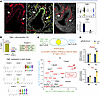 Figure 6
TGF-β–activating cells are key determinants of the distinct response of AJ to prenatal disruption of RA signaling. (A) Image panels: Tgfbi transcript distribution in distal E14.5 mesenchyme, and p-SMAD2/3 IF signals in the stalk mesenchyme associated with the emergence of Sox2+ airway epithelial progenitors. Violin plot: Tgfbi enrichment in Clusters 2 and 7. White arrows depict pSMAD2/3+ (left and middle panels) and Tgfbi+ regions (right panel). Graph: Tgfbi expression in lung explants cultured under control or SB431542-containing (SB4-containing) media. Marked downregulation by disruption of endogenous TGF-β signaling. qPCR: Mean ± SEM, 2-tailed Student’s t test, Ctr (n = 7), SB4 (n = 8). (B) Quantitative analysis of the Tgfbi+ cell proportion in Cluster 2 compared to all other clusters and effect of BMS. Highest Tgfbi expression in Clusters 2 and 7. (C) Top: Venn diagram depicting Cluster 2–enriched genes among the DEGs upregulated by BMS in Tgfbi+ cells of AJ lungs (Padj < 0.05 and |log2[fold change]| > 0). Bottom: Differences in gene expression in BMS versus control Tgfbi+ cells (|log2[fold change]|; x-axis) against the |log2(fold change)| of genes selectively enriched in Cluster 2 (y-axis). Genes carrying SMAD2, SMAD3, or SMAD4 binding sites (marked in red) found upregulated by BMS in Tgfbi+ cells from AJ lungs. (D) qPCR analysis of lung homogenates from E14.5 embryos. Significant upregulation by BMS only in AJ (2-tailed Student’s t test, n = 3 per group). *P < 0.05. All images: ×20 magnification.
Embryonic lungs from AJ and B6 mice were cultured under well-established conditions in control or BMS-containing media and analyzed after 48 hours (12). Efficient disruption of RA signaling was confirmed by downregulation of RA targets (Rarb and Cyp26b1) in both AJ and B6 lung cultures. IF of aSMA confirmed ectopic induction of airway SM in AJ, but not in B6-derived lung cultures, confirming the strain-dependent differential response we observed in vivo (Supplemental Figure 12, A and B). Control cultures showed the typical distribution of p-SMAD2/3 signals in the distal mesenchyme associated with lung bud stalks also observed in vivo (Supplemental Figure 12C and Figure 6A). By contrast, we found p-SMAD2/3 signals markedly increased and sharper in the distal mesenchyme of BMS-treated AJ lung cultures. No difference in p-SMAD2/3 staining was observed between control and BMS-treated lung cultures from B6 mice (Supplemental Figure 12D). The data strongly supported the idea of a strain-dependent regulation of TGF-β activation by RA in a subpopulation of progenitor cells undergoing SM fate specification in developing airways. The overactive TGF-β signaling was evident by the increased p-SMAD2/3 signals and upregulation of Tnc, Pdgfra, and Plxna4, known targets of TGF-β signaling, in BMS-treated AJ cultures compared with controls (Supplemental Figure 12E).
There is accumulated evidence that Tgfbi identifies sites of canonical TGF-β activation (28, 29). In the E14.5 lung, we found Tgfbi transcript distribution was closely associated with sites of SMAD2/3 phosphorylation in the distal mesenchyme, and overall levels were markedly downregulated when endogenous TGF-β signaling was inhibited by SB431542 in lung embryonic explant cultures (Figure 6A). Given the evidence of the differential upregulation of TGF-β pathway genes by BMS in AJ lungs, we raised the possibility that Tgfbi+ cells may harbor key mediators of this differential response. Although largely represented in the Cluster 2 population, Tgfbi was also found to a lesser extent in other clusters (66% of 1473 Cluster 2 cells, 13% of 8742 non–Cluster 2 cells) (Figure 6, A and B, and Supplemental Figure 14A). Thus, we computationally filtered all Tgfbi+ cells and collectively investigated their gene expression signature in each experimental condition. Tgfbi+ cells were filtered by selecting cells with normalized Tgfbi expression greater than 0.1. Analysis of control lungs from both strains showed a relatively high proportion of Tgfbi+ cells in AJ (27%) compared with B6 (16%). This proportion was largely unchanged by BMS when compared to controls from respective strain (AJ BMS: 25%; B6 BMS: 14%). By contrast, the number of DEGs in BMS versus control Tgfbi+ cells was remarkably different in AJ (n = 337) compared with B6 lungs (n = 6) (Padj < 0.05) (Supplemental Figure 11A and Supplemental Table 13). Notably, while we found 79 DEGs upregulated by BMS in Tgfbi+ cells from AJ, only 3 DEGs were upregulated in BMS-B6 lungs (Supplemental Figure 14B). Given that the proportion of Tgfbi+ cells was unchanged by BMS in both strains, we conclude that disruption of RA signaling resulted in overly activated TGF-β signaling selectively in this cell population of AJ lungs. In searching for a potential reason for this discrepancy, we examined a panel of markers of RA activation in Tgfbi+ cells from both strains. Surprisingly, although Rarb and Cyp7b1 were consistently expressed in Tgfbi+ cells from controls in both AJ and B6 lungs, these genes were downregulated by BMS only in AJ lungs (Supplemental Figure 14C). This suggested that the Tgfbi+ cell population of B6 was selectively insensitive to BMS and maintained its normal program, while the same population in AJ lungs under the same conditions responded aberrantly derepressing genes whose levels would be normally controlled by endogenous RA in these cells. Their identification as TGF-β–activating cells in both strains and their distinct response to an RA-deficient environment are consistent with the idea that the Tgfbi+ cells represented the key cellular target of the SM phenotype observed in BMS AJ lungs.
To further refine our analysis and enrich genes with a greater chance of being mediators of the BMS aberrant phenotype, we first identified DEGs from cluster 2 that were most upregulated by BMS in AJ Tgfbi+ cells (Padj < 0.05 and absolute log2[fold change] > 0). This ensured a greater representation in genes predicted by GSEA/GO analysis of Cluster 2 to be regulators of the SM program. Second, given that Tgfbi+ cells were identified for their differential activation of TGF-β signaling, we used the public dataset (ChEA Transcription Factor Targets 2022; https://maayanlab.cloud/Harmonizome/dataset/ChEA+Transcription+Factor+Targets+2022 accessed March 2025) to search for targets of SMAD2, SMAD3, or SMAD4 among these genes. As predicted, Tgfbi featured as one of the top DEGs in this cell population, further supporting this gene as a direct target of TGF-β signaling in these cells. Top differentially upregulated SMAD2/3 targets in Tgfbi+ AJ cells included Tnc, Pdgfra, Plxna4, Nrxn3, and Nnat (Figure 6C). Targets such as Pdgfra and Tnc are particularly relevant in the context of the question addressed here. During lung morphogenesis, Pdgfra+ cells are reported to be abundant in the mesenchymal compartment, including the domain of putative SM progenitors we identified in Cluster 2. Pdgfra+ cells in peribronchiolar and alveolar regions are known to differentiate into contractile aSMA+ cells in the developing lung and later shown to be crucial for alveolar septation (18, 30–32). Various lines of evidence support interactions of Pdgf, Tgfb, and Tnc in myofibroblast/fibroblast differentiation in different tissue contexts. For example, a functional TGF-β/PDGFRA signaling crosstalk regulates myofibroblast migration and differentiation (33). A mechanism in which PDGFRA and TGF-β signaling promotes TNC expression in intestinal myofibroblasts has been described in a mouse model of intestinal injury (34). This crosstalk also involved SMAD3 and SMAD4, which were shown to activate TNC promoter activity, and their overexpression further enhanced the effect of TGF-β (35). In addition, we found Plxna4 among the most upregulated genes in Tgfbi+ cells (Figure 6, C and D). This gene encodes a receptor for semaphorins, known regulators of migration and neuron guidance. Plxna4 is a TGF-β/SMAD3 target upregulated in SM cells and fibroblasts by Tgfb1 (36, 37) (Figure 6D). None of the Cluster 2 genes upregulated by BMS in Tgfbi+ cells in B6 carried SMAD2, SMAD3, or SMAD4 binding sites (Supplemental Figure 11C). We confirmed the relevance of these findings and showed that the impact of RA signaling disruption on regulators of the SM program, PDGFRA and TNC, could be faithfully recapitulated in vivo embryos exposed to maternal BMS or control regimens as described below in subsequent experiments.
Next, we investigated whether blocking TGF-β overactivation could prevent the aberrant airway SM program of BMS-exposed embryos in vivo. We selected from a list of pharmacological compounds those proven to inhibit TGF-β signaling, with some evidence that they could be well tolerated during pregnancy, and able to cross the placental barrier without inducing embryonic lethality. Then different doses were tested to identify those that effectively interfered with TGF-β signaling in the embryo during organogenesis with minimal impact on overall embryo and organ growth. Special attention was given to identify maternal dosing regimens compatible with the developmental window of our study (E9.5–E14.5), applying criteria analogous to those we chose in our analysis of BMS in the lung (overall preserved epithelial patterning and growth).
Although none of the compounds screened were able to fulfil all criteria, we selected galunisertib (Gal) (LY2157299), a selective TGF-βRI kinase inhibitor, for its effectiveness, ability to cross the placental barrier, and interfere with TGF-β signaling in the embryo. Gal was administered to both AJ and B6 mothers at concentrations of 20, 75, and 150 mg/kg BW on gestation days 9.5–14.5, and embryos were analyzed subsequently (Supplemental Figure 15A). E14.5 embryos from AJ and B6 mothers exposed to 20 and 75 mg/kg/BW Gal were viable, but 150 mg/kg Gal consistently resulted in fetal death. Lungs from AJ embryos exposed to 20 mg/kg/BW Gal showed no obvious gross morphological defects compared to controls, but differed from those at 75 mg/kg/BW Gal, which appeared developmentally less mature (Supplemental Figure 15A). Disruption of TGF-β signaling by Gal was confirmed by downregulation of TGF-β targets Tgfbi, Pdgfra, Tnc, and Myh11, prominently at the highest concentration (Supplemental Figure 15B). Given the relatively preserved morphology and epithelial patterning of lungs with 20 mg/kg/BW Gal (no significant changes in Epcam, Sftpc, and Sox2/Sox9) compared to controls, we selected this dose for subsequent studies (Supplemental Figure 15, A and B).
Thus, we examined how simultaneous disruption of RA signaling and TGF-β inactivation influenced the airway SM program emerging in stalks of growing lung buds. AJ mothers were administered BMS (3.75 μg/g) plus Gal (BMS+Gal; 20 μg/g), BMS alone (3.75 μg/g), or oil (control) starting on gestation day 9.5, and embryonic lungs were analyzed on E14.5 (Figure 7A). IF of PDGFRA and TNC in controls confirmed their local signals distribution in the distal niche associated with initiation of the airway SM program (Figure 7A), consistent with their pattern of mRNA expression (Figure 4C and Supplemental Figure 10B). This contrasted with the marked increase in PDGFRA and TNC expression and ectopic expansion of their domain to adjacent areas found in BMS-exposed AJ lungs (Figure 7A). These changes were not found in BMS-exposed B6 embryos, further supporting the strain-related differences in RA regulation of the airway SM program (Supplemental Figure 16, A and B).
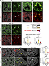 Figure 7
The aberrant airway SM program of prenatal RA signaling disruption in AJ lungs is prevented by TGF-β pathway inactivation in uterus. (A) Analyses of E14.5 AJ lungs exposed to maternal BMS, BMS+Gal, or control conditions. IF of PDGFRA and TNC in Control AJ showing typical expression pattern of these markers in distal mesenchyme associated with bud stalks (white arrows), distinct from BMS-exposed lungs, in which signals are much stronger and widely distributed (yellow arrowheads). Concomitant disruption of RA signaling and TGF-β inactivation in uterus prevents the aberrant PDGFRA and TNC expression of BMS-exposed lungs, as shown by their distinct pattern in BMS+Gal lungs comparable with controls. Graphs: Relative area of marker expression per field in AJ distal lung. Mean ± SEM (1-way ANOVA followed by Tukey’s post hoc test, n = 3 measurements per group). *P < 0.05. (B) IF of aSMA and SM22 in E14.5 AJ lungs showing that exposure to BMS+Gal prevented the aberrant ectopic SM formation in distal airways of embryos exposed to BMS alone. Graphs: Quantitative analysis of marker expression. Mean ± SEM (1-way ANOVA followed by Tukey’s post hoc test, n = 3–4 measurements per group). *P < 0.05. Scale bars: 20 μm.
Notably, IF analysis of E14.5 lungs from mothers administered BMS+Gal showed a pattern of PDGFRA and TNC expression strikingly different from that of the BMS group, being rather comparable to that typically found in control lungs (Figure 7A). The data suggested that the aberrant expression and spatial distribution of regulators of the SM program (such as PDGFRA and TNC) that resulted from RA signaling disruption in AJ lungs could be at least partially prevented by restricting TGF-β signaling activation. Consistent with this, lungs from BMS+Gal-treated embryos showed no evidence of the increased and ectopic expression of SM markers (aSMA and SM22) seen in BMS-AJ lungs (Figure 7B).
Based on these findings, we asked whether the offspring from AJ mothers administered BMS+Gal, if followed through adulthood, would have normal pulmonary function and no AHR, the phenotype characteristic of the adult AJ mice exposed to BMS (alone) prenatally. However, in contrast with the high survival of AJ prenatally exposed to BMS, we found that many of the BMS+Gal AJ embryos were already not viable by E14.5 (only 4 embryos survived out of 6 litters) (Supplemental Figure 15C). Their minimal survival still at an early stage — (a) prior to crucial events that allow postnatal life, such as sacculation and alveolarization, combined with (b) the overly large-scale breeding required to generate enough surviving adults, (c) the need to perform pulmonary function tests, and (d) the need for appropriate replicates to reach meaningful statistical significance — altogether made these experiments unrealistic. We hypothesize that the high lethality observed likely resulted from a crucial but still undetermined requirement for TGF-β signaling in the embryo, since it was infrequent in embryos exposed to BMS alone.
Whether the regulatory network and mechanisms identified here extend beyond AJ mice to other genetic contexts remains unclear. We performed similar experiments in the outbred CD-1 mouse strain that we previously reported to develop AHR in adulthood if exposed prenatally to RA signaling disruption (10). Analysis of E14.5 CD-1 embryos from mothers administered BMS, Gal, or BMS+Gal under the same regimens showed a remarkable resemblance to what we observed in AJ mice. Consistent with our findings in AJ mice, E14.5 lungs from CD-1 embryos showed the distinctive BMS aberrant SM phenotype (e.g., increased ectopic PDGFRA and aSMA), which was not seen when TGF-β was inactivated by concomitant BMS+Gal exposure (Supplemental Figure 15D). These findings provide proof of principle that the RA/TGF-β regulatory network identified in AJ mice is not strain restricted but can also operate in distinct genetic backgrounds, including outbred populations.
Our data identify a mesenchymal cell niche exquisitely sensitive to RA during initiation of the SM program in the developing lung of AJ mice. We show that differences in genetic background profoundly influence this mesenchymal niche, as seen by the distinct strain response to RA disruption. Collectively, these observations emphasize the effect of genetic background on the impact of developmental perturbations on adult AHR, highlighting mechanisms in the SM program that mediate such responses.


