
Pulmonology
Abstract
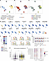
Systemic sclerosis (SSc) is characterized by fibrosis and vasculopathy affecting the skin and internal organs, leading to multiorgan dysfunction. Injury of microvascular endothelial cells (ECs) in SSc impairs blood flow and causes tissue ischemia, leading to vascular complications such as Raynaud’s, digital ulcers, and pulmonary hypertension (PH). PH in SSc presents as group 1 pulmonary arterial hypertension or as group 3 PH related to hypoxia and interstitial lung disease (ILD), both major causes of mortality. Analysis of multiome data from SSc ILD-PH lungs inferred transcription factors regulating EC phenotype, including FOSL2. Overexpression of FOSL2 in transgenic mice (Fosl2tg) leads to vascular changes mirroring human SSc-PH, such as intimal thickening and fibrosis. scRNA-Seq analysis of altered EC gene expression in Fosl2tg mice showed strong overlap with altered EC gene expression in SSc-ILD-PH. Overlapping as well as discrete EC gene expression in Sugen/hypoxia- and hypoxia-treated mice suggested that FOSL2 regulates both hypoxia-dependent and -independent pathways in Fosl2tg mice and SSc-ILD-PH. A deep learning model, ChromBPNet, inferred increased AP-1 binding at base pair resolution in SSc-ILD-PH ECs, and binding to the same motifs was found upon FOSL2 overexpression in primary vascular ECs, highlighting FOSL2’s key role in driving the pathological changes seen in SSc-ILD-PH.
Authors
Rithika Behera, Yuechen Zhou, Peter H. Gerges, Jingyu Fan, Tracy Tabib, Alyxzandria M. Gaydosik, Mengqi Huang, Jishnu Das, Elena Pachera, Amela Hukara, Ying Tang, Florian Renoux, Miranda Tai, Oliver Distler, Gabriela Kania, Stephen Y. Chan, Harinder Singh, Eleanor Valenzi, Robert Lafyatis

Abstract
Mutations in the cystic fibrosis transmembrane conductance regulator (CFTR) gene, which encodes a cAMP-activated chloride channel, cause cystic fibrosis (CF), the most common life-threatening inherited disorder among White individuals. Current CFTR correctors and potentiators, such as elexacaftor-tezacaftor-ivacaftor (ETI), only partially restore the function of the most prevalent mutant, F508del-CFTR, resulting in residual disease in people with CF. Here, we demonstrate that a mimetic peptide targeting the A-kinase–anchoring protein (AKAP) function of PI3Kγ (PI3Kγ MP), and driving localized cAMP elevation, enhances F508del-CFTR membrane localization, maximizing ETI efficacy in restoring chloride secretion. Mechanistically, PI3Kγ MP activates an AKAP-Lbc–anchored pool of PKD1, a known regulator of membrane trafficking. Consistently, PKD1 inhibition prevents PI3Kγ MP from enhancing the membrane expression of ETI-corrected F508del-CFTR. Overall, our findings reveal a regulatory pathway controlling CFTR membrane abundance via the AKAP function of PI3Kγ, which can be targeted to overcome the limitations of current CFTR modulator therapies.
Authors
Alessandra Murabito, Marco Mergiotti, Valeria Capurro, Alessia Loffreda, Mingchuan Li, Paola Peretto, Kai Ren, Andrea Raimondi, Carlo Tacchetti, Dario Diviani, Nicoletta Pedemonte, Emilio Hirsch, Alessandra Ghigo
Abstract
The molecular mechanisms responsible for the “atopic march” of allergic skin disease to allergic airway disease are incompletely understood. Secreted phospholipase A2 group X (sPLA2-X) is implicated in human asthma and modulates airway hyperresponsiveness (AHR) and inflammation in murine models of allergic asthma. We developed a complete proteolytic allergen model of dermal sensitization followed by airway challenge to mimic the “atopic march” and examined the role of sPLA2-X in regulating peripheral allergen sensitization, AHR, and airway inflammation. Pla2g10-/- mice receiving both house dust mite (HDM) peripheral sensitization and airway challenge had attenuated AHR relative to WT mice and lower airway eosinophils. Transgenic C57BL/6 PLA2G10 mice (only expressing the human sPLA2-X gene) receiving treatment with a small molecule inhibitor of sPLA2-X (ROC0929) during the dermal sensitization phase demonstrated attenuated AHR and a reduction in lung tissue dust mite-specific tissue resident memory CD4+ T cells. Thus, sPLA2-X acts as an endogenous adjuvant to facilitate allergic sensitization in the periphery, which leads to AHR and airway inflammation following inhalation of the allergen. These results provide proof of concept that inhibition of sensitization in the periphery with a sPLA2-X inhibitor modulates subsequent allergen-induced airway dysfunction.
Authors
Ryan C. Murphy, Ying Lai, Yu-Hua Chow, Matt Liu, Brian D. Hondowicz, Dowon An, Marion Pepper, William A. Altemeier, Teal S. Hallstrand
Abstract
Moderate hyperoxia (30–60% O₂) in premature infants promotes bronchial airway hyperresponsiveness (AHR) via airway smooth muscle (ASM), a key regulator of bronchoconstriction, bronchodilation, and remodeling. Understanding how O2 exposure drives long-term bronchial changes in prematurity is critical for developing therapies for airway disease across the lifespan. Premature lungs have immature antioxidant defenses, potentially due to disrupted mitochondrial dynamics, increasing susceptibility to O2-induced oxidative stress. Thus, mitochondrial homeostasis is highly relevant to ASM dysfunction and airway disease. We propose that hyperoxia in prematurity promotes mitochondrial dysfunction, and that the gasotransmitter hydrogen sulfide (H₂S) mitigates O2-induced mitochondrial damage in developing ASM. Human fetal ASM (fASM) were exposed to moderate hyperoxia to investigate the effects of exogenous H₂S donors (GYY4137, AP39) and stabilization of cystathionine β-synthase (CBS), an H₂S biosynthetic enzyme, on mitochondrial structure and function. Hyperoxia impaired fASM mitochondrial integrity, while H₂S donors in particular, or CBS stabilization attenuated adverse O2 effects on mitochondrial morphology, reactive oxygen species, respiration, calcium regulation, and contractility. These findings highlight the therapeutic potential of H₂S in the premature lung exposed to moderate hyperoxia.
Authors
Colleen M. Bartman, Michael Thompson, Samantha K. Hamrick, Niyati A. Borkar, Daniel Pfeffer-Kleemann, Preetham Ravi, Marta Schiliro, Yak Nak, Christian Vivar Ramon, Li Drake, Y. S. Prakash, Christina Pabelick
Abstract
Viral lower respiratory tract infections are common early in life and are associated with long-term development of asthma, a chronic condition defined by reversible airflow obstruction secondary to inflammation. Understanding the immunologic mechanism connecting these two pathologies observed early in life becomes imperative to guide therapeutic measures. To investigate this connection, neonatal (day of life 4-6) or adult mice were infected with human metapneumovirus (HMPV) followed by a secondary HMPV infection 6 weeks later. Mice initially infected as neonates demonstrate increased mucus production, eosinophil recruitment, airway hyperresponsiveness, and Th2 T-cell differentiation following re-challenge compared to adult mice rechallenged with HMPV. Neonatal HMPV infection led to formation of Th2 clonally expanded tissue resident memory (TRM) T cells that were absent after adult HMPV. FTY720-mediated disruption of lymphocyte circulation demonstrated TRMs contribute to pathology. Local depletion of lung CD4+ T cells and JAK2-inhibition mitigated pathology. These findings suggest TRMs uniquely generated after early life viral infection can contribute to Th2-driven asthma pathology.
Authors
Emma E. Brown, Jie Lan, Olivia B. Parks, Li Fan, Dequan Lou, Alysia McCray, Lisa Mathews, Alexander J. Wardropper, Anna Shull, Michelle L. Manni, Hēth R. Turnquist, Kong Chen, Taylor Eddens

Abstract
Mycobacterium tuberculosis (Mtb) survives within multiple macrophage populations during infection, including alveolar macrophages (AMs) and recruited inflammatory macrophages. In mice, itaconate, produced in macrophages by ACOD1-mediated decarboxylation of aconitate, has direct antimicrobial activity, modulates inflammatory cytokines, and is required for resistance to Mtb infection. The role of itaconate in human macrophages is less clear, and it is unknown whether itaconate mediates distinct effects in macrophage subtypes. Here, we investigated the role of itaconate in macrophages derived from human induced pluripotent stem cells (iPSCs), induced by either GM-CSF to resemble AMs (AM-like cells, hereafter ipAM-Ls) or M-CSF to resemble monocyte-derived macrophages (MDM-like cells, hereafter ipMDM-Ls). Both human macrophage types produced substantially less itaconate than mouse macrophages, and ipAM-Ls produced 4-fold less itaconate than ipMDM-Ls. Surprisingly, ACOD1-deficient ipAM-Ls, but not ipMDM-Ls, were permissive for Mtb growth. Moreover, itaconate functioned to dampen the Mtb-induced inflammatory response in ipMDM-Ls, but not ipAM-Ls, affecting both the type I IFN and TNF pathways. These results indicate that itaconate is involved in human macrophage responses to tuberculosis, with distinct roles in different macrophage subsets. These results also show that genetically tractable iPSC-derived macrophages are a useful model to dissect cellular host-pathogen interactions in human macrophages.
Authors
Adam S. Krebs, Tomi Lazarov, Anthony T. Reynolds, Kimberly A. Dill-McFarland, Abigail Xie, James M. Bean, Muxue Du, Olivier Levy, John A. Buglino, Aaron Zhong, Anna-Lena Neehus, Stéphanie Boisson-Dupuis, Jean-Laurent Casanova, Elouise E. Kroon, Marlo Möller, Thomas R. Hawn, Ting Zhou, Lydia W.S. Finley, Marc Antoine Jean Juste, Dan W. Fitzgerald, Frederic Geissmann, Michael S. Glickman
Abstract
Aberrant neutrophil (PMN) accumulation in the tissue induces chronic vascular diseases. Endothelial cells (ECs) regulate the access of PMNs into the tissue from the blood. However, the mechanisms that prevent PMNs from being activated and accumulating in the tissue, a hallmark of acute lung injury (ALI), remain elusive. We demonstrate that conditional deletion of Erg in ECs spontaneously alters the PMN transcriptome, which is enriched with genes that induce PMN recruitment, adhesion, activation, and 'do not eat me' signals due to impaired synthesis of the deubiquitinase, A20. Decreased A20 levels, in turn, activated the transcription factor NFκB and the secretion of MIP2α (human homolog of IL8) in ECs. EC-secreted MIP2α/IL8 engaged the CXCR2 cascade on PMNs, leading to their activation and inflammatory injury. These findings were recapitulated in the lungs and blood of PMNs from patients dying of ALI. Overexpression of the A20 gene in EC or pharmacological inhibition of CXCR2 on PMNs in iEC-Erg–/– mice rescued EC control of PMNs and tissue homeostasis, and enhanced mouse survival after pneumonia. Thus, the EC-Erg-A20 axis regulates PMN accumulation and hyperactivation in the lungs by inhibiting EC-mediated IL-8 activation of PMN-CXCR2, thereby providing a potential target for neutrophilic inflammatory vascular diseases.
Authors
Vigneshwaran Vellingiri, Vijay Avin Balaji Ragunathrao, Jagdish Chandra Joshi, Md Zahid Akhter, Mumtaz Anwar, Somenath Banerjee, Sayanti Datta, Viktor Pinneker, Steven M. Dudek, Yoshikazu Tsukasaki, Sandra Pinho, Dolly Mehta
Abstract
Alveolar macrophages (AMs) catabolize lipid-rich pulmonary surfactant to support gas exchange and have anti-inflammatory programming to limit tissue damage in response to minor challenges. GATA transcription factors (TFs) shape immune cell fates and GATA2 is expressed in a lung-specific manner in macrophages. GATA2 mutations and lung macrophage downregulation of GATA2 have been associated with chronic pulmonary pathologies in humans, but the role of GATA2 in coordinating AM function is not well defined. Using mice with myeloid-specific deletion of the GATA2 DNA binding C-terminal zinc finger domain, we show that GATA2 deficiency promotes enhanced inflammatory gene expression and metabolic dysfunction in AMs in response to type 2 stimuli. While homeostatic functions of AMs remain largely intact, GATA2 deficiency increases expression of type 2 response genes during IL-33-induced inflammation. Coincident with GATA2-dependent expression of genes in metabolic pathways, seahorse metabolic flux analysis indicates that AM metabolism is compromised in the absence of GATA2. AM GATA2-dependent gene networks are enriched for targets of TFs previously demonstrated to interact with GATA2 in other cellular contexts, including PU.1, PPARγ, and other regulators of AM function. Our data suggest that GATA2 modulates AM metabolic and transcriptomic programming to restrain responses and maintain AM identity during inflammation.
Authors
Morgan Jackson-Strong, Satarupa Ganguly, Aaron Francis, Flavia Rago, Jitendra Kanshana, Brandon A. Michalides, Lihong Teng, Omkar S. Betsur, Sonia Kruszelnicki, Karsen E. Shoger, Aaron Kim, Kay Bajpai, Amina Suleyman, Abigail Sekyere, Mika Hara, Varsha Sriram, Alok Kumar, Greg M. Delgoffe, Niranjana Natarajan, John F. Alcorn, Alison B. Kohan, Rachel A. Gottschalk
Abstract
BACKGROUND. To construct multi-trait polygenic scores (PRS) predicting chronic obstructive pulmonary disease (COPD) and exacerbations, validate their performance in diverse cohorts, and identify PRS-related proteins for potential therapeutic targeting. METHODS. PRSmix+, a multi-trait PRS framework, is used to train a composite PRS (PRSmulti) in COPDGene non-Hispanic white participants (n=6,647). Associations of PRSmulti with COPD status (GOLD 2-4 vs. GOLD 0 or ICD) and exacerbation frequency were tested in COPDGene African American (n=2,466), ECLIPSE (n=1,858), MassGeneral Brigham Biobank (n=15,152), and All of Us (n=118,566). Protein prediction models were applied to GWAS summary statistics from traits contributing to PRSmulti and were validated with proteomic data in COPDGene (n=5,173) and UK Biobank (n=5,012). RESULTS. PRSmix+ selected 7 traits for PRSmulti. In multivariable models, PRSmulti was associated with COPD status (meta-analysis random effects (RE) OR 1.58 [95% CI: 1.28-1.94]) and exacerbation frequency (meta-analysis RE beta 0.21 [95% CI: 0.11-0.31]), with higher effect sizes observed in smoking-enriched cohorts. PRSmulti outperformed traditional single-trait PRS in all tested cohorts. Using protein prediction models, we identified 73 proteins associated with the PRS that were also validated with measured protein levels in COPDGene and UK biobank. Of these proteins, 25 were linked to approved or investigational drugs. Notable targets include RAGE/sRAGE, IL1RL1, and SCARF2, all implicated in COPD pathogenesis and exacerbations. CONCLUSIONS. Multi-trait PRS improves prediction of COPD and exacerbation risk. Integration with proteomic data identifies druggable protein targets, offering a promising avenue for precision medicine in COPD management. TRIAL REGISTRATION. COPDGene: NCT00608764; ECLIPSE: NCT00292552.
Authors
Chengyue Zhang, Iain R. Konigsberg, Yixuan He, Jingzhou Zhang, Tinashe Chikowore, William B. Feldman, Xiaowei Hu, Yi Ding, Bogdan Pasaniuc, Diana Chang, Qingwen Chen, Jessica A. Lasky-Su, Julian Hecker, Martin D. Tobin, Jing Chen, Sean Kalra, Katherine A. Pratte, Hae Kyung Im, Emily S. Wan, Ani Manichaikul, Edwin K. Silverman, Russell P. Bowler, Leslie A. Lange, Victor E. Ortega, Alicia R. Martin, Michael H. Cho, Matthew R. Moll
Abstract
Systemic sclerosis (SSc) is a complex and heterogeneous condition characterized by progressive fibrosis in multiple organs. Recent studies implicate plasminogen activator inhibitor 1 (PAI-1) in the pathogenesis of SSc, and PAI-1 is considered as a potential target for therapy. Here, using single-cell and spatial RNA-seq analysis of skin biopsies from 18 healthy individuals and 22 SSc patients, we found elevated PAI-1 co-localizing to myofibroblasts with enriched extracellular matrix-associated biological processes. Treatment of SSc dermal fibroblasts with the small molecule PAI-1 inhibitor MDI-2517 reduced the expression of the profibrotic markers COL1A1 and ACTA2. To investigate the therapeutic potential of MDI-2517, we evaluated its efficacy in reducing fibrosis in a preclinical model of SSc. Treatment of mice with MDI-2517 significantly reduced both skin and lung fibrosis and was superior to treatment with either pirfenidone or mycophenolate mofetil. Additionally, MDI-2517 attenuated weight loss and significantly reduced the expression of key profibrotic markers. Compared to tiplaxtinin, another PAI-1 inhibitor previously shown to be effective in a model of SSc, MDI-2517 was found to have superior efficacy at a 10-fold lower dose. These findings highlight the role of PAI-1 in the pathogenesis of SSc, and the potential of MDI-2517 for the treatment of SSc.
Authors
Enming J. Su, Pei-Suen Tsou, Mark Warnock, Natalya Subbotina, Kris Mann, Sirapa Vichaikul, Alyssa Rosek, Lisa Leung, Xianying Xing, Enze Xing, Olesya Plazyo, Rachael Bogle, Lam C. Tsoi, Cory D. Emal, Dinesh Khanna, John Varga, Thomas H. Sisson, Johann E. Gudjonsson, Daniel A. Lawrence
No posts were found with this tag.


