
Go to The Journal of Clinical Investigation
  • About
  • Editors
  • Consulting Editors
  • For authors
  • Journal stats
  • Publication ethics
  • Publication alerts by email
  • Transfers
  • Advertising
  • Job board
  • Contact
  • Physician-Scientist Development
  • Current issue
  • Past issues
  • By specialty
    • COVID-19
    • Cardiology
    • Immunology
    • Metabolism
    • Nephrology
    • Oncology
    • Pulmonology
    • All ...
  • Videos
  • Collections
    • In-Press Preview
    • Resource and Technical Advances
    • Clinical Research and Public Health
    • Research Letters
    • Editorials
    • Perspectives
    • Physician-Scientist Development
    • Reviews
    • Top read articles

  • Current issue
  • Past issues
  • Specialties
  • In-Press Preview
  • Resource and Technical Advances
  • Clinical Research and Public Health
  • Research Letters
  • Editorials
  • Perspectives
  • Physician-Scientist Development
  • Reviews
  • Top read articles
  • About
  • Editors
  • Consulting Editors
  • For authors
  • Journal stats
  • Publication ethics
  • Publication alerts by email
  • Transfers
  • Advertising
  • Job board
  • Contact
Top
  • View PDF
  • Download citation information
  • Send a comment
  • Terms of use
  • Standard abbreviations
  • Need help? Email the journal
  • Top
  • Abstract
  • Introduction
  • Results
  • Discussion
  • Methods
  • Manuscript information
  • Supplemental material
  • Footnotes
  • References
  • Version history
  • Article usage
  • Citations to this article
Advertisement

Research ArticleImmunologyInflammationOphthalmology Open Access | 10.1172/jci.insight.204483

IL-1R2+ neutrophils define an early sterile injury-expanded subset that restrains inflammation and promotes repair

Hyun Ju Lee,1 Jung Hwa Ko,1 and Joo Youn Oh1,2

1Laboratory of Ocular Regenerative Medicine and Immunology, Biomedical Research Institute, Seoul National University Hospital, Seoul, Korea.

2Department of Ophthalmology, Seoul National University College of Medicine, Seoul, Korea.

Address for correspondence. Joo Youn Oh, Department of Ophthalmology, Seoul National University College of Medicine, 103 Daehak-ro, Jongno-gu, Seoul 03080, Korea. Phone: 82.2.2072.0027; Email: bonzoo1@snu.ac.kr.

Find articles by Lee, H. in: PubMed | Google Scholar

1Laboratory of Ocular Regenerative Medicine and Immunology, Biomedical Research Institute, Seoul National University Hospital, Seoul, Korea.

2Department of Ophthalmology, Seoul National University College of Medicine, Seoul, Korea.

Address for correspondence. Joo Youn Oh, Department of Ophthalmology, Seoul National University College of Medicine, 103 Daehak-ro, Jongno-gu, Seoul 03080, Korea. Phone: 82.2.2072.0027; Email: bonzoo1@snu.ac.kr.

Find articles by Ko, J. in: PubMed | Google Scholar

1Laboratory of Ocular Regenerative Medicine and Immunology, Biomedical Research Institute, Seoul National University Hospital, Seoul, Korea.

2Department of Ophthalmology, Seoul National University College of Medicine, Seoul, Korea.

Address for correspondence. Joo Youn Oh, Department of Ophthalmology, Seoul National University College of Medicine, 103 Daehak-ro, Jongno-gu, Seoul 03080, Korea. Phone: 82.2.2072.0027; Email: bonzoo1@snu.ac.kr.

Find articles by Oh, J. in: PubMed | Google Scholar |

Published September 22, 2026 - More info

Published in Volume 11, Issue 21 on November 9, 2026
JCI Insight. 2026;11(21):e204483. https://doi.org/10.1172/jci.insight.204483.
© 2026 Lee et al. This work is licensed under the Creative Commons Attribution 4.0 International License. To view a copy of this license, visit http://creativecommons.org/licenses/by/4.0/.
Published September 22, 2026 - Version history
Received: January 12, 2026; Accepted: September 14, 2026
View PDF
Abstract

Sterile tissue injury triggers a rapid neutrophil response that can be either pathogenic or protective, reflecting substantial functional heterogeneity of neutrophils; however, the neutrophil subsets underlying these divergent functions remain poorly defined. Here, using a well-established sterile corneal injury model, we delineate the time-dependent functional and transcriptional heterogeneity of neutrophils following sterile injury. Temporal neutrophil depletion revealed that neutrophils recruited at day 1, but not day 7, are essential for suppressing inflammation, promoting epithelial healing, and preserving nerve density. Single-cell RNA-seq uncovered substantial transcriptional heterogeneity among circulating neutrophils and their rapid reprogramming within 24 hours after injury. Specifically, Il1r2 was highly enriched in these early injury-responsive neutrophils. Functional validation demonstrated that adoptive transfer of IL-1R2+ neutrophils markedly attenuated inflammation and accelerated epithelial and nerve repair, restoring tissue integrity, whereas IL-1R2– neutrophils exacerbated inflammatory responses. Together, these findings identify IL-1R2+ neutrophils as an early protective neutrophil subset expanded by sterile injury that restrains excessive inflammation and preserves tissue homeostasis, providing mechanistic insight into injury-induced neutrophil reprogramming and highlighting a potential therapeutic target for enhancing tissue repair.

Introduction

Tissue repair following injury is a complex and dynamic process that restores homeostasis through coordinated interactions among diverse cell types, signaling molecules, and extracellular matrix components (1). In recent years, immune cells have emerged as central regulators of tissue injury and repair, extending their roles beyond debris and pathogen clearance to the active modulation of inflammation and regeneration (2). Among these, neutrophils — the most abundant immune cell population in circulation — dominate the early response to tissue damage by rapidly infiltrating injured sites in response to damage-associated molecular patterns (DAMPs) released from compromised cells and nerves (3).

Neutrophils have traditionally been considered short-lived, “suicidal” effector cells that amplify inflammation and cause collateral tissue damage. However, accumulating evidence indicates that neutrophils also exert tissue-protective and reparative functions (4–7). Indeed, we previously demonstrated that neutrophils infiltrate the cornea in 2 distinct phases following sterile injury: an immediate influx occurring within minutes, driven by the neuropeptide secretoneurin released from damaged sensory nerves, followed by a later wave peaking at 24–48 hours that is mediated by the small heat shock protein HSPB4, a major DAMP derived from injured keratocytes (8). In a subsequent study, we further showed that neutrophil depletion during the first week after injury exacerbates inflammation and impairs corneal nerve regeneration, suggesting that early-mobilized neutrophils are essential for restoring immune homeostasis and promoting tissue repair after sterile injury (9).

The contradictory roles of neutrophils in health and disease reflect their substantial phenotypic and functional heterogeneity and their capacity for plastic responses to tissue microenvironments and disease-specific cues (10, 11). Historically, neutrophil heterogeneity has been defined based on nuclear morphology, density gradients, and surface marker expression. More recently, advances in single-cell RNA-seq (scRNA-seq) have enabled high-resolution dissection of immune cell diversity, uncovering transient activation states, injury-imprinted transcriptional programs, and tissue- or injury-specific phenotypes that are obscured in bulk analyses (12–19). Despite these advances, neutrophil subset diversity remains comparatively underexplored, particularly in the context of sterile injury. Identifying the specific neutrophil subsets that promote tissue protection and regeneration could open therapeutic avenues that harness early neutrophil responses for regenerative medicine.

In the present study, we employed temporal neutrophil depletion and scRNA-seq to delineate the time-dependent functional and transcriptional heterogeneity of neutrophils following sterile corneal injury. We found that distinct neutrophil subsets are enriched on day 1 after injury and play crucial roles in repressing excessive inflammation and facilitating corneal epithelial and nerve regeneration, thereby preserving corneal transparency, whereas neutrophils recruited on day 7 are largely dispensable for these processes. Transcriptional profiling identified IL-1R2 (IL-1 receptor type 2) as a defining biomarker of these early protective neutrophils. Notably, adoptive transfer of IL-1R2+CD11b+Ly6G+ neutrophils abrogated inflammation and accelerated epithelial and nerve recovery, leading to restoration of corneal clarity, whereas IL-1R2–CD11b+Ly6G+ neutrophils aggravated inflammatory responses. Together, these findings establish IL-1R2+ neutrophils as an early injury-expanded immune subset that confers tissue protection and reveal a mechanistic link between early neutrophil reprogramming and the restoration of tissue homeostasis following sterile injury.

Results

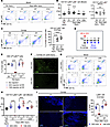
Neutrophils expand during the first week after injury, and day 1 depletion triggers a later surge. We first confirmed that neutrophils are the predominant myeloid cell population expanding during the early phase of injury. Using a mouse model of sterile corneal injury, we longitudinally tracked the numbers of CD11b+Ly6G+Ly6C– neutrophils and CD11b+Ly6G–Ly6C+ monocytes in peripheral blood over a 28-day period after injury. CD11b+Ly6G+Ly6C– neutrophils increased significantly on days 1 and 7 after injury and returned to baseline levels by day 14 (Figure 1, A and B). In contrast, the number of CD11b+Ly6G–Ly6C+ monocytes remained unchanged throughout the first week after injury (Figure 1B). Consistent with these findings, CD11b+Ly6G+ neutrophils, examined by flow cytometry, significantly increased in the cornea on both days 1 and 7 after injury (Figure 1C).
Neutrophils, but not monocytes, increase in the blood and cornea during theFigure 1

Neutrophils, but not monocytes, increase in the blood and cornea during the first week after sterile corneal injury. (A and B) Flow cytometric analysis of peripheral blood showing CD11b+Ly6G+Ly6C– neutrophils and CD11b+Ly6G–Ly6C+ monocytes at the indicated time points after sterile corneal injury. Representative flow cytometry plots (A) and quantification (B) are shown. (C) Flow cytometric analysis of the cornea showing CD11b+Ly6G+ neutrophils at the indicated time points after injury. (D) Study schematic. Anti-Ly6G Ab (1A8) or control IgG (2A3) was administered i.p. immediately after corneal injury, and peripheral blood and corneas were analyzed on days 1, 7, 14, and 28 after injury. (E) Flow cytometric analysis of peripheral blood for SSC-AhiLy6Cint cells, showing depletion of neutrophils on day 1, but not on day 7, after 1A8 injection. (F) Immunostaining of corneal flat mounts for S100, showing depletion of neutrophils on day 1 after injury and 1A8 injection on day 0. Images are representative of 3 mice. Scale bar: 100 μm. (G and H) Representative flow cytometry plots (G) and quantification (H) of CD11b+Ly6G+Ly6C– neutrophils in peripheral blood at the indicated time points after injury and treatment with 1A8 or 2A3 on day 0 (D0). (I) Immunostaining of corneal flat mounts for Ly6G and quantification of the Ly6G-stained area on days 1 and 7 after injury and treatment with 1A8 or 2A3 on day 0 (D0). Images are representative of 6 mice. Scale bar: 200 μm. Data are presented as mean ± SD, with each dot representing an individual mouse (n = 5–41 per group). **P < 0.01, ***P < 0.001, ****P < 0.0001; by 1-way ANOVA with Dunnett’s (B), Tukey’s multiple-comparison test (C), or 2-tailed Student’s t test (E, H, and I). In B, asterisks indicate significant differences compared with the day 0 (D0, No injury) group.

To investigate the functional contribution of early neutrophils, we depleted neutrophils by i.p. administration of anti-Ly6G (1A8) or isotype control Ab (2A3) immediately after injury on day 0 and then temporally analyzed neutrophil proportions in the blood and cornea over 28 days (Figure 1D). Neutrophil depletion was confirmed in the blood (identified as SSC-AhiLy6Cint cells) of both uninjured and injured mice (Figure 1E and Supplemental Figure 1, A–C; supplemental material available online with this article; https://doi.org/10.1172/jci.insight.204483DS1) and in the cornea (assessed by S100 immunostaining) of injured mice (Figure 1F) 1 day after injection (day 1), although this effect did not persist through day 7 (Figure 1E and Supplemental Figure 1, A and B). Consistent with the results in Figure 1B, circulating neutrophils increased significantly on days 1 and 7 after injury in 2A3-injected mice and gradually returned to baseline levels by day 28 (Figure 1, G and H). As expected, few neutrophils were detected in the blood on day 1 after injury in 1A8-injected mice, reflecting effective neutrophil depletion (Figure 1, G and H). Interestingly, however, a pronounced rebound in circulating neutrophils occurred in 1A8-injected mice by day 7, surpassing the levels in the 2A3-injected control group and remaining elevated through day 28 (Figure 1, G and H). A similar pattern was observed for cornea-infiltrating neutrophils. Ly6G+ neutrophil infiltration, assessed by Ly6G immunostaining, was effectively suppressed on day 1 after injury in 1A8-injected mice but markedly exceeded the levels in 2A3-injected control mice by day 7 (Figure 1I).

These results demonstrate that neutrophils are the primary myeloid responders during the early phase of corneal injury and that early neutrophil depletion on day 1 after injury leads to delayed accumulation of neutrophils both in the circulation and at the injury site.

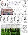
Early neutrophils reduce inflammation and promote regeneration. To examine the time-dependent functions of neutrophils, we next performed temporal depletion experiments. Based on our observation that neutrophils increase during the first week after injury (Figure 1, A–C), we selectively depleted neutrophils either on day 1 (by i.p. injection of 1A8 immediately after injury on day 0) or on day 7 (by i.p. injection of 1A8 on day 6) and monitored the cornea and peripheral blood over 28 days (Figure 2A). Isotype control Ab (2A3) was i.p. administered to control mice.
Depletion of day 1, but not day 7, neutrophils exacerbates corneal damage aFigure 2

Depletion of day 1, but not day 7, neutrophils exacerbates corneal damage after sterile injury. (A) Schematic of the study design. Anti-Ly6G Ab (1A8) was administered i.p. either immediately after sterile corneal injury (D0) or on day 6 (D6) after injury. Control IgG (2A3) was administered to control groups. Corneas were examined by slit-lamp biomicroscopy on days 1, 7, 14, and 28, and corneas and peripheral blood were collected on day 28 for analysis. (B) Representative flow cytometry plots and quantification of CD11b+Ly6G+Ly6C– neutrophils in peripheral blood on day 28 after injury. 1A8 (D0) indicates depletion of early (D1) neutrophils, whereas 1A8 (D6) indicates depletion of late (D7) neutrophils. (C) Representative corneal photographs before and after lissamine green vital dye staining on day 28 after injury, quantified in D and E. Green-stained areas indicate regions of corneal epithelial damage. (D and E) Quantification of corneal neovascularization (D) and epithelial defects (E) based on standardized clinical scoring systems. (F) qPCR analysis of proinflammatory cytokine and chemokine mRNA expression in corneas collected on day 28 after injury. Values are shown relative to those in uninjured control corneas. (G) Gross examination of corneal flat mounts showing new vessel ingrowth (top) and representative micrographs of corneal nerves stained for β-tubulin III (middle and bottom) on day 28 after injury; higher-magnification images of the central cornea are shown in the yellow boxes. Scale bars: 500 μm (middle) and 200 μm (bottom). (H) Quantification of G: β-tubulin III+ areas representing corneal nerve density. Data are presented as mean ± SD, with each circle representing an individual mouse (n = 9–16 per group). *P < 0.05, **P < 0.01, ***P < 0.001, ****P < 0.0001; by 1-way ANOVA with Tukey’s multiple-comparison test (B, D, F, and H) or 2-tailed Student’s t test (E).

Depletion of neutrophils on day 1 induced a marked increase in circulating CD11b+Ly6G+Ly6C– neutrophils on day 7, which persisted through day 28 (Figure 2B), consistent with our earlier findings (Figure 1, G and H). In contrast, neutrophil depletion on day 7 did not result in a subsequent rebound of circulating neutrophils (Figure 2B).

More prominent results were obtained in the cornea. Day 1 neutrophil depletion significantly aggravated corneal neovascularization (NV) and epithelial defects, as assessed on day 28 by slit-lamp biomicroscopy (Figure 2, C–E) and gross examination of corneal flat mounts (Figure 2G, top), whereas day 7 neutrophil depletion produced no detectable changes in clinical outcomes (Figure 2, C–E). Consistently, mRNA levels of Il1b, Il6, Cxcl2, and Mmp9 were markedly elevated in mice depleted of day 1 neutrophils but were unaffected by day 7 neutrophil depletion (Figure 2F). Similar findings were observed for corneal nerve density. Day 1 neutrophil depletion significantly aggravated nerve loss, as demonstrated by β-tubulin III immunostaining of corneal whole mounts on day 28, whereas day 7 neutrophil depletion resulted in nerve density comparable with that observed in 2A3-injected control mice (Figure 2, G and H).

Collectively, these findings indicate that day 1 neutrophils play an essential role in restraining inflammation and promoting epithelial and nerve repair after sterile corneal injury, whereas day 7 neutrophils are largely dispensable for these processes.

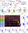
scRNA-seq reveals neutrophil heterogeneity following injury. To elucidate the molecular mechanisms underlying early neutrophil-mediated protection, we performed scRNA-seq on CD11b+Ly6G+ neutrophils isolated from the peripheral blood of mice on day 1 after sterile corneal injury (day 1 neutrophils) or from uninjured control mice (control neutrophils) using the 10x Genomics Chromium platform (Figure 3, A and B). Following quality-control filtering (200–4,000 detected genes per cell and < 15% mitochondrial gene content), the data were integrated and analyzed using the Seurat v5.0 workflow.
Single-cell RNA-seq reveals transcriptional heterogeneity of circulating neFigure 3

Single-cell RNA-seq reveals transcriptional heterogeneity of circulating neutrophils under steady-state and postinjury conditions. (A) Schematic of the experimental design. CD11b+Ly6G+ neutrophils were isolated by FACS from the peripheral blood of uninjured control mice (no injury) and mice on day 1 after sterile corneal injury (D1 postinjury) and processed for scRNA-seq. (B) Sorting strategy. Live FSC/SSC granulocytes were gated for CD11b+Ly6G+ neutrophils, and the morphology of sorted cells was confirmed by Wright-Giemsa staining. (C) Integrated UMAP of control and D1 postinjury neutrophils, colored by condition, showing substantial overlap between the 2 conditions. (D) UMAP and t-SNE projections of each condition, colored by cluster, showing the 6 shared neutrophil clusters (C0–C5). (E) Heatmap of the top differentially expressed genes across the 6 neutrophil clusters (C0–C5). (F and G) Feature plots showing the expression of representative cluster markers — Nampt and Wfdc17 (F), and Ptpn22, Rsad2, Ngp, and Pf4 (G) — projected onto the integrated UMAP.

Following integration, control and day 1 neutrophils overlapped substantially in the shared embedding, as visualized by Uniform Manifold Approximation and Projection (UMAP) and t-distributed stochastic neighbor embedding (t-SNE) dimensionality reduction (Figure 3, C and D). Unbiased clustering (resolution = 0.5) of the integrated dataset identified shared neutrophil clusters present across both conditions (Figure 3, D and E, and Supplemental Figure 2A). Of a total of 8 clusters (C0–C7; Supplemental Figure 2A), 2 clusters (C6 and C7) were excluded as low-abundance contaminating cell types: C6 as monocytes/macrophages, characterized by Ms4a4a, Ms4a6d, Ms4a8a, and Cfh, and C7 as a lymphocyte population comprising both T and B cells, characterized by Trbc1/2, Themis, Il7r, Ccr7, Pax5, and Ebf1 (Supplemental Table 1). Among the 6 major neutrophil clusters (C0–C5), injury shifted the relative proportions of the shared clusters: C2 and C5 expanded from 23.8% to 27.4% and from 0.3% to 3.1%, respectively, whereas C3 and C4 contracted from 8.7% to 7.0% and from 7.3% to 2.7%, respectively (Figure 3D and Supplemental Figure 2B). Notably, every shared cluster contained cells from both conditions, indicating that injury shifted the proportions of preexisting neutrophil states rather than generating an injury-exclusive population.

We further characterized neutrophil heterogeneity by examining the 6 major clusters (C0–C5), each defined by a distinct transcriptional signature (Figure 3, E–G, and Supplemental Table 1). Cluster C0 was enriched for metabolic and homeostatic genes, including Nampt (NAD biosynthesis), Msra (oxidative-stress defense), and Lrrk2, consistent with a metabolically active, baseline state. Cluster C1 was characterized by granule and alarmin genes such as S100a8, Wfdc17, Stfa2l1, and Lrg1. Cluster C2 expressed NF-κB regulators and stress-responsive immediate-early genes, including Tnfaip3 (encoding the NF-κB regulator A20), Atf3, Ptgs2, Ppp1r15a, Ccrl2 (encoding an atypical receptor that forms heterodimers with CXCR2 on neutrophils) (20), and Ptpn22 (a regulator of Fcγ receptor signaling) (21), indicative of an inflammation-activated state. Cluster C3 displayed a prominent IFN-stimulated gene (ISG) signature, including Ifit1, Ifit3b, Isg15, Rsad2, Usp18, and Oasl2. Cluster C4 was marked by mature neutrophil granule genes such as Ngp (encoding neutrophilic granule protein), Ly6g, Serpinb1a, and Stfa2/3. Cluster C5 expressed platelet-associated genes, including Ppbp, Pf4, and Gng11; however, it retained robust expression of canonical neutrophil markers, including Cxcr2, the neutrophil granule protein Mmp9, and the G-CSF receptor Csf3r, particularly among day 1 cells (Supplemental Figure 2C). C5 was therefore classified as a neutrophil population corresponding to neutrophil-platelet aggregates (22) and was retained for analysis.

Collectively, these data reveal extensive transcriptional heterogeneity among circulating neutrophils under both steady-state and injury conditions and indicate that sterile injury to the cornea rapidly reprograms the circulating neutrophil transcriptome within 24 hours, shifting the composition of preexisting neutrophil states rather than generating an entirely discrete injury-specific population.

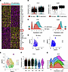
Il1r2 is enriched in early injury-responsive neutrophils. We next sought to identify surface markers defining the neutrophil subset responsible for corneal protection. To this end, we performed differential gene expression analysis comparing day 1 neutrophils with control neutrophils. Among the top 25 differentially expressed genes (DEGs) that were upregulated in day 1 neutrophils, 2 encoded surface proteins: Csf3r and Il1r2 (Figure 4A).
Il1r2 is enriched in early injury-responsive neutrophils.Figure 4

Il1r2 is enriched in early injury-responsive neutrophils. (A) Heatmap of the top differentially expressed genes (DEGs) between control (no injury) and D1 postinjury neutrophils. Csf3r and Il1r2, the 2 genes encoding surface proteins among the top 25 DEGs, are highlighted (red boxes). (B and C) Violin plots (B) and ridge plots (C) comparing Il1r2 and Csf3r expression between control and D1 postinjury neutrophils. (D) Feature plots of Il1r2 expression projected onto the integrated UMAP for the No injury and D1 postinjury conditions. (E) UMAP of the 6 neutrophil clusters (C0–C5) used for cluster-level analysis. (F and G) Violin plots (F) and ridge plots (G) of Il1r2 expression across the 6 neutrophil clusters (C0–C5), showing high expression in C2 and C5 and low expression in C4.

Csf3r was excluded as a candidate marker for the injury-responsive subset because it was constitutively and robustly expressed across all neutrophil clusters in both control and day 1 postinjury conditions (Figure 4, B and C, and Supplemental Figure 2C). This broad expression pattern of Csf3r is consistent with the well-defined role of CSF3R (granulocyte colony-stimulating factor receptor) in neutrophil biology, encompassing granulopoiesis, maturation, survival, mobilization, and effector functions.

In contrast, Il1r2 expression was significantly enriched in day 1 neutrophils relative to control neutrophils (Figure 4, B–D). Among clusters, Il1r2 expression was high in clusters C2 and C5, which expanded after injury, and low in C4, which shrank after injury (Figure 4, E–G, and Supplemental Figure 2, B and D). The encoded protein, IL-1R2, acts as both a decoy receptor and a scavenger for IL-1 ligands (23, 24), thereby attenuating IL-1–driven inflammatory responses (25, 26). This functional property suggests a potential antiinflammatory role for IL-1R2–expressing neutrophils in the context of sterile corneal injury. Notably, cluster C2 coexpressed both proinflammatory genes (Ptgs2, Ccrl2) and negative-feedback regulators of inflammation (Tnfaip3 encoding A20, Atf3) (Figure 3E and Supplemental Table 1), suggesting an activated yet self-regulating state in which Il1r2 induction may contribute to restraining excessive inflammation.

Together, these findings led us to hypothesize that IL-1R2+ neutrophils constitute an injury-expanded subset that contributes to corneal protection following sterile injury.

IL-1R2+ neutrophils increase in the blood and cornea after injury. To validate the scRNA-seq findings, we quantified IL-1R2+CD11b+Ly6G+ cells by flow cytometry using a fluorescence-minus-one–based (FMO-based) gating strategy (Figure 5A). We confirmed that the frequency of IL-1R2+CD11b+Ly6G+ neutrophils in peripheral blood was significantly increased on day 1 after injury compared with the no-injury condition (24.3% ± 6.5% versus 42.4% ± 10.2%; Figure 5B). By day 7 after injury, the proportion of IL-1R2+CD11b+Ly6G+ neutrophils returned to a level (26.1% ± 11.7%) comparable with that of the no-injury condition and significantly lower than the day 1 peak (Figure 5B). These findings indicate that the expansion of circulating IL-1R2+ neutrophils is an early and transient response to sterile corneal injury.

IL-1R2+ neutrophils increase in the blood and cornea after sterile cornealFigure 5

IL-1R2+ neutrophils increase in the blood and cornea after sterile corneal injury. (A) Fluorescence-minus-one (FMO) gating strategy for IL-1R2+CD11b+Ly6G+ neutrophils. FMO controls lacking anti-CD11b, anti-Ly6G, or anti-IL-1R2 were used to define positivity thresholds. Representative dot plots and IL-1R2 histograms are shown for the no-injury and D1 postinjury conditions, quantified in B. (B) Flow cytometric analysis of peripheral blood for IL-1R2+CD11b+Ly6G+ neutrophils, showing a representative histogram and quantification in the no-injury condition and on days 1 and 7 after injury. (C) Representative histogram and quantification of IL-1R2+CD11b+Ly6G+ cells in the cornea, assessed by flow cytometry, in the no-injury condition and on day 1 after injury. (D) Immunofluorescence staining of corneal flat mounts for Ly6G (blue) and IL-1R2 (green) in the no-injury condition and on D1 after injury; boxed regions (pink) are shown at higher magnification. Images are representative of 5 mice. Scale bars: 100 μm (top) and 50 μm (middle and bottom). Data are presented as mean ± SD, with each dot representing an individual mouse (n = 5–17 per group). ***P < 0.001, ****P < 0.0001; by 1-way ANOVA with Tukey’s multiple-comparison test (B) or 2-tailed Student’s t test (C).

We next examined whether IL-1R2+ neutrophils infiltrate the injured cornea using both flow cytometry and immunostaining. IL-1R2+CD11b+Ly6G+ cells were undetectable in uninjured corneas but were significantly increased on day 1 after injury, as assessed by flow cytometry of corneal tissue (Figure 5C). This finding was corroborated by immunofluorescence staining of corneal flat-mounts, which revealed IL-1R2+ cells colocalizing with the neutrophil marker Ly6G in the day 1 cornea, whereas neither Ly6G+ nor IL-1R2+ signals were detected in uninjured corneas (Figure 5D). Together, these results demonstrate that IL-1R2+ neutrophils expand in the circulation following sterile injury and infiltrate the injured cornea during the early phase.

IL-1R2+ neutrophils suppress inflammation and promote tissue recovery. To directly test whether IL-1R2+ neutrophils confer corneal protection, we performed adoptive transfer experiments (Figure 6A). IL-1R2+CD11b+Ly6G+ or IL-1R2–CD11b+Ly6G+ neutrophils were isolated from the peripheral blood of donor mice on day 1 after injury by FACS, and each population was transferred into separate cohorts of recipient mice via subconjunctival injection immediately after injury (1 × 10⁵ cells in 7 μL of HBSS). Control mice received an equivalent volume of vehicle (HBSS). Wright-Giemsa staining revealed no discernible morphological differences between the 2 neutrophil populations (Figure 6A). Corneas were analyzed on day 7 after injury and transfer.
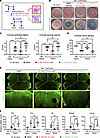
Adoptive transfer of IL-1R2+ neutrophils suppresses inflammation and promotFigure 6

Adoptive transfer of IL-1R2+ neutrophils suppresses inflammation and promotes corneal repair. (A) Schematic of the adoptive transfer experiment. IL-1R2+CD11b+Ly6G+ or IL-1R2–CD11b+Ly6G+ neutrophils were sorted from the peripheral blood of donor mice on day 1 after injury and transferred subconjunctivally into recipient mice immediately after injury. Recipient corneas were analyzed on day 7. Sorted-cell morphology was confirmed by Wright-Giemsa staining (IL-1R2+CD11b+Ly6G+ cells in the red box and IL-1R2–CD11b+Ly6G+ in the green box). (B) Representative corneal photographs before and after lissamine green vital dye staining on day 7 after injury in mice receiving vehicle (HBSS), IL-1R2+, or IL-1R2– neutrophils, compared with uninjured controls. (C) Clinical scores of corneal epithelial defects and stromal opacity. (D) Quantification of corneal nerve density (β-tubulin III+ area). (E) Immunofluorescence staining of corneal flat mounts for β-tubulin III on day 7 after injury, quantified in D; yellow-boxed regions are shown at higher magnification. Scale bars: 500 μm (top) and 200 μm (bottom). (F) qPCR analysis of Il1b, Il1a, Il6, Mmp9, and Cxcl2 mRNA levels in corneas on day 7 after injury, shown relative to uninjured controls. Data are presented as mean ± SD, with each dot representing an individual mouse (n = 10 per group in C; n = 5 per group in D and F). *P < 0.05, **P < 0.01, ***P < 0.001, ****P < 0.0001; by 1-way ANOVA with Tukey’s multiple-comparison test.

Adoptive transfer of IL-1R2+CD11b+Ly6G+ neutrophils significantly reduced corneal epithelial defects and stromal opacity compared with vehicle-treated controls, as assessed by slit-lamp biomicroscopy (Figure 6, B and C). In contrast, transfer of IL-1R2–CD11b+Ly6G+ neutrophils tended to aggravate epithelial damage and stromal opacity relative to vehicle controls (Figure 6, B and C). A similar pattern was observed for corneal nerve recovery. IL-1R2+CD11b+Ly6G+ neutrophils markedly preserved corneal nerve density compared with vehicle-treated controls, as demonstrated by β-tubulin III immunostaining of corneal flat-mounts, whereas IL-1R2–CD11b+Ly6G+ neutrophils did not promote nerve recovery and tended to reduce nerve density further (Figure 6, D and E).

Consistent with these clinical outcomes, IL-1R2+CD11b+Ly6G+ neutrophils markedly suppressed corneal inflammation, reducing the mRNA levels of Il1b, Il1a, Il6, Mmp9, and Cxcl2 to near-baseline values (Figure 6F). Conversely, transfer of IL-1R2–CD11b+Ly6G+ neutrophils increased the expression of Il1b, Mmp9, and Cxcl2 compared with vehicle treatment (Figure 6F). Notably, both Il1a and Il1b were concurrently reduced by IL-1R2+CD11b+Ly6G+ neutrophils.

Collectively, these results demonstrate that IL-1R2+ neutrophils represent an injury-expanded subset with potent antiinflammatory and tissue-protective properties that are essential for restoring tissue homeostasis and preserving corneal transparency following sterile injury, whereas IL-1R2– neutrophils lack these protective functions and instead exacerbate inflammation.
Discussion

Neutrophils were previously regarded as a terminally differentiated and largely homogeneous immune cell population with limited transcriptional activity. However, accumulating evidence over the past decade has established that neutrophils are highly heterogeneous, exhibiting substantial phenotypic diversity and functional plasticity. These cells can rapidly adapt to microenvironmental cues and exert a wide range of context-dependent functions, ranging from tissue-protective to pathogenic roles (10, 12, 14, 27, 28). In our model of sterile corneal injury, neutrophils expanded in the circulation and accumulated in the cornea during the first week after injury, returning to baseline levels by 2 weeks. Importantly, neutrophils present on day 1 after injury were essential for suppressing inflammation, supporting epithelial repair, and preserving corneal nerve density, whereas neutrophils recruited on day 7 were dispensable for these functions. This functional divergence was associated with rapid transcriptomic reprogramming of the early day 1 neutrophil population, as revealed by scRNA-seq. Specifically, IL-1R2+ neutrophils within the day 1 compartment were identified as the injury-expanded subset mediating antiinflammatory and tissue-protective effects, whereas IL-1R2– neutrophils exacerbated inflammation. Collectively, our findings demonstrate marked functional, transcriptional, and phenotypic heterogeneity among neutrophils, both at steady state and during the early phase of injury.

Recent advances in single-cell profiling techniques have enabled high-resolution dissection of neutrophil heterogeneity and facilitated the identification of transcriptional signatures, biomarkers, and potential therapeutic targets (10, 11). In line with our findings, Kwok et al. reported enrichment of IL1R2+ immature neutrophils in the blood of patients with sepsis and poor clinical outcomes using combined scRNA-seq and scATAC-seq, linking this population to an immunosuppressive neutrophil state (15). Similarly, Ding et al. observed increased Il1r2 expression in lung-infiltrating neutrophils in a mouse model of LPS-induced acute lung injury by scRNA-seq and demonstrated that neutrophil-specific Il1r2 overexpression attenuated lung inflammation, supporting a regulatory role for neutrophil-derived IL-1R2 in immune responses (29). Moreover, hydrocortisone, an antiinflammatory agent, has been shown to upregulate IL-1R2 expression on peripheral blood neutrophils, whereas LPS, a proinflammatory mediator, induces shedding of IL-1R2 from the neutrophil surface (30). Given that IL-1R2, a decoy receptor and scavenger for IL-1α and IL-1β (23, 24), is predominantly expressed by neutrophils (30–32), IL-1R2+ neutrophils are well positioned to limit excessive inflammation by negatively regulating IL-1–mediated immune responses. Accordingly, IL-1R2+ neutrophils may serve both as a biomarker of immunoregulatory neutrophil subsets and as a promising target for therapeutic exploitation.

Mechanistically, IL-1R2 functions as a decoy receptor that competes with the primary signaling receptor IL-1R1 for IL-1 binding, thereby preventing activation of multiple downstream proinflammatory pathways, including NF-κB, c-Jun N-terminal kinase, p38 and other mitogen-activated protein kinases, extracellular signal-regulated kinases, and members of the IFN-regulatory factor signaling cascades (33). In addition, IL-1R2 rapidly scavenges free IL-1 in the extracellular milieu, further limiting IL-1 bioavailability (26). Given the central role of IL-1 signaling in inflammation and immune responses (34), IL-1R2 has emerged as a critical regulatory molecule and a promising therapeutic target in diverse pathological contexts, including sepsis (32, 35), autoimmune diseases (24, 36), cancer (26, 37), and sterile injury (38, 39).

In the present study, we observed that IL-1R2 expression was robustly upregulated in neutrophils following sterile corneal injury. Functionally, adoptive transfer of IL-1R2+ neutrophils suppressed inflammation and promoted corneal epithelial and nerve regeneration, whereas IL-1R2– neutrophils failed to confer such benefits and instead exacerbated inflammatory responses. Mechanistically, IL-1R2+ neutrophils reduced the corneal mRNA levels of both Il1a and Il1b, together with the downstream mediators Il6, Mmp9, and Cxcl2. Although IL-1R2 acts posttranslationally as a decoy receptor rather than as a transcriptional regulator, this concurrent reduction of Il1a and Il1b transcripts is consistent with disruption of the IL-1 autocrine/paracrine feedback loop, in which IL-1 signaling through IL-1R1 normally sustains NF-κB–dependent transcription of Il1a, Il1b, and downstream proinflammatory genes (25, 26, 33–36). By sequestering IL-1, IL-1R2+ neutrophils interrupt this circuit. These findings identify IL-1R2 as a key molecular determinant underlying the antiinflammatory and tissue-protective functions of neutrophils in sterile injury.

Consistent with our observations, Mattos et al. demonstrated that neutrophils upregulated Il1r2 following acetaminophen-induced sterile liver injury and that adoptive transfer of these neutrophils reduced hepatic inflammation and accelerated tissue repair (38). Furthermore, Zheng et al. showed that sterile inflammation is regulated in a cell type–specific manner that largely depends on IL-1R2 expression (39). Cells expressing high levels of IL-1R2 release little active IL-1α, a potent DAMP (40), thereby limiting inflammation. In contrast, IL-1R2–deficient cells fully activate IL-1α, resulting in severe inflammatory responses Although it remains unclear whether IL-1R2 is expressed by corneal resident cells, both IL-1α and IL-1β are well-established mediators of the corneal response to sterile injury (41–43). Thus, these findings, along with our results, support the notion that IL-1R2 and IL-1R2+ neutrophils play a central role in maintaining and restoring corneal homeostasis by constraining excessive IL-1 signaling following sterile injury.

The clinical relevance of these findings is underscored by the availability of IL-1–targeting agents suitable for clinical application. Indeed, topical IL-1 receptor antagonists have already been evaluated in ocular surface inflammatory disease (44–46). However, whether IL-1 blockade alone accounts for the protective functions of early-infiltrating neutrophils, including IL-1R2+ neutrophils, remains unclear. In the present study, we focused on surface markers distinguishing early expanding neutrophils from steady-state neutrophils primarily for practical reasons related to cell isolation. A comprehensive pathway-level analysis of the injury-responsive neutrophil transcriptome, together with functional validation of additional candidate subsets, would provide further insight into the diverse protective neutrophil populations beyond IL-1R2+ neutrophils. In addition, although the present study investigated the early innate phase, mononuclear phagocytes and T cells become the predominant immune cell types in the cornea at later stages of injury, contributing to inflammation resolution and tissue reconstruction. Investigation of these later-infiltrating cells will offer a more comprehensive understanding of the homeostatic tissue-repair process.

In conclusion, our study demonstrates that neutrophils expanding during the early phase of sterile corneal injury exhibit pronounced functional and transcriptional heterogeneity, providing a framework for understanding the time-dependent protective roles of neutrophils during sterile inflammation. Within this early population, we identify distinct neutrophil subsets, among which IL-1R2+ neutrophils represent an injury-expanded subset that mediates antiinflammatory and tissue-protective responses. Collectively, these findings establish IL-1R2 as a biomarker of a tissue-protective neutrophil subset and highlight IL-1R2+ neutrophils and their associated molecular pathways as promising therapeutic targets for sterile injury in the cornea. Given the central role of neutrophils in a wide range of pathological conditions characterized by sterile inflammation, including trauma, surgery, ischemia-reperfusion injury, autoimmune diseases, neurodegenerative disorders, and aging-related tissue dysfunction, our findings extend beyond the cornea and have broad implications for regenerative medicine and immunomodulatory therapies.

Methods

Sex as a biological variable. Our study examined male and female animals, and similar findings are reported for both sexes.

Animal model. BALB/c mice (6–8 weeks old) were purchased from KOATECH and maintained under specific pathogen–free conditions with a 12-hour light/dark cycle.

For induction of sterile corneal injury, mice were anesthetized with an intramuscular injection of zolazepam-tiletamine (Zoletil, Virbac). After topical application of 0.5% proparacaine hydrochloride (Hanmi Pharm.), absolute ethanol was applied to the whole cornea for 15 seconds, followed by thorough rinsing with 2 mL sterile HBSS. The whole corneal epithelium, including the limbal area, was then mechanically removed with a #11 surgical blade under an operating microscope. This procedure induces a reproducible, severe, noninfectious corneal injury with consequent inflammatory NV and stromal opacity, as demonstrated in our previous studies (8, 9, 47). The day of injury was designated as day 0.

Neutrophil depletion. Neutrophils were depleted by i.p. injection of 100 μg anti–mouse Ly6G mAb (clone 1A8, BP0075-1, BioXCell) either immediately after corneal injury on day 0 (day 1 depletion) or on day 6 after injury (day 7 depletion). Control mice received an equivalent dose of isotype-matched control antibody (clone 2A3, BP0089, BioXCell). Because the anti-Ly6G clone used for depletion (1A8) is identical to the clone used for Ly6G detection by flow cytometry, depletion efficiency was verified using clone-independent approaches to exclude epitope masking. Specifically, circulating neutrophils were quantified by flow cytometry as SSC-AhiLy6Cint granulocytes, and corneal neutrophil infiltration was assessed by immunostaining with an anti-S100A9 antibody (clone D3U8M, 73425, Cell Signaling Technology; 1:1,000).

Clinical assessment of corneal injury. Clinical manifestations of corneal damage were evaluated in 3 aspects using a slit-lamp biomicroscope (Haag-Streit): corneal NV, corneal epithelial defects, and corneal stromal opacity.

Corneal NV was assessed by measuring the extent of new vessel ingrowth from the limbus toward the center and scored on a 0–3 scale for each corneal quadrant (scale 0: no new vessels; scale 1: new vessels at corneal limbus; scale 2: new vessels spanning limbus and approaching corneal center; scale 3: new vessels spanning center). The overall corneal NV score was then determined by summing the scales of the 4 quadrants, resulting in a total score of 0–12 (48, 49).

For assessment of epithelial defects, corneas were visualized with 1% lissamine green vital dye staining (Sigma-Aldrich), and the extent of the stained area was graded according to a standardized clinical grading system (score 0: no staining; score 1: less than one-third; score 2: less than two-thirds; score 3: more than two-thirds staining of cornea) (50).

Corneal stromal opacity was evaluated based on the opacification scoring system (score 0: no opacity; score 1: slight superficial opacity, iris details visible; score 2: moderate stromal opacity, pupillary margin visible, iris details not visible; score 3: significant stromal opacity, pupillary margin not visible; score 4: completely opaque, pupil and anterior chamber not visible) (51).

All clinical scoring for corneal NV, epithelial defects, and stromal opacity was performed independently by 2 investigators who were blinded to the treatment conditions.

Flow cytometry. Peripheral blood was collected via cardiac puncture into EDTA-containing tubes. After rbc lysis using RBC lysis buffer (Invitrogen), the remaining cells were washed with PBS and stained with the following fluorochrome-conjugated Abs: anti-CD11b (clone M1/70, 25-0112-82, Invitrogen), anti-Ly6G (clone 1A8, 17-9668-82, Invitrogen), anti-Ly6C (clone HK1.4, 45-5932-82, Invitrogen), or anti-IL-1R2 (bs-2595R, Bioss). For flow cytometric analysis of corneal cells, corneas were excised and cut into 6–8 pieces, before being digested with 5 mg/mL collagenase type I (1,325 U; Worthington Biochemical Corporation) at 37°C for 30 minutes. The tissue was dissociated by gentle pipetting, filtered through a 70 μm cell strainer to obtain single-cell suspensions, washed, and stained as above. For quantification of IL-1R2+CD11b+Ly6G+ neutrophils, FMO controls — staining panels lacking anti-IL-1R2, anti-CD11b, or anti-Ly6G — were used to define positivity thresholds for each marker and to account for spectral spillover from the other channels. The stained cells were assessed for fluorescence using an Aurora Flow Cytometer (CYTEK Biosciences). Data were analyzed using the FlowJo software program (Tree Star). Neutrophils were defined as CD11b+Ly6G+Ly6C– cells, and monocytes were defined as CD11b+Ly6G–Ly6C+ cells. For clone-independent confirmation of neutrophil depletion, neutrophils were alternatively identified as SSC-AhiLy6Cint cells.

IHC of corneal flat mounts. Corneas were excised and fixed in acetone/methanol (1:1, v/v) at room temperature (RT) for 10 minutes. After blocking with 2% bovine serum albumin for 1 hour, tissues were incubated overnight at 4°C with the following Abs: anti-β III tubulin (AB15708A4, MilliporeSigma), anti-Ly6G (562737, BD Horizon), anti–IL-1R2 (bs-2595R, Bioss), or anti-S100A9 (clone D3U8M, 73425, Cell Signaling Technology). After PBS washing, corneas were transferred onto glass slides, incised radially into 4 quadrants, and mounted flat using Dako Faramount Aqueous Mounting Medium (Agilent).

Stained corneal whole-mounts were examined and imaged using fluorescence confocal microscopy (BX53-DP74, Olympus). Quantification of immunofluorescence signals was performed using ImageJ software (NIH). After background subtraction and thresholding, the stained area was quantified as the percentage of the total corneal area positive for β-tubulin III or Ly6G in whole-mount images by 2 independent investigators.

Cell sorting and scRNA-seq. CD11b+Ly6G+ neutrophils were isolated from the peripheral blood of uninjured control mice or mice on day 1 after corneal injury by FACS using a FACSAria III cell sorter (BD Biosciences) and were gated sequentially on FSC/SSC granulocytes and CD11b+Ly6G+ cells. Post-sort cell viability was greater than 85% as determined using the Countess II Automated Cell Counter (Thermo Fisher Scientific). The control and day 1 samples were prepared on separate days (before injury and 24 hours after injury) with independent library preparation and sequencing runs. Single-cell suspensions were loaded onto the 10x Genomics Chromium platform (10x Genomics) for library preparation using the Chromium Single Cell 3′ Reagent Kit. Libraries were then sequenced on a NovaSeq 6000 system (Illumina).

Raw sequencing reads were processed using Cell Ranger software (10x Genomics). In total, 4,000 single cells per group were sequenced at a mean depth of 41,914 reads per cell for the day 1 neutrophil group and 44,486 reads per cell for the control group, as assessed by the Cell Ranger count pipeline. The median gene and unique molecular identifier counts per cell were 1,539 and 4,276, respectively, in day 1 neutrophils, and 1,490 and 4,385, respectively, in control cells. The total numbers of genes detected were 21,097 in day 1 neutrophils and 20,625 in control cells.

Quality control, normalization, dimensionality reduction, and clustering were performed using Seurat (version 5.0) in R. Because the 2 samples were prepared on separate days, the datasets were integrated, not merged, to correct for batch effects. Quality-control thresholds were adjusted to accommodate the characteristically low RNA content of neutrophils: cells with fewer than 200 (nFeature_RNA < 200) or more than 4,000 detected genes, or with mitochondrial gene content exceeding 15%, were excluded. Following log-normalization, 2,000 highly variable features were identified using the variance-stabilizing transformation (VST) method, and the data were scaled and subjected to principal component analysis (30 principal components). The 2 datasets were then integrated using the reciprocal PCA (RPCA) method implemented in Seurat v5, in which the RNA assay counts were split by sample into separate layers prior to integration and rejoined (JoinLayers) prior to differential expression analysis. Shared nearest-neighbor graph construction (FindNeighbors, dims = 1:30) and clustering (FindClusters, resolution = 0.5) were then performed on the integrated embedding, and UMAP and t-SNE projections were generated. Clustering and dimensionality-reduction embeddings (UMAP and t-SNE) were computed on the RPCA-integrated reduction, whereas all gene-expression visualizations were generated from the RNA assay using log-normalized expression values. DEGs were identified using standard Seurat workflows.

Adoptive transfer of neutrophil subsets. Peripheral blood was collected from BALB/c mice on day 1 after corneal injury, and IL-1R2+CD11b+Ly6G+ or IL-1R2−CD11b+Ly6G+ neutrophils were sorted by FACS. Postsort cell viability was greater than 80%. Sorted cells (1 × 10⁵ cells in 7 μL of HBSS) were injected subconjunctivally into recipient mice immediately after corneal injury. Control mice received the same volume of HBSS alone. Corneas were analyzed on day 7 after adoptive transfer.

Wright-Giemsa staining. Freshly isolated IL-1R2+CD11b+Ly6G+ and IL-1R2−CD11b+Ly6G+ neutrophils were cytocentrifuged onto glass slides at 254 × g using a Shandon Cytospin 4 centrifuge (Thermo Fisher Scientific). Slides were air-dried and stained with Wright-Giemsa solution (Sigma-Aldrich) for 45 minutes. After rinsing with deionized water and air drying, the cells were examined under a bright-field light microscope (BX53, Olympus Corporation), and representative images were captured for morphological assessment.

qPCR. For RNA extraction, corneal tissues were homogenized on ice in RNA isolation reagent (RNA Bee, Tel-Test Inc.) using an ultrasound sonicator (Ultrasonic Processor, Cole Parmer Instruments). Total RNA was purified using the RNeasy Mini kit (Qiagen) and reverse-transcribed into cDNA using the High-Capacity cDNA Reverse Transcription Kit (Applied Biosystems). qPCR was performed on an ABI 7500 Real Time PCR System (Applied Biosystems) using gene-specific TaqMan probes (TaqMan Gene Expression Assay kits, Applied Biosystems) and TaqMan Universal PCR Master Mix (Applied Biosystems). Gene expression levels were normalized to Gapdh and calculated using the 2−ΔΔCt method. Results are presented as fold changes in mRNA expression relative to uninjured control samples.

Statistics. Statistical analyses were performed using GraphPad Prism software (version 10.6.1, GraphPad Software). Data are presented as mean ± SD. Comparisons among multiple groups were conducted using 1-way ANOVA with Dunnett’s or Tukey’s multiple-comparison test. Two-group comparisons were performed using 2-tailed Student’s t test. A P value less than 0.05 was considered significant.

Study approval. All animal experiments were approved by the IACUC of Seoul National University Hospital Biomedical Research Institute and performed in accordance with the Association for Research in Vision and Ophthalmology (ARVO) Statement for the Use of Animals in Ophthalmic and Vision Research.

Data availability. Values for all data points shown in graphs and values behind any reported means are provided in the Supporting Data Values file. The scRNA-seq datasets generated in this study have been deposited in the Gene Expression Omnibus (GEO) under accession no. GSE338556 and are publicly available.

Manuscript information

Author contributions. HJL conducted experiments, acquired and analyzed data, and wrote the manuscript. JHK conducted experiments and analyzed data. JYO designed the study, acquired funding, conducted experiments, acquired and analyzed data, and wrote the manuscript.

AI disclosure. The authors declare that no AI tools were used in the original research for or writing of this article.

Conflict of interest. The authors have declared that no conflict of interest exists.

Funding. This work was supported by the following:

  • National Research Foundation of Korea (NRF) grant funded by the Korean Ministry of Science and ICT, No. RS-2025-00514435 (JYI).
Supplemental material

View Supplemental data

Footnotes

Copyright: © 2026, Lee et al. This is an open access article published under the terms of the Creative Commons Attribution 4.0 International License.

Reference information: JCI Insight. 2026;11(21):e204483.https://doi.org/10.1172/jci.insight.204483.

Address for correspondence. Joo Youn Oh, Department of Ophthalmology, Seoul National University College of Medicine, 103 Daehak-ro, Jongno-gu, Seoul 03080, Korea. Phone: 82.2.2072.0027; Email: bonzoo1@snu.ac.kr.

References
  1. Forbes SJ, Rosenthal N. Preparing the ground for tissue regeneration: from mechanism to therapy. Nat Med. 2014;20(8):857–869.
    View this article via: CrossRef PubMed Google Scholar
  2. Julier Z, et al. Promoting tissue regeneration by modulating the immune system. Acta Biomater. 2017;53:13–28.
    View this article via: CrossRef PubMed Google Scholar
  3. Pittman K, Kubes P. Damage-associated molecular patterns control neutrophil recruitment. J Innate Immun. 2013;5(4):315–323.
    View this article via: CrossRef PubMed Google Scholar
  4. Herro R, Grimes HL. The diverse roles of neutrophils from protection to pathogenesis. Nat Immunol. 2024;25(12):2209–2219.
    View this article via: CrossRef PubMed Google Scholar
  5. Peiseler M, Kubes P. More friend than foe: the emerging role of neutrophils in tissue repair. J Clin Invest. 2019;129(7):2629–2639.
    View this article via: JCI CrossRef PubMed Google Scholar
  6. Rizo-Téllez SA, Filep JG. Beyond host defense and tissue injury: the emerging role of neutrophils in tissue repair. Am J Physiol Cell Physiol. 2024;326(3):C661–C683.
    View this article via: CrossRef PubMed Google Scholar
  7. Wang J. Neutrophils in tissue injury and repair. Cell Tissue Res. 2018;371(3):531–539.
    View this article via: CrossRef PubMed Google Scholar
  8. Oh JY, et al. Identification of the HSPB4/TLR2/NF-κB axis in macrophage as a therapeutic target for sterile inflammation of the cornea. EMBO Mol Med. 2012;4(5):435–448.
    View this article via: CrossRef PubMed Google Scholar
  9. Lee HJ, et al. Myeloid cells protect corneal nerves against sterile injury through negative-feedback regulation of TLR2-IL-6 axis. J Neuroinflammation. 2023;20(1):27.
    View this article via: CrossRef PubMed Google Scholar
  10. Ng LG, et al. From complexity to consensus: a roadmap for neutrophil classification. Immunity. 2025;58(8):1890–1903.
    View this article via: CrossRef PubMed Google Scholar
  11. Montaldo E, et al. Cellular and transcriptional dynamics of human neutrophils at steady state and upon stress. Nat Immunol. 2022;23(10):1470–1483.
    View this article via: CrossRef PubMed Google Scholar
  12. Xie X, et al. Single-cell transcriptome profiling reveals neutrophil heterogeneity in homeostasis and infection. Nat Immunol. 2020;21(9):1119–1133.
    View this article via: CrossRef PubMed Google Scholar
  13. Grieshaber-Bouyer R, et al. The neutrotime transcriptional signature defines a single continuum of neutrophils across biological compartments. Nat Commun. 2021;12(1):2856.
    View this article via: CrossRef PubMed Google Scholar
  14. Nishide M, et al. Neutrophil single-cell analysis identifies a type II interferon-related subset for predicting relapse of autoimmune small vessel vasculitis. Nat Commun. 2025;16(1):3581.
    View this article via: CrossRef PubMed Google Scholar
  15. Kwok AJ, et al. Neutrophils and emergency granulopoiesis drive immune suppression and an extreme response endotype during sepsis. Nat Immunol. 2023;24(5):767–779.
    View this article via: CrossRef PubMed Google Scholar
  16. Wigerblad G, et al. Single-cell analysis reveals the range of transcriptional states of circulating human neutrophils. J Immunol. 2022;209(4):772–782.
    View this article via: CrossRef PubMed Google Scholar
  17. Zhao Y, et al. Neutrophils resist ferroptosis and promote breast cancer metastasis through aconitate decarboxylase 1. Cell Metab. 2023;35(10):1688–1703.
    View this article via: CrossRef PubMed Google Scholar
  18. Gong Z, et al. Immunosuppressive reprogramming of neutrophils by lung mesenchymal cells promotes breast cancer metastasis. Sci Immunol. 2023;8(80):eadd5204.
    View this article via: CrossRef PubMed Google Scholar
  19. Ng MSF, et al. Deterministic reprogramming of neutrophils within tumors. Science. 2024;383(6679):eadf6493.
    View this article via: CrossRef PubMed Google Scholar
  20. Del Prete A, et al. The atypical receptor CCRL2 is required for CXCR2-dependent neutrophil recruitment and tissue damage. Blood. 2017;130(10):1223–1234.
    View this article via: CrossRef PubMed Google Scholar
  21. Vermeren S, et al. PTPN22 is a critical regulator of Fcγ receptor-mediated neutrophil activation. J Immunol. 2016;197(12):4771–4779.
    View this article via: CrossRef PubMed Google Scholar
  22. Lecot P, et al. Gene signature of circulating platelet-bound neutrophils is associated with poor prognosis in cancer patients. Int J Cancer. 2022;151(1):138–152.
    View this article via: CrossRef PubMed Google Scholar
  23. Bourke E, et al. IL-1 beta scavenging by the type II IL-1 decoy receptor in human neutrophils. J Immunol. 2003;170(12):5999–6005.
    View this article via: CrossRef PubMed Google Scholar
  24. Pyrillou K, et al. Loss of T follicular regulatory cell-derived IL-1R2 augments germinal center reactions via increased IL-1. JCI Insight. 2024;9(5):e174005.
    View this article via: JCI Insight CrossRef PubMed Google Scholar
  25. Supino D, et al. Negative regulation of the IL-1 system by IL-1R2 and IL-1R8: relevance in pathophysiology and disease. Front Immunol. 2022;13:804641.
    View this article via: CrossRef PubMed Google Scholar
  26. Supino D, et al. Beyond negative regulation: IL-1R8 and IL-1R2 as novel immune checkpoints. Eur J Immunol. 2025;55(9):e70050.
    View this article via: CrossRef PubMed Google Scholar
  27. Khoyratty TE, et al. Distinct transcription factor networks control neutrophil-driven inflammation. Nat Immunol. 2021;22(9):1093–1106.
    View this article via: CrossRef PubMed Google Scholar
  28. Das S, et al. Neutrophils and galectin-3 defend mice from lethal bacterial infection and humans from acute respiratory failure. Nat Commun. 2024;15(1):4724.
    View this article via: CrossRef PubMed Google Scholar
  29. Ding W, et al. Neutrophil-derived Il1r2 modulates inflammation and alleviates acute lung injury by promoting M2 macrophage polarization. Sci Rep. 2025;15(1):39204.
    View this article via: CrossRef PubMed Google Scholar
  30. Martin P, et al. Mouse neutrophils express the decoy type 2 interleukin-1 receptor (IL-1R2) constitutively and in acute inflammatory conditions. J Leukoc Biol. 2013;94(4):791–802.
    View this article via: CrossRef PubMed Google Scholar
  31. Shimizu K, et al. IL-1 receptor type 2 suppresses collagen-induced arthritis by inhibiting IL-1 signal on macrophages. J Immunol. 2015;194(7):3156–3168.
    View this article via: CrossRef PubMed Google Scholar
  32. Tan C, et al. Decoding IL-1 receptor 1 and 2 expression profiles across organs in sepsis. Front Cell Dev Biol. 2025;13:1675870.
    View this article via: CrossRef PubMed Google Scholar
  33. Dinarello CA. Immunological and inflammatory functions of the interleukin-1 family. Annu Rev Immunol. 2009;27:519–550.
    View this article via: CrossRef PubMed Google Scholar
  34. Mantovani A, et al. Interleukin-1 and related cytokines in the regulation of inflammation and immunity. Immunity. 2019;50(4):778–795.
    View this article via: CrossRef PubMed Google Scholar
  35. Dave K, Munteanu CR. IL-1R2 as a precision therapeutic target in sepsis: molecular insights into immune regulation. Curr Issues Mol Biol. 2025;47(6):429.
    View this article via: CrossRef PubMed Google Scholar
  36. Zhang Y, et al. Negative regulator IL-1 receptor 2 (IL-1R2) and its roles in immune regulation of autoimmune diseases. Int Immunopharmacol. 2024;136:112400.
    View this article via: CrossRef PubMed Google Scholar
  37. Xia J, et al. IL1R2 blockade alleviates immunosuppression and potentiates anti-PD-1 efficacy in triple-negative breast cancer. Cancer Res. 2024;84(14):2282–2296.
    View this article via: CrossRef PubMed Google Scholar
  38. Mattos MS, et al. Prolonged neutrophil survival at necrotic sites is a fundamental feature for tissue recovery and resolution of hepatic inflammation. J Leukoc Biol. 2020;108(4):1199–1213.
    View this article via: CrossRef PubMed Google Scholar
  39. Zheng Y, et al. Intracellular interleukin-1 receptor 2 binding prevents cleavage and activity of interleukin-1α, controlling necrosis-induced sterile inflammation. Immunity. 2013;38(2):285–295.
    View this article via: CrossRef PubMed Google Scholar
  40. Chen CJ, et al. Identification of a key pathway required for the sterile inflammatory response triggered by dying cells. Nat Med. 2007;13(7):851–856.
    View this article via: CrossRef PubMed Google Scholar
  41. Wilson SE, Esposito A. Focus on molecules: interleukin-1: a master regulator of the corneal response to injury. Exp Eye Res. 2009;89(2):124–125.
    View this article via: CrossRef PubMed Google Scholar
  42. Wilson SE. Interleukin-1 and transforming growth factor beta: commonly opposing, but sometimes supporting, master regulators of the corneal wound healing response to injury. Invest Ophthalmol Vis Sci. 2021;62(4):8.
    View this article via: CrossRef PubMed Google Scholar
  43. Shimizu H, et al. Pro-inflammatory role of NLRP3 inflammasome in experimental sterile corneal inflammation. Sci Rep. 2019;9(1):9596.
    View this article via: CrossRef PubMed Google Scholar
  44. Kovalchin J, et al. Preclinical development of EBI-005: an IL-1 receptor-1 inhibitor for the topical ocular treatment of ocular surface inflammatory diseases. Eye Contact Lens. 2018;44(3):170–181.
    View this article via: CrossRef PubMed Google Scholar
  45. Amparo F, et al. Topical interleukin 1 receptor antagonist for treatment of dry eye disease: a randomized clinical trial. JAMA Ophthalmol. 2013;131(6):715–723.
    View this article via: CrossRef PubMed Google Scholar
  46. Solomon A, et al. Pro- and anti-inflammatory forms of interleukin-1 in the tear fluid and conjunctiva of patients with dry-eye disease. Invest Ophthalmol Vis Sci. 2001;42(10):2283–2292.
    View this article via: PubMed Google Scholar
  47. Oh JY, et al. Anti-inflammatory protein TSG-6 reduces inflammatory damage to the cornea following chemical and mechanical injury. Proc Natl Acad Sci U S A. 2010;107(39):16875–16880.
    View this article via: CrossRef PubMed Google Scholar
  48. Ko JH, et al. Toll-like receptor 2 deficiency exacerbates corneal angiogenesis in injury by impairing regulatory T cells. Theranostics. 2025;15(13):6082–6099.
    View this article via: CrossRef PubMed Google Scholar
  49. Lee HJ, et al. Ocular microbiota promotes pathological angiogenesis and inflammation in sterile injury-driven corneal neovascularization. Mucosal Immunol. 2022;15(6):1350–1362.
    View this article via: CrossRef PubMed Google Scholar
  50. Oh JY, et al. The Link module of human TSG-6 (Link_TSG6) promotes wound healing, suppresses inflammation and improves glandular function in mouse models of Dry Eye Disease. Ocul Surf. 2022;24:40–50.
    View this article via: CrossRef PubMed Google Scholar
  51. Choi H, et al. Absence of therapeutic benefit of the anti-inflammatory protein TSG-6 for corneal alkali injury in a rat model. Curr Eye Res. 2019;44(8):873–881.
    View this article via: CrossRef PubMed Google Scholar
Version history
  • Version 1 (September 22, 2026): In-Press Preview
  • Version 2 (November 9, 2026): Electronic publication

Article tools

  • View PDF
  • Download citation information
  • Send a comment
  • Terms of use
  • Standard abbreviations
  • Need help? Email the journal

Metrics

  • Article usage
  • Citations to this article

Go to

  • Top
  • Abstract
  • Introduction
  • Results
  • Discussion
  • Methods
  • Manuscript information
  • Supplemental material
  • Footnotes
  • References
  • Version history
Advertisement
Advertisement

Copyright © 2026 American Society for Clinical Investigation
ISSN 2379-3708

Sign up for email alerts