
Research ArticleImmunologyInflammationPulmonology
Open Access |  10.1172/jci.insight.202201
Activation of lung megakaryocyte α7 nAChR exacerbates allergic airway inflammation via IL-33/p38 MAPK signaling
Hang Wu,1,2 Rujia Tao,3 Shitao Xie,2 Yao Zhou,1 Jin-Fu Xu,4 Zhenwei Xia,1 and Xiao Su2,5
1Department of Pediatrics, Ruijin Hospital, Shanghai Jiao Tong University School of Medicine, Shanghai, China.
2Key Laboratory of Immune Response and Immunotherapy, Shanghai Institute of Materia Medica, Chinese Academy of Sciences, Shanghai, China.
3Department of Respiratory Medicine, Hunan Provincial People’s Hospital, The First Affiliated Hospital of Hunan Normal University, Changsha, China.
4Department of Respiratory and Critical Care Medicine, Tongji Hospital, School of Medicine, Tongji University, Shanghai, China.
5Shanghai Key Laboratory of Lung Inflammation and Injury, Department of Pulmonary Medicine, Zhongshan Hospital, Fudan University, Shanghai, China.
Address correspondence to: Xiao Su, 320 Yue Yang Road, B514 Life Science Research Building, Shanghai, China. Email: xsu@ips.ac.cn. Or to: Zhenwei Xia, 197 Ruijin Second Road, Shanghai, China. Email: xzw10484@rjh.com.cn. Or to: Jinfu Xu, 389 Xincun Road, Shanghai, China. Email: jfxucn@163.com.
Authorship note: HW and RT are co–first authors.
Find articles by Wu, H. in: PubMed | Google Scholar
1Department of Pediatrics, Ruijin Hospital, Shanghai Jiao Tong University School of Medicine, Shanghai, China.
2Key Laboratory of Immune Response and Immunotherapy, Shanghai Institute of Materia Medica, Chinese Academy of Sciences, Shanghai, China.
3Department of Respiratory Medicine, Hunan Provincial People’s Hospital, The First Affiliated Hospital of Hunan Normal University, Changsha, China.
4Department of Respiratory and Critical Care Medicine, Tongji Hospital, School of Medicine, Tongji University, Shanghai, China.
5Shanghai Key Laboratory of Lung Inflammation and Injury, Department of Pulmonary Medicine, Zhongshan Hospital, Fudan University, Shanghai, China.
Address correspondence to: Xiao Su, 320 Yue Yang Road, B514 Life Science Research Building, Shanghai, China. Email: xsu@ips.ac.cn. Or to: Zhenwei Xia, 197 Ruijin Second Road, Shanghai, China. Email: xzw10484@rjh.com.cn. Or to: Jinfu Xu, 389 Xincun Road, Shanghai, China. Email: jfxucn@163.com.
Authorship note: HW and RT are co–first authors.
Find articles by Tao, R. in: PubMed | Google Scholar
1Department of Pediatrics, Ruijin Hospital, Shanghai Jiao Tong University School of Medicine, Shanghai, China.
2Key Laboratory of Immune Response and Immunotherapy, Shanghai Institute of Materia Medica, Chinese Academy of Sciences, Shanghai, China.
3Department of Respiratory Medicine, Hunan Provincial People’s Hospital, The First Affiliated Hospital of Hunan Normal University, Changsha, China.
4Department of Respiratory and Critical Care Medicine, Tongji Hospital, School of Medicine, Tongji University, Shanghai, China.
5Shanghai Key Laboratory of Lung Inflammation and Injury, Department of Pulmonary Medicine, Zhongshan Hospital, Fudan University, Shanghai, China.
Address correspondence to: Xiao Su, 320 Yue Yang Road, B514 Life Science Research Building, Shanghai, China. Email: xsu@ips.ac.cn. Or to: Zhenwei Xia, 197 Ruijin Second Road, Shanghai, China. Email: xzw10484@rjh.com.cn. Or to: Jinfu Xu, 389 Xincun Road, Shanghai, China. Email: jfxucn@163.com.
Authorship note: HW and RT are co–first authors.
Find articles by Xie, S. in: PubMed | Google Scholar
1Department of Pediatrics, Ruijin Hospital, Shanghai Jiao Tong University School of Medicine, Shanghai, China.
2Key Laboratory of Immune Response and Immunotherapy, Shanghai Institute of Materia Medica, Chinese Academy of Sciences, Shanghai, China.
3Department of Respiratory Medicine, Hunan Provincial People’s Hospital, The First Affiliated Hospital of Hunan Normal University, Changsha, China.
4Department of Respiratory and Critical Care Medicine, Tongji Hospital, School of Medicine, Tongji University, Shanghai, China.
5Shanghai Key Laboratory of Lung Inflammation and Injury, Department of Pulmonary Medicine, Zhongshan Hospital, Fudan University, Shanghai, China.
Address correspondence to: Xiao Su, 320 Yue Yang Road, B514 Life Science Research Building, Shanghai, China. Email: xsu@ips.ac.cn. Or to: Zhenwei Xia, 197 Ruijin Second Road, Shanghai, China. Email: xzw10484@rjh.com.cn. Or to: Jinfu Xu, 389 Xincun Road, Shanghai, China. Email: jfxucn@163.com.
Authorship note: HW and RT are co–first authors.
Find articles by Zhou, Y. in: PubMed | Google Scholar
1Department of Pediatrics, Ruijin Hospital, Shanghai Jiao Tong University School of Medicine, Shanghai, China.
2Key Laboratory of Immune Response and Immunotherapy, Shanghai Institute of Materia Medica, Chinese Academy of Sciences, Shanghai, China.
3Department of Respiratory Medicine, Hunan Provincial People’s Hospital, The First Affiliated Hospital of Hunan Normal University, Changsha, China.
4Department of Respiratory and Critical Care Medicine, Tongji Hospital, School of Medicine, Tongji University, Shanghai, China.
5Shanghai Key Laboratory of Lung Inflammation and Injury, Department of Pulmonary Medicine, Zhongshan Hospital, Fudan University, Shanghai, China.
Address correspondence to: Xiao Su, 320 Yue Yang Road, B514 Life Science Research Building, Shanghai, China. Email: xsu@ips.ac.cn. Or to: Zhenwei Xia, 197 Ruijin Second Road, Shanghai, China. Email: xzw10484@rjh.com.cn. Or to: Jinfu Xu, 389 Xincun Road, Shanghai, China. Email: jfxucn@163.com.
Authorship note: HW and RT are co–first authors.
Find articles by Xu, J. in: PubMed | Google Scholar
1Department of Pediatrics, Ruijin Hospital, Shanghai Jiao Tong University School of Medicine, Shanghai, China.
2Key Laboratory of Immune Response and Immunotherapy, Shanghai Institute of Materia Medica, Chinese Academy of Sciences, Shanghai, China.
3Department of Respiratory Medicine, Hunan Provincial People’s Hospital, The First Affiliated Hospital of Hunan Normal University, Changsha, China.
4Department of Respiratory and Critical Care Medicine, Tongji Hospital, School of Medicine, Tongji University, Shanghai, China.
5Shanghai Key Laboratory of Lung Inflammation and Injury, Department of Pulmonary Medicine, Zhongshan Hospital, Fudan University, Shanghai, China.
Address correspondence to: Xiao Su, 320 Yue Yang Road, B514 Life Science Research Building, Shanghai, China. Email: xsu@ips.ac.cn. Or to: Zhenwei Xia, 197 Ruijin Second Road, Shanghai, China. Email: xzw10484@rjh.com.cn. Or to: Jinfu Xu, 389 Xincun Road, Shanghai, China. Email: jfxucn@163.com.
Authorship note: HW and RT are co–first authors.
Find articles by Xia, Z. in: PubMed | Google Scholar
1Department of Pediatrics, Ruijin Hospital, Shanghai Jiao Tong University School of Medicine, Shanghai, China.
2Key Laboratory of Immune Response and Immunotherapy, Shanghai Institute of Materia Medica, Chinese Academy of Sciences, Shanghai, China.
3Department of Respiratory Medicine, Hunan Provincial People’s Hospital, The First Affiliated Hospital of Hunan Normal University, Changsha, China.
4Department of Respiratory and Critical Care Medicine, Tongji Hospital, School of Medicine, Tongji University, Shanghai, China.
5Shanghai Key Laboratory of Lung Inflammation and Injury, Department of Pulmonary Medicine, Zhongshan Hospital, Fudan University, Shanghai, China.
Address correspondence to: Xiao Su, 320 Yue Yang Road, B514 Life Science Research Building, Shanghai, China. Email: xsu@ips.ac.cn. Or to: Zhenwei Xia, 197 Ruijin Second Road, Shanghai, China. Email: xzw10484@rjh.com.cn. Or to: Jinfu Xu, 389 Xincun Road, Shanghai, China. Email: jfxucn@163.com.
Authorship note: HW and RT are co–first authors.
Find articles by Su, X. in: PubMed | Google Scholar
Published September 8, 2026 - More info
JCI Insight. 2026;11(17):e202201. https://doi.org/10.1172/jci.insight.202201.
© 2026 Wu et al. This work is licensed under the Creative Commons Attribution 4.0 International License. To view a copy of this license, visit http://creativecommons.org/licenses/by/4.0/.
Received: November 6, 2025; Accepted: July 28, 2026

-
Results
Mks worsen allergic airway inflammation. It has been reported that Mks and platelets play various important roles in many respiratory diseases, such as acute respiratory distress syndrome (32), pulmonary fibrosis (33), and chronic obstructive pulmonary diseases (34). Takeda et al. found that platelets can modulate eosinophilic airway inflammation (17). However, whether Mks themselves impact allergic airway inflammation is still unknown. In our study, we generated Pf4cretdTomato mice to label Mks in lung. Then we challenged Pf4cretdTomato mice with PBS or papain for 5 days via intratracheal instillation. The results showed that the proportions of tdTomato+ and CD41+ double-positive Mks in lung tissues of the papain group were significantly higher than those of the PBS group (Figure 1, A and B). Similar results were seen in ovalbumin-induced (OVA-induced) and house dust mite–induced (HDM-induced) allergic airway inflammation models; we found the percentage of lung Mks increased in the asthma group (Supplemental Figure 1A; supplemental material available online with this article; https://doi.org/10.1172/jci.insight.202201DS1).
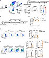 Figure 1
Mks participate in allergic airway inflammation. (A) The flow cytometric gating strategy for labeling lung Mks in Pf4cretdTomato mice. (B) The percentages of Mks in papain and control groups. (C) Schematic diagram of Mk depletion and platelet supplementation in papain-induced allergic airway inflammation by using Pf4creiDTR mice. (D) Determination of platelets in blood by peripheral blood test in each group. (E) The percentage of CD41+ cells in FSC-high/SSC-high lung cells between papain and control groups. (F) Determination of total cells and eosinophils (CD45+Siglec-F+CD11c– cells) in BALF by flow cytometry. Data are representative of at least 3 independent experiments and are presented as mean ± SD (n = 5). **P < 0.01; ***P < 0.001; ****P < 0.0001 by 2-sided t test (B) or 1-way ANOVA with Tukey’s post hoc analysis (D–F). NS, not significant.
Next, we generated Pf4creiDTR mice to conditionally deplete Mks after diphtheria toxin (DT) injection (Figure 1C). To eliminate the potential impact of platelet depletion by DT injection, each Pf4creiDTR mouse received donor platelets 3 times. Blood test verified the platelet replenishment was effective (Figure 1D). Flow cytometric analysis showed that the percentage of CD41+ Mks in FSC-high/SSC-high cells in lung was markedly decreased after DT injection in Pf4creiDTR mice (Figure 1E), but the injection of DT did not affect the numbers of DCs, monocytes, neutrophils, CD4+ T cells, and CD8+ T cells in lung (Supplemental Figure 1, B and C). We also evaluated the impact of DT injection on myeloid cells and lymphocytes in the peripheral blood of Pf4creiDTR mice. Our results indicated that under our experimental conditions, DT-treated Pf4creiDTR mice exhibited no significant fluctuations in peripheral blood lymphocyte counts between day 0 and day 9 (Supplemental Figure 1D). Furthermore, no significant differences were observed in myeloid cells (such as neutrophils, monocytes, and eosinophils) across the groups (Supplemental Figure 1E). We found that depletion of Mks significantly relieved papain-induced airway inflammation, including the decreased total cell numbers in bronchoalveolar lavage fluid (BALF), eosinophil infiltration in BALF, and eosinophil and ILC2 numbers in lung. At the same time, it had no effect on the number of neutrophils in BALF (Figure 1F and Supplemental Figure 1F). We also examined the effect of Mk depletion on other important parameters in peripheral blood. We found Mk depletion did not affect the number of white blood cells, red blood cells (RBCs), hemoglobin, mean platelet volume, and mean corpuscular hemoglobin concentration in peripheral blood (Supplemental Figure 1G). These observations revealed that Mks participated in and accelerated the development of allergic airway inflammation, independently of platelets.
Lung Mks express α7 nAChR. α7 nAChR is present on various cells, including T cells, macrophages, epithelial cells, and others (18, 23). Here, we investigated the expression of α7 nAChR on Mks. In Pf4cretdTomato mice, we discovered that Mks from both the BM and lung expressed α7 nAChR; however, the expression of α7 nAChR on lung Mks was much greater than that on BM Mks (Figure 2A). Then, we tested the excitability of the vagus nerve in papain-induced allergic airway inflammation. We found that in papain-induced airway inflammation, the vagus nerve was activated (Figure 2B) and the number of compound action potentials (CAPs) was significantly increased (Figure 2C). Additionally, we found that the expression of α7 nAChR on lung Mks was upregulated in vivo after papain challenge for 5 days (Figure 2D). Similar results were observed in OVA- or HDM-induced airway inflammation (Figure 2E). These findings indicated that lung Mks expressed α7 nAChR, which could be regulated by vagal signals during allergic airway inflammation. Previous studies have shown that there are differences in the expression of immune molecules such as MHC-II, CD40, and CD11c between lung and BM Mks. In our study, we also found that CD11c and ST2 (IL-33 receptor) were expressed at high levels on α7 nAChR+ lung Mks relative to α7 nAChR– lung Mks. However, MHC-II showed the opposite expression (Figure 2F), suggesting their different immune functions and timing. Most importantly, we confirmed that lung Mks express α7 nAChR at relatively high levels, and that α7 nAChR on lung Mks may play a potential role in allergic airway inflammation.
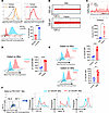 Figure 2
Lung Mks are modulated by vagus nerve/α7 nAChR axis. (A) The expression of α7 nicotinic acetylcholine receptor (α7 nAChR) on Mks from BM and lung using Alexa Fluor 647–conjugated α-bungarotoxin, a nicotinic acetylcholine receptor blocker. (B) Vagus nerve electrical activity after PBS and papain stimulation for 5 days in vivo. (C) Vagus nerve compound action potential (CAP) rate and CAP counts after PBS and papain stimulation for 5 days in vivo. (D) Upregulated expression of α7 nAChR on lung Mks after papain stimulation for 5 days in vivo. (E) Upregulated expression of α7 nAChR on lung Mks in OVA and HDM models in vivo. (F) The difference in CD11c, MHC-II, and ST2 expression between α7 nAChR+ and α7 nAChR– lung Mks. Data are representative of at least 3 independent experiments and are presented as mean ± SD (n = 4). *P < 0.05, **P < 0.01, ***P < 0.001, ****P < 0.0001 by 2-sided t test (A and C–E). NS, not significant.
Deletion of Chrna7 in Mks alleviates allergic airway inflammation. Aiming to confirm the role of α7 nAChR on Mks in allergic airway inflammation, we generated Pf4creChrna7fl/fl mice to conditionally delete α7 nAChR on Mks. We then challenged Chrna7fl/fl mice and Pf4creChrna7fl/fl mice with PBS or papain for 5 days and assessed airway inflammation (Figure 3A). We also confirmed that platelets did not express α7 nAChR (Supplemental Figure 2A). Our experiments revealed that conditional deletion of α7 nAChR on Mks alleviated papain-induced allergic airway inflammation, including the decreased percentage and numbers of eosinophils in BALF (Figure 3B), decreased percentage and numbers of ILC2s (Figure 3C), alleviated airway inflammation (Figure 3, D and E), and reduced relative mRNA expression of Il33, Il5, and Il13 in lung tissue (Figure 3F). Given that α7 nAChR+ Mks exhibited higher expression of ST2, we also tested the expression of ST2 on Mks after conditional knockout of α7 nAChR. We found that with the knockout of α7 nAChR, the expression of ST2 on Mks was correspondingly downregulated (Figure 3G). To exclude the effects of Pf4-cre activity on other immune cell subsets, we also quantified lymphocytes and other myeloid cells in lung tissue from both control and experimental groups. The numbers of CD4+ T cells, CD8+ T cells, DCs, monocytes, and neutrophils, as well as the proportion of Mks (which could not be accurately counted due to the FSC-high gating strategy) in lung tissue were not affected by Pf4-cre activity in either the control or inflammation model groups (Supplemental Figure 2B). In addition, the deletion of Chrna7 on Mks did not affect the number of platelets, leukocytes, erythrocytes, hemoglobin, mean platelet volume, and mean corpuscular hemoglobin concentration in peripheral blood (Supplemental Figure 2C). In order to verify our findings, we challenged Chrna7fl/fl mice and Pf4creChrna7fl/fl mice with HDM extracts for 14 days or IL-33 for 3 days to induce other types of allergic airway inflammation (Figure 4A and Figure 5A). We found less inflammatory cell infiltration (Figure 4, B and C), lower percentages of Th2 cells in mediastinal lymph nodes (MLNs), and alleviated lung injury in Pf4creChrna7fl/fl mice compared with Chrna7fl/fl mice (Figure 4, D–G). However, knockout of α7 nAChR on Mks did not affect Th17 cells in MLNs (Figure 4E). These data suggest that α7 nAChR on Mks play an important role in the pathogenesis of HDM-induced type 2 airway inflammation in vivo. We have found that lung Mks express ST2 (Figure 2F), which indicated that Mks were regulated by IL-33/ST2 signaling, so we tested protein expression of the alarmin receptor ST2 on lung Mks. As shown in Figure 5B, we found ST2 expression was significantly higher than that of the thymic stromal lymphopoietin (TSLP) receptor (TSLPR) and IL-17RA (IL-25 receptor) on lung Mks by flow cytometry. Similar results were observed in mouse models of allergic airway inflammation induced by IL-33 (Figure 5, C–F); knockout of α7 nAChR on Mks alleviated IL-33–induced allergic airway inflammation.
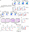 Figure 3
Deletion of α7 nAChR on Mks alleviates papain-induced allergic airway inflammation. (A) Schematic diagram of intratracheal PBS or papain delivery to Chrna7fl/fl and Pf4creChrna7fl/fl mice for 5 days. (B) Determination of total cells and eosinophils in BALF by flow cytometry. (C) Determination of ILC2s in lung tissue by flow cytometry. (D) Representative H&E staining of lung histology. Scale bar: 200 μm. (E) Severity score of lung tissue. (F) The relative mRNA expression of Il33, Il5, and Il13 in lung tissue homogenate tested by RT-qPCR. (G) Expression of ST2 on Mks was tested by flow cytometry. Data are representative of at least 3 independent experiments and are presented as mean ± SD (n = 3–5). *P < 0.05; **P < 0.01; ***P < 0.001; ****P < 0.0001 by 1-way ANOVA with Tukey’s post hoc analysis (B, C, and E–G). NS, not significant.
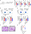 Figure 4
Deletion of α7 nAChR on Mks alleviates HDM-induced type 2 airway inflammation. (A) Schematic diagram of intratracheal HDM delivery to Chrna7fl/fl and Pf4creChrna7fl/fl mice for 14 days. (B) Total cells and eosinophils in BALF determined by flow cytometry. (C) Total DCs in lung determined by flow cytometry. (D) Th2 cells (CD4+IL-13+) in MLNs determined by flow cytometry. (E) Th17 cells (CD4+IL-17+) in MLNs determined by flow cytometry. (F) Representative H&E staining of lung tissue. Scale bar: 200 μm. (G) Severity score of lung tissue. Data are representative of at least 3 independent experiments and are presented as mean ± SD (n = 5). *P < 0.05, **P < 0.01 by 2-sided t test (B–E, and G). NS, not significant.
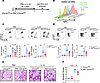 Figure 5
Deletion of α7 nAChR on Mks attenuates IL-33–induced airway inflammation. (A) Schematic diagram of intratracheal PBS or IL-33 delivery to Chrna7fl/fl and Pf4creChrna7fl/fl mice for 5 days. (B) Expression of TSLPR, IL-17RA, and ST2 in lung Mks was tested by flow cytometry. (C) Determination of total cells and eosinophils in BALF by flow cytometry. (D) Determination of ILC2s in lung tissue by flow cytometry. (E) Representative H&E staining. Scale bar: 200 μm. (F) Severity score of lung tissue. Data are representative of at least 3 independent experiments and are presented as mean ± SD (n = 3–5). *P < 0.05; **P < 0.01; ***P < 0.001; ****P < 0.0001 by 1-way ANOVA with Tukey’s post hoc analysis (C, D, and F). NS, not significant.
To further demonstrate that activation of α7 nAChR on lung Mks aggravates allergic airway inflammation, we treated WT mice with PBS, GTS-21, papain, or GTS-21 plus papain for 5 days (Figure 6A). As expected, we noticed that activation of α7 nAChR amplified papain-induced allergic inflammation. GTS-21 increased the percentage and numbers of eosinophils in BALF and ILC2s in lung (Figure 6, B–E) and promoted the Il33 expression of lung tissue in papain-challenged groups (Figure 6F). We also tested the number of peripheral blood platelets in this experiment, and no differences were observed in the 4 groups (Supplemental Figure 3A). As shown in Supplemental Figure 3, B and C, α7 nAChR on Mks had no effect on relative gene expression of Tslp and Il25 in lung tissue. Given that knockout of α7 nAChR on Mks alleviates papain-induced airway inflammation and GTS-21 aggravates airway inflammation, we next treated Pf4creChrna7fl/fl mice with PBS plus papain or GTS-21 plus papain for 5 days (Supplemental Figure 4A). Interestingly, we found that GTS-21 failed to amplify airway inflammation in Pf4creChrna7fl/fl mice (Supplemental Figure 4B). These results demonstrated that the proinflammatory effect of GTS-21 was dependent on Mks. To verify the proinflammatory role of α7 nAChR+ Mks, we sorted α7 nAChR– and α7 nAChR+ lung Mks and transfused them back into Pf4creChrna7fl/fl mice before papain challenge for 3 days (Figure 6, G and H). We found α7 nAChR+ Mk reinfusion aggravated papain-induced airway inflammation compared with α7 nAChR– Mk reinfusion, characterized by more eosinophil and ILC2 infiltration (Figure 6, I and J). Collectively, these results indicate that activation of α7 nAChR on Mks amplifies allergic airway inflammation.
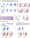 Figure 6
α7 nAChR agonist GTS-21 aggravates papain-induced airway inflammation. (A) Schematic diagram of GTS-21 intervention in papain-induced allergic airway inflammation model. (B) Determination of total cells and eosinophils in BALF by flow cytometry. (C) Determination of ILC2s in lung tissue by flow cytometry. (D) Representative H&E staining and severity score of lung histology. Scale bar: 200 μm. (E) Severity score of lung histology. (F) The relative mRNA expression of Il33 in lung tissue homogenate tested by RT-qPCR. (G) The flow cytometric gating strategy for sorting α7 nAChR– Mks and α7 nAChR+ Mks. (H) α7 nAChR– and α7 nAChR+ Mks were intratracheally delivered to Pf4creChrna7fl/fl mice (1 × 10⁶ Mks per mouse) in the papain-induced allergic airway inflammation model. (I) Determination of total cells, eosinophils, and neutrophils in BALF by flow cytometry. (J) Determination of ILC2s in lung tissue by flow cytometry. Data are representative of at least 3 independent experiments and are presented as mean ± SD (n = 3–5). *P < 0.05; **P < 0.01; ***P < 0.001; ****P < 0.0001 by 1-way ANOVA with Tukey’s post hoc analysis (B, C, E, and F) or 2-sided t test (I and J). NS, not significant.
Activation of α7 nAChR promotes Mk IL-33 expression and release through p38 MAPK signaling pathway. To determine how α7 nAChR on Mks affects airway inflammation, we cultured the MEG-01 cell line in vitro with PBS, GTS-21, papain, or GTS-21 plus papain. Considering the decreased mRNA expression of Il33 in Pf4creChrna7fl/fl mice, we hypothesized that α7 nAChR had a regulatory effect on IL-33 expression in Mks. As we expected, Mks cultured with GTS-21 upregulated relative mRNA and protein expression of IL-33 compared with Mks cultured with papain alone (Figure 7, A and B). Through Western blot and enzyme-linked immunosorbent assay (ELISA) detection of secretory proteins in the supernatant of the culture system, we also found that activation of α7 nAChR promoted IL-33 release from Mks (Figure 7, C and D). IL-33 is an epithelial cell–derived cytokine that is predominantly expressed in lung AECs during allergic airway inflammation (31). To eliminate the impact of AEC-derived IL-33 on the results, we sorted AEC adhesion molecule–positive (EpCAM+) AECs and CD41+, FSC-high megakaryocytes from murine lung tissues to detect IL-33 expression levels (Figure 7E). We found that compared with Chrna7fl/fl mice, IL-33 expression in lung epithelial cells of Pf4creChrna7fl/fl mice remained unchanged, whereas IL-33 expression in lung Mks was significantly reduced during airway inflammation (Figure 7F). To further demonstrate the importance of IL-33 in Mks during airway inflammation, we sorted lung Mks from Il33–/– or WT mice and administered them via intratracheal instillation into Pf4creiDTR mice (Figure 7G), whose Mks had been depleted by DT (1 × 10⁶ cells per mouse). We then reestablished the inflammation model to assess airway inflammation. Compared with WT controls, inflammation levels were significantly milder in mice receiving lung Mks from Il33–/– mice (Figure 7H). Furthermore, GTS-21 did not upregulate Il33 expression in BEAS-2B, an AEC cell line (Supplemental Figure 5). Considering the ability of Mks to process antigens such as OVA, we used DQ-OVA (Thermo Fisher D120553) to test whether activation of α7 nAChR affects OVA processing. We incubated MEG-01 with or without GTS-21 (50 μM) for 2 hours, then treated the cells with DQ-OVA (200 μg/mL) and 30 minutes later determined fluorescence by flow cytometry. Consistent with previous reports, Mks significantly internalized DQ-OVA. However, the GTS-21 had no effect on this process (Supplemental Figure 6A). We also tested Mk exosomes after GTS-21 treatment. As shown, activation of α7 nAChR does not affect the release of exosomes by Mks (Supplemental Figure 6B).
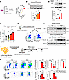 Figure 7
Activation of α7 nAChR on lung Mks aggravates airway inflammation via IL-33 release. (A) The relative IL33 mRNA expression in MEG-01 cells cultured with GTS-21 and papain was tested by RT-qPCR and Western blotting. (B) The expression of IL-33 (35 kDa) protein in MEG-01 was tested by Western blotting. WCL, whole-cell lysate. (C) The IL-33 (25 kDa) protein in supernatant of MEG-01 was tested by Western blotting. (D) The release of IL-33 in supernatant of MEG-01 was tested by ELISA. (E) The flow cytometric gating strategy for sorting lung epithelial cells and Mks. (F) The expression of IL-33 (35 kDa) protein in lung epithelial cells and Mks was tested by Western blotting. (G) Schematic diagram of intratracheal Mk reinfusion into Pf4creiDTR mice. (H) Determination of eosinophils in BALF and ILC2s in lung tissue by flow cytometry. Data are representative of at least 3 independent experiments and are presented as mean ± SD (n = 3–5). *P < 0.05; **P < 0.01; ***P < 0.001; ****P < 0.0001 by 1-way ANOVA with Tukey’s post hoc analysis (A–D and H). NS, not significant.
Next, we investigated which signaling pathway mediates the proinflammatory effect of α7 nAChR on Mks. Via Western blot, we found that GTS-21 promoted phosphorylation of p38 MAPK in Mks. For comparison, we also examined the NF-κB signaling pathway and found that GTS-21 did not affect the phosphorylation level of p65 (Figure 8A). To verify our findings, we tested the phosphorylation of p38 MAPK in lung Mks. Consistent with the cell line results, GTS-21 promoted phosphorylation of p38 MAPK in Mks, while knockout of α7 nAChR inhibited this process (Figure 8, B and C). Then we utilized a selective p38 MAPK inhibitor (SB203580) to suppress the phosphorylation of p38 MAPK in sorted lung Mks and MEG-01. We found that SB203580 treatment restrained the increased gene expression of Il33 and IL-33 protein release in lung Mks caused by GTS-21 (Figure 8, D and E). These findings were further confirmed in MEG-01 cells (Figure 8, F and G). In summary, these results suggest that overexpression of IL-33 caused by α7 nAChR activation on Mks is dependent on phosphorylation of p38 MAPK.
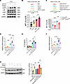 Figure 8
Activation of α7 nAChR promotes Mk IL-33 expression and release through p38 MAPK signaling pathway. (A) The phosphorylation of p38 and p65 was assessed by Western blotting. (B) The phosphorylation of p38 in lung Mks was evaluated in a papain-induced allergic airway inflammation model with GTS-21 intervention by flow cytometry. (C) The phosphorylation of p38 in lung Mks was tested in Chrna7fl/fl and Pf4creChrna7fl/fl mice with papain challenge. (D) The relative mRNA expression of Il33 in lung Mks cultured with GTS-21, papain, and SB203580 was tested by RT-qPCR. (E) The release of IL-33 in supernatant of lung Mks was assessed by ELISA. (F) The relative mRNA expression of IL33 in MEG-01 cultured with GTS-21, papain, and SB203580 was evaluated by RT-qPCR. (G) The expression of IL-33 (35 kDa) protein in MEG-01 cultured with GTS-21, papain, and SB203580 was assessed by Western blotting. Data are representative of at least 3 independent experiments and are presented as mean ± SD (n = 3–5). *P < 0.05; **P < 0.01; ***P < 0.001; ****P < 0.0001 by 1-way ANOVA with Tukey’s post hoc analysis (B–G). NS, not significant.


