
Research ArticleMetabolismVascular biology
Open Access |  10.1172/jci.insight.201311
BAT-derived miR-378a-3p facilitates endothelial angiogenic function and promotes wound healing
Hongyan Deng,1,2 Yuyu Xie,1,2 Jiadai Liu,1,2 Jing Ge,1,2 Qianqian Kang,2 Rui He,1,2 Zhihan Wang,1,2 Xuemin Peng,1,2 Zengzhe Zhu,1,2 Wenshe Wang,1,2 Yulian Liu,1,2 Ronghui Gao,1,2 Ruping Pan,2,3 Min Yang,4 and Yong Chen1,2,5,6
1Division of Endocrinology, Department of Internal Medicine, Tongji Hospital, Tongji Medical College and State Key Laboratory for Diagnosis and Treatment of Severe Zoonotic Infectious Diseases, Huazhong University of Science and Technology, Wuhan, China.
2Laboratory of Endocrinology and Metabolism, Ministry of Education Key Laboratory of Vascular Aging, Tongji Hospital, Tongji Medical College, Huazhong University of Science and Technology, Wuhan, China.
3Department of Nuclear Medicine, Tongji Hospital, Tongji Medical College, Huazhong University of Science and Technology, Wuhan, China.
4Department of Endocrinology, Fifth People’s Hospital of Shanghai, Fudan University, Shanghai, China.
5Branch of National Clinical Research Center for Metabolic Diseases, Wuhan, China.
6The Center for Biomedical Research, Tongji Hospital, Tongji Medical College, Huazhong University of Science and Technology, Wuhan, China.
Address correspondence to: Min Yang, Division of Endocrinology, The Fifth People’s Hospital of Shanghai, Fudan University, 801 Heqing Road, Minhang District, Shanghai 200240, China. Phone: 86.18915951712; Email: yangmin56789@163.com. Or to: Yong Chen, Division of Endocrinology, Tongji Medical College, Huazhong University of Science and Technology, 13 Hangkong Road, Qiaokou District, Wuhan 430030, China. Phone: 86.18986059966; Email: tj.y.chen@vip.163.com.
Authorship note: HD, YX, JL, and JG are co-first authors.
Find articles by Deng, H. in: PubMed | Google Scholar
1Division of Endocrinology, Department of Internal Medicine, Tongji Hospital, Tongji Medical College and State Key Laboratory for Diagnosis and Treatment of Severe Zoonotic Infectious Diseases, Huazhong University of Science and Technology, Wuhan, China.
2Laboratory of Endocrinology and Metabolism, Ministry of Education Key Laboratory of Vascular Aging, Tongji Hospital, Tongji Medical College, Huazhong University of Science and Technology, Wuhan, China.
3Department of Nuclear Medicine, Tongji Hospital, Tongji Medical College, Huazhong University of Science and Technology, Wuhan, China.
4Department of Endocrinology, Fifth People’s Hospital of Shanghai, Fudan University, Shanghai, China.
5Branch of National Clinical Research Center for Metabolic Diseases, Wuhan, China.
6The Center for Biomedical Research, Tongji Hospital, Tongji Medical College, Huazhong University of Science and Technology, Wuhan, China.
Address correspondence to: Min Yang, Division of Endocrinology, The Fifth People’s Hospital of Shanghai, Fudan University, 801 Heqing Road, Minhang District, Shanghai 200240, China. Phone: 86.18915951712; Email: yangmin56789@163.com. Or to: Yong Chen, Division of Endocrinology, Tongji Medical College, Huazhong University of Science and Technology, 13 Hangkong Road, Qiaokou District, Wuhan 430030, China. Phone: 86.18986059966; Email: tj.y.chen@vip.163.com.
Authorship note: HD, YX, JL, and JG are co-first authors.
Find articles by Xie, Y. in: PubMed | Google Scholar
1Division of Endocrinology, Department of Internal Medicine, Tongji Hospital, Tongji Medical College and State Key Laboratory for Diagnosis and Treatment of Severe Zoonotic Infectious Diseases, Huazhong University of Science and Technology, Wuhan, China.
2Laboratory of Endocrinology and Metabolism, Ministry of Education Key Laboratory of Vascular Aging, Tongji Hospital, Tongji Medical College, Huazhong University of Science and Technology, Wuhan, China.
3Department of Nuclear Medicine, Tongji Hospital, Tongji Medical College, Huazhong University of Science and Technology, Wuhan, China.
4Department of Endocrinology, Fifth People’s Hospital of Shanghai, Fudan University, Shanghai, China.
5Branch of National Clinical Research Center for Metabolic Diseases, Wuhan, China.
6The Center for Biomedical Research, Tongji Hospital, Tongji Medical College, Huazhong University of Science and Technology, Wuhan, China.
Address correspondence to: Min Yang, Division of Endocrinology, The Fifth People’s Hospital of Shanghai, Fudan University, 801 Heqing Road, Minhang District, Shanghai 200240, China. Phone: 86.18915951712; Email: yangmin56789@163.com. Or to: Yong Chen, Division of Endocrinology, Tongji Medical College, Huazhong University of Science and Technology, 13 Hangkong Road, Qiaokou District, Wuhan 430030, China. Phone: 86.18986059966; Email: tj.y.chen@vip.163.com.
Authorship note: HD, YX, JL, and JG are co-first authors.
Find articles by Liu, J. in: PubMed | Google Scholar
1Division of Endocrinology, Department of Internal Medicine, Tongji Hospital, Tongji Medical College and State Key Laboratory for Diagnosis and Treatment of Severe Zoonotic Infectious Diseases, Huazhong University of Science and Technology, Wuhan, China.
2Laboratory of Endocrinology and Metabolism, Ministry of Education Key Laboratory of Vascular Aging, Tongji Hospital, Tongji Medical College, Huazhong University of Science and Technology, Wuhan, China.
3Department of Nuclear Medicine, Tongji Hospital, Tongji Medical College, Huazhong University of Science and Technology, Wuhan, China.
4Department of Endocrinology, Fifth People’s Hospital of Shanghai, Fudan University, Shanghai, China.
5Branch of National Clinical Research Center for Metabolic Diseases, Wuhan, China.
6The Center for Biomedical Research, Tongji Hospital, Tongji Medical College, Huazhong University of Science and Technology, Wuhan, China.
Address correspondence to: Min Yang, Division of Endocrinology, The Fifth People’s Hospital of Shanghai, Fudan University, 801 Heqing Road, Minhang District, Shanghai 200240, China. Phone: 86.18915951712; Email: yangmin56789@163.com. Or to: Yong Chen, Division of Endocrinology, Tongji Medical College, Huazhong University of Science and Technology, 13 Hangkong Road, Qiaokou District, Wuhan 430030, China. Phone: 86.18986059966; Email: tj.y.chen@vip.163.com.
Authorship note: HD, YX, JL, and JG are co-first authors.
Find articles by Ge, J. in: PubMed | Google Scholar
1Division of Endocrinology, Department of Internal Medicine, Tongji Hospital, Tongji Medical College and State Key Laboratory for Diagnosis and Treatment of Severe Zoonotic Infectious Diseases, Huazhong University of Science and Technology, Wuhan, China.
2Laboratory of Endocrinology and Metabolism, Ministry of Education Key Laboratory of Vascular Aging, Tongji Hospital, Tongji Medical College, Huazhong University of Science and Technology, Wuhan, China.
3Department of Nuclear Medicine, Tongji Hospital, Tongji Medical College, Huazhong University of Science and Technology, Wuhan, China.
4Department of Endocrinology, Fifth People’s Hospital of Shanghai, Fudan University, Shanghai, China.
5Branch of National Clinical Research Center for Metabolic Diseases, Wuhan, China.
6The Center for Biomedical Research, Tongji Hospital, Tongji Medical College, Huazhong University of Science and Technology, Wuhan, China.
Address correspondence to: Min Yang, Division of Endocrinology, The Fifth People’s Hospital of Shanghai, Fudan University, 801 Heqing Road, Minhang District, Shanghai 200240, China. Phone: 86.18915951712; Email: yangmin56789@163.com. Or to: Yong Chen, Division of Endocrinology, Tongji Medical College, Huazhong University of Science and Technology, 13 Hangkong Road, Qiaokou District, Wuhan 430030, China. Phone: 86.18986059966; Email: tj.y.chen@vip.163.com.
Authorship note: HD, YX, JL, and JG are co-first authors.
Find articles by Kang, Q. in: PubMed | Google Scholar
1Division of Endocrinology, Department of Internal Medicine, Tongji Hospital, Tongji Medical College and State Key Laboratory for Diagnosis and Treatment of Severe Zoonotic Infectious Diseases, Huazhong University of Science and Technology, Wuhan, China.
2Laboratory of Endocrinology and Metabolism, Ministry of Education Key Laboratory of Vascular Aging, Tongji Hospital, Tongji Medical College, Huazhong University of Science and Technology, Wuhan, China.
3Department of Nuclear Medicine, Tongji Hospital, Tongji Medical College, Huazhong University of Science and Technology, Wuhan, China.
4Department of Endocrinology, Fifth People’s Hospital of Shanghai, Fudan University, Shanghai, China.
5Branch of National Clinical Research Center for Metabolic Diseases, Wuhan, China.
6The Center for Biomedical Research, Tongji Hospital, Tongji Medical College, Huazhong University of Science and Technology, Wuhan, China.
Address correspondence to: Min Yang, Division of Endocrinology, The Fifth People’s Hospital of Shanghai, Fudan University, 801 Heqing Road, Minhang District, Shanghai 200240, China. Phone: 86.18915951712; Email: yangmin56789@163.com. Or to: Yong Chen, Division of Endocrinology, Tongji Medical College, Huazhong University of Science and Technology, 13 Hangkong Road, Qiaokou District, Wuhan 430030, China. Phone: 86.18986059966; Email: tj.y.chen@vip.163.com.
Authorship note: HD, YX, JL, and JG are co-first authors.
Find articles by He, R. in: PubMed | Google Scholar
1Division of Endocrinology, Department of Internal Medicine, Tongji Hospital, Tongji Medical College and State Key Laboratory for Diagnosis and Treatment of Severe Zoonotic Infectious Diseases, Huazhong University of Science and Technology, Wuhan, China.
2Laboratory of Endocrinology and Metabolism, Ministry of Education Key Laboratory of Vascular Aging, Tongji Hospital, Tongji Medical College, Huazhong University of Science and Technology, Wuhan, China.
3Department of Nuclear Medicine, Tongji Hospital, Tongji Medical College, Huazhong University of Science and Technology, Wuhan, China.
4Department of Endocrinology, Fifth People’s Hospital of Shanghai, Fudan University, Shanghai, China.
5Branch of National Clinical Research Center for Metabolic Diseases, Wuhan, China.
6The Center for Biomedical Research, Tongji Hospital, Tongji Medical College, Huazhong University of Science and Technology, Wuhan, China.
Address correspondence to: Min Yang, Division of Endocrinology, The Fifth People’s Hospital of Shanghai, Fudan University, 801 Heqing Road, Minhang District, Shanghai 200240, China. Phone: 86.18915951712; Email: yangmin56789@163.com. Or to: Yong Chen, Division of Endocrinology, Tongji Medical College, Huazhong University of Science and Technology, 13 Hangkong Road, Qiaokou District, Wuhan 430030, China. Phone: 86.18986059966; Email: tj.y.chen@vip.163.com.
Authorship note: HD, YX, JL, and JG are co-first authors.
Find articles by Wang, Z. in: PubMed | Google Scholar
1Division of Endocrinology, Department of Internal Medicine, Tongji Hospital, Tongji Medical College and State Key Laboratory for Diagnosis and Treatment of Severe Zoonotic Infectious Diseases, Huazhong University of Science and Technology, Wuhan, China.
2Laboratory of Endocrinology and Metabolism, Ministry of Education Key Laboratory of Vascular Aging, Tongji Hospital, Tongji Medical College, Huazhong University of Science and Technology, Wuhan, China.
3Department of Nuclear Medicine, Tongji Hospital, Tongji Medical College, Huazhong University of Science and Technology, Wuhan, China.
4Department of Endocrinology, Fifth People’s Hospital of Shanghai, Fudan University, Shanghai, China.
5Branch of National Clinical Research Center for Metabolic Diseases, Wuhan, China.
6The Center for Biomedical Research, Tongji Hospital, Tongji Medical College, Huazhong University of Science and Technology, Wuhan, China.
Address correspondence to: Min Yang, Division of Endocrinology, The Fifth People’s Hospital of Shanghai, Fudan University, 801 Heqing Road, Minhang District, Shanghai 200240, China. Phone: 86.18915951712; Email: yangmin56789@163.com. Or to: Yong Chen, Division of Endocrinology, Tongji Medical College, Huazhong University of Science and Technology, 13 Hangkong Road, Qiaokou District, Wuhan 430030, China. Phone: 86.18986059966; Email: tj.y.chen@vip.163.com.
Authorship note: HD, YX, JL, and JG are co-first authors.
Find articles by Peng, X. in: PubMed | Google Scholar
1Division of Endocrinology, Department of Internal Medicine, Tongji Hospital, Tongji Medical College and State Key Laboratory for Diagnosis and Treatment of Severe Zoonotic Infectious Diseases, Huazhong University of Science and Technology, Wuhan, China.
2Laboratory of Endocrinology and Metabolism, Ministry of Education Key Laboratory of Vascular Aging, Tongji Hospital, Tongji Medical College, Huazhong University of Science and Technology, Wuhan, China.
3Department of Nuclear Medicine, Tongji Hospital, Tongji Medical College, Huazhong University of Science and Technology, Wuhan, China.
4Department of Endocrinology, Fifth People’s Hospital of Shanghai, Fudan University, Shanghai, China.
5Branch of National Clinical Research Center for Metabolic Diseases, Wuhan, China.
6The Center for Biomedical Research, Tongji Hospital, Tongji Medical College, Huazhong University of Science and Technology, Wuhan, China.
Address correspondence to: Min Yang, Division of Endocrinology, The Fifth People’s Hospital of Shanghai, Fudan University, 801 Heqing Road, Minhang District, Shanghai 200240, China. Phone: 86.18915951712; Email: yangmin56789@163.com. Or to: Yong Chen, Division of Endocrinology, Tongji Medical College, Huazhong University of Science and Technology, 13 Hangkong Road, Qiaokou District, Wuhan 430030, China. Phone: 86.18986059966; Email: tj.y.chen@vip.163.com.
Authorship note: HD, YX, JL, and JG are co-first authors.
Find articles by Zhu, Z. in: PubMed | Google Scholar
1Division of Endocrinology, Department of Internal Medicine, Tongji Hospital, Tongji Medical College and State Key Laboratory for Diagnosis and Treatment of Severe Zoonotic Infectious Diseases, Huazhong University of Science and Technology, Wuhan, China.
2Laboratory of Endocrinology and Metabolism, Ministry of Education Key Laboratory of Vascular Aging, Tongji Hospital, Tongji Medical College, Huazhong University of Science and Technology, Wuhan, China.
3Department of Nuclear Medicine, Tongji Hospital, Tongji Medical College, Huazhong University of Science and Technology, Wuhan, China.
4Department of Endocrinology, Fifth People’s Hospital of Shanghai, Fudan University, Shanghai, China.
5Branch of National Clinical Research Center for Metabolic Diseases, Wuhan, China.
6The Center for Biomedical Research, Tongji Hospital, Tongji Medical College, Huazhong University of Science and Technology, Wuhan, China.
Address correspondence to: Min Yang, Division of Endocrinology, The Fifth People’s Hospital of Shanghai, Fudan University, 801 Heqing Road, Minhang District, Shanghai 200240, China. Phone: 86.18915951712; Email: yangmin56789@163.com. Or to: Yong Chen, Division of Endocrinology, Tongji Medical College, Huazhong University of Science and Technology, 13 Hangkong Road, Qiaokou District, Wuhan 430030, China. Phone: 86.18986059966; Email: tj.y.chen@vip.163.com.
Authorship note: HD, YX, JL, and JG are co-first authors.
Find articles by Wang, W. in: PubMed | Google Scholar
1Division of Endocrinology, Department of Internal Medicine, Tongji Hospital, Tongji Medical College and State Key Laboratory for Diagnosis and Treatment of Severe Zoonotic Infectious Diseases, Huazhong University of Science and Technology, Wuhan, China.
2Laboratory of Endocrinology and Metabolism, Ministry of Education Key Laboratory of Vascular Aging, Tongji Hospital, Tongji Medical College, Huazhong University of Science and Technology, Wuhan, China.
3Department of Nuclear Medicine, Tongji Hospital, Tongji Medical College, Huazhong University of Science and Technology, Wuhan, China.
4Department of Endocrinology, Fifth People’s Hospital of Shanghai, Fudan University, Shanghai, China.
5Branch of National Clinical Research Center for Metabolic Diseases, Wuhan, China.
6The Center for Biomedical Research, Tongji Hospital, Tongji Medical College, Huazhong University of Science and Technology, Wuhan, China.
Address correspondence to: Min Yang, Division of Endocrinology, The Fifth People’s Hospital of Shanghai, Fudan University, 801 Heqing Road, Minhang District, Shanghai 200240, China. Phone: 86.18915951712; Email: yangmin56789@163.com. Or to: Yong Chen, Division of Endocrinology, Tongji Medical College, Huazhong University of Science and Technology, 13 Hangkong Road, Qiaokou District, Wuhan 430030, China. Phone: 86.18986059966; Email: tj.y.chen@vip.163.com.
Authorship note: HD, YX, JL, and JG are co-first authors.
Find articles by Liu, Y. in: PubMed | Google Scholar
1Division of Endocrinology, Department of Internal Medicine, Tongji Hospital, Tongji Medical College and State Key Laboratory for Diagnosis and Treatment of Severe Zoonotic Infectious Diseases, Huazhong University of Science and Technology, Wuhan, China.
2Laboratory of Endocrinology and Metabolism, Ministry of Education Key Laboratory of Vascular Aging, Tongji Hospital, Tongji Medical College, Huazhong University of Science and Technology, Wuhan, China.
3Department of Nuclear Medicine, Tongji Hospital, Tongji Medical College, Huazhong University of Science and Technology, Wuhan, China.
4Department of Endocrinology, Fifth People’s Hospital of Shanghai, Fudan University, Shanghai, China.
5Branch of National Clinical Research Center for Metabolic Diseases, Wuhan, China.
6The Center for Biomedical Research, Tongji Hospital, Tongji Medical College, Huazhong University of Science and Technology, Wuhan, China.
Address correspondence to: Min Yang, Division of Endocrinology, The Fifth People’s Hospital of Shanghai, Fudan University, 801 Heqing Road, Minhang District, Shanghai 200240, China. Phone: 86.18915951712; Email: yangmin56789@163.com. Or to: Yong Chen, Division of Endocrinology, Tongji Medical College, Huazhong University of Science and Technology, 13 Hangkong Road, Qiaokou District, Wuhan 430030, China. Phone: 86.18986059966; Email: tj.y.chen@vip.163.com.
Authorship note: HD, YX, JL, and JG are co-first authors.
Find articles by Gao, R. in: PubMed | Google Scholar
1Division of Endocrinology, Department of Internal Medicine, Tongji Hospital, Tongji Medical College and State Key Laboratory for Diagnosis and Treatment of Severe Zoonotic Infectious Diseases, Huazhong University of Science and Technology, Wuhan, China.
2Laboratory of Endocrinology and Metabolism, Ministry of Education Key Laboratory of Vascular Aging, Tongji Hospital, Tongji Medical College, Huazhong University of Science and Technology, Wuhan, China.
3Department of Nuclear Medicine, Tongji Hospital, Tongji Medical College, Huazhong University of Science and Technology, Wuhan, China.
4Department of Endocrinology, Fifth People’s Hospital of Shanghai, Fudan University, Shanghai, China.
5Branch of National Clinical Research Center for Metabolic Diseases, Wuhan, China.
6The Center for Biomedical Research, Tongji Hospital, Tongji Medical College, Huazhong University of Science and Technology, Wuhan, China.
Address correspondence to: Min Yang, Division of Endocrinology, The Fifth People’s Hospital of Shanghai, Fudan University, 801 Heqing Road, Minhang District, Shanghai 200240, China. Phone: 86.18915951712; Email: yangmin56789@163.com. Or to: Yong Chen, Division of Endocrinology, Tongji Medical College, Huazhong University of Science and Technology, 13 Hangkong Road, Qiaokou District, Wuhan 430030, China. Phone: 86.18986059966; Email: tj.y.chen@vip.163.com.
Authorship note: HD, YX, JL, and JG are co-first authors.
Find articles by Pan, R. in: PubMed | Google Scholar
1Division of Endocrinology, Department of Internal Medicine, Tongji Hospital, Tongji Medical College and State Key Laboratory for Diagnosis and Treatment of Severe Zoonotic Infectious Diseases, Huazhong University of Science and Technology, Wuhan, China.
2Laboratory of Endocrinology and Metabolism, Ministry of Education Key Laboratory of Vascular Aging, Tongji Hospital, Tongji Medical College, Huazhong University of Science and Technology, Wuhan, China.
3Department of Nuclear Medicine, Tongji Hospital, Tongji Medical College, Huazhong University of Science and Technology, Wuhan, China.
4Department of Endocrinology, Fifth People’s Hospital of Shanghai, Fudan University, Shanghai, China.
5Branch of National Clinical Research Center for Metabolic Diseases, Wuhan, China.
6The Center for Biomedical Research, Tongji Hospital, Tongji Medical College, Huazhong University of Science and Technology, Wuhan, China.
Address correspondence to: Min Yang, Division of Endocrinology, The Fifth People’s Hospital of Shanghai, Fudan University, 801 Heqing Road, Minhang District, Shanghai 200240, China. Phone: 86.18915951712; Email: yangmin56789@163.com. Or to: Yong Chen, Division of Endocrinology, Tongji Medical College, Huazhong University of Science and Technology, 13 Hangkong Road, Qiaokou District, Wuhan 430030, China. Phone: 86.18986059966; Email: tj.y.chen@vip.163.com.
Authorship note: HD, YX, JL, and JG are co-first authors.
Find articles by Yang, M. in: PubMed | Google Scholar
1Division of Endocrinology, Department of Internal Medicine, Tongji Hospital, Tongji Medical College and State Key Laboratory for Diagnosis and Treatment of Severe Zoonotic Infectious Diseases, Huazhong University of Science and Technology, Wuhan, China.
2Laboratory of Endocrinology and Metabolism, Ministry of Education Key Laboratory of Vascular Aging, Tongji Hospital, Tongji Medical College, Huazhong University of Science and Technology, Wuhan, China.
3Department of Nuclear Medicine, Tongji Hospital, Tongji Medical College, Huazhong University of Science and Technology, Wuhan, China.
4Department of Endocrinology, Fifth People’s Hospital of Shanghai, Fudan University, Shanghai, China.
5Branch of National Clinical Research Center for Metabolic Diseases, Wuhan, China.
6The Center for Biomedical Research, Tongji Hospital, Tongji Medical College, Huazhong University of Science and Technology, Wuhan, China.
Address correspondence to: Min Yang, Division of Endocrinology, The Fifth People’s Hospital of Shanghai, Fudan University, 801 Heqing Road, Minhang District, Shanghai 200240, China. Phone: 86.18915951712; Email: yangmin56789@163.com. Or to: Yong Chen, Division of Endocrinology, Tongji Medical College, Huazhong University of Science and Technology, 13 Hangkong Road, Qiaokou District, Wuhan 430030, China. Phone: 86.18986059966; Email: tj.y.chen@vip.163.com.
Authorship note: HD, YX, JL, and JG are co-first authors.
Find articles by Chen, Y. in: PubMed | Google Scholar
Published April 21, 2026 - More info
JCI Insight. 2026;11(11):e201311. https://doi.org/10.1172/jci.insight.201311.
© 2026 Deng et al. This work is licensed under the Creative Commons Attribution 4.0 International License. To view a copy of this license, visit http://creativecommons.org/licenses/by/4.0/.
Received: October 17, 2025; Accepted: March 31, 2026


-
Results
Ablation of BAT impairs vascular repair and wound healing. To investigate the association between BAT activity and angiogenesis, WT mice were exposed to 4°C for 3 days to activate BAT. Cold exposure significantly increased UCP1 expression in BAT (Figure 1A, Figure 1C, and Supplemental Figure 1A; supplemental material available online with this article; https://doi.org/10.1172/jci.insight.201311DS1), along with a marked upregulation of CD31 (encoded by Pecam1), an EC marker (Figure 1, B and C, and Supplemental Figure 1A), indicating a striking correlation between BAT activation and increased local vascularization. To investigate the role of BAT in angiogenesis, we analyzed cold-induced gene expression changes in mouse BAT using dataset GSE280981. By focusing on upregulated genes, we found substantial enrichment of pathways related to vesicle-mediated transport and angiogenesis (Supplemental Figure 1B). Next, to directly assess the role of BAT in vascular repair, we established 2 models: surgical removal of interscapular BAT (BATectomy) and pharmacological activation of BAT using the β3-adrenergic receptor agonist CL316,243 (Supplemental Figure 1C). Endothelial angiogenic function was assessed by monitoring subcutaneous angiogenesis in a dorsal skin wound model and reendothelialization after femoral artery injury, as previously described (14, 15). Compared with sham-operated controls, BATectomy mice exhibited a significantly reduced reendothelialization rate in injured femoral arteries, as demonstrated by Evans blue staining (Supplemental Figure 1D and Figure 1D). CD31 IHC also revealed fewer ECs in the femoral arteries of BATectomy mice (Figure 1E). In addition, CD31+ immunofluorescence in wound tissue was reduced in BATectomized mice relative to controls, indicating impaired angiogenesis (Figure 1F). Notably, BATectomy mice showed delayed wound healing compared with sham mice at day 5 (Figure 1, G and H). Consistently, BATectomy significantly reduced microvascular perfusion at the wound site (Figure 1I). Moreover, activation of BAT by β3-adrenergic receptor agonist CL316,243, as evidenced by the marked upregulation of Ucp1 mRNA and UCP1 protein levels (Supplemental Figure 1, E and F), significantly accelerated wound closure by day 3 compared with vehicle-treated mice (Figure 1, J and K). Also, this coincided with enhanced microvascular perfusion after CL316,243 injection (Figure 1L and Supplemental Figure 1, G and H). Interestingly, serum CD63 concentration was significantly reduced after BAT ablation, as revealed by ELISA (Supplemental Figure 1I). Collectively, these results demonstrate that BAT positively regulates angiogenesis and promotes wound healing, likely via modulation of local vascular density and perfusion.
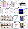 Figure 1
Ablation of BAT impairs vascular repair and wound healing. (A) H&E and UCP1 IHC staining of BAT from cold-exposed WT mice. Scale bar: 50 μm. (B) CD31 IHC staining in BAT after cold exposure with quantification of CD31+ area. Scale bar: 100 μm (top), 50 μm (bottom). (C) Western blot of UCP1 and CD31 in BAT after cold exposure. (D) Representative images of Evans blue staining of injured femoral artery in the sham and BATectomy mice (n = 3). Quantification data of percentage of reendothelialization are shown on the right. (E) Representative images of femoral arteries with CD31 IHC staining in the sham and BATectomy mice (n = 3). Scale bar: 50 μm. Right: quantification of the data. (F) Immunofluorescence staining of CD31 (red) in wounds of BATectomy and sham mice (n = 3). Scale bar: 50 μm. Right: quantification of the data. (G) Picture of back skin wounds in BATectomy and sham mice. (H) Quantification data of wound closure rate over time in BATectomy and sham mice (n = 3–4). (I) Representative images of blood flow at the wounds in BATectomy and sham mice. Calculated wound microvascular perfusion is shown on the right (n = 3). (J) Representative pictures of skin wounds in control and CL316,243-injected mice (n = 3–4). (K) Quantification data of wound closure rate over time in control and CL316,243-injected mice (n = 3–4). (L) Representative images of blood flow at the wounds in control and CL316,243-injected mice. Calculated wound microvascular perfusion is shown on the right (n = 3). The value n represents the number of biologically independent samples, from which all experimental data were obtained. The data are expressed as mean ± SEM (analyzed by 2-tailed Student’s t test). *P < 0.05, **P < 0.01, ****P < 0.0001.
BATexos facilitate vascular repair and wound healing. Activated BAT is known to secrete exosomes into the circulation (5, 16). To investigate whether BATexos contribute to wound healing and vascular repair, we locally injected the exosome secretion inhibitor GW4869 (100 μL) into the BAT of WT mice to block exosome release (Figure 2A). In addition, we isolated exosomes from cultured brown adipocytes of WT mice in vitro and administered them to the BATectomized mice either via tail vein injection or direct injection into the wound area (Figure 2A). To demonstrate that circulating BATexos can reach the wound site and exert their effects, we i.v. injected PKH26-labeled BATexos via the tail vein and detected the presence of PKH26-labeled exosomes at the wound site. PKH26-labeled BATexos were detected in the wound tissue, indicating that these exosomes reached the wound site via the circulatory system to exert their biological functions (Supplemental Figure 2A). The extracted exosomes showed an atypical cup-shaped morphology under a transmission electron microscope (Figure 2B). Nanoparticle tracking analysis showed particle diameters ranging from 100 to 170 nm (Figure 2C). These particles expressed exosomal protein markers such as CD63 and tumor susceptibility gene 101 (TSG101), whose expression was markedly upregulated in serum exosomes extracted from cold-exposed mice (Figure 2D). Functionally, GW4869-mediated inhibition of exosome secretion in BAT significantly reduced reendothelialization of injured femoral arteries, as evidenced by increased Evans blue staining (Figure 2E). Conversely, the impaired reendothelialization observed in BATectomized mice was rescued by systemic administration of BATexos from WT mice (Figure 2E). Similarly, BAT GW4869 treatment reduced CD31 expression in wound tissues, indicating compromised angiogenesis (Figure 2F). This effect was reversed by local administration of BATexos in BATectomized mice, which restored CD31+ EC levels in the skin wound (Figure 2F), indicating improved angiogenesis. Consistent with these findings, GW4869 injection in BAT significantly impaired wound healing (Figure 2G), whereas topical BATexos delivery, but not the PBS control, effectively restored wound healing in BATectomized mice (Figure 2G). We next investigated the role of BATexos in wound perfusion. Local injection of GW4869 into BAT significantly reduced blood perfusion at the wound site (Figure 2H). Importantly, BATexos restored wound perfusion in BATectomized mice (Figure 2H). It has been reported that BATexos play a role in systemic metabolic regulation and energy homeostasis (12, 17). To determine whether the pro-angiogenic effect of BATexos is secondary to altered systemic metabolism, we performed a series of metabolic assessments in BATectomized mice receiving BATexos or vehicle during the active wound healing phase (days 3–5). Indirect calorimetry revealed no significant differences between the BATectomized mice receiving BATexos or the vehicle in terms of either oxygen consumption (VO2) (Supplemental Figure 2, B and C) or the respiratory exchange ratio (Supplemental Figure 2, D and E). Furthermore, there was no significant difference in food intake between the 2 groups (Supplemental Figure 2F). Consistent with these findings, an i.p. glucose tolerance test showed no improvement in glucose homeostasis upon BATexos administration (Supplemental Figure 2, G and H). Collectively, these data demonstrate that the observed enhancement in wound angiogenesis and perfusion mediated by BATexos occurs independently of changes in whole-body energy metabolism or glucose tolerance. It is widely recognized that Rab27a plays a critical role in regulating exosome release. Thus, we measured the RAB27A protein level in interscapular BAT of mice after cold exposure and found that RAB27A expression was significantly elevated in response to cold exposure (Supplemental Figure 3A). Consequently, we established Rab27a global KO mice (Rab27a-KO) and confirmed successful ablation of RAB27A protein expression (Supplemental Figure 3B). BATexos isolated from Rab27a-KO mouse BAT showed markedly reduced levels of the exosome hallmark proteins CD63 and TSG101, confirming a defect in exosome biogenesis or secretion upon Rab27a loss (Supplemental Figure 3C). Meanwhile, our analysis showed impaired angiogenesis in wounds of Rab27a-KO mice, evidenced by decreased CD31 immunostaining (Supplemental Figure 3D). Consistent with this result, Rab27a-KO mice exhibited impaired wound healing (Supplemental Figure 3, E and F). Angiogenesis-related genes such as Vegf and Pecam1 were also decreased in Rab27a-KO mice (Supplemental Figure 3G). Collectively, these results provide compelling evidence that BAT-derived exosomes play an essential role in promoting endothelial regeneration and accelerating wound repair.
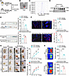 Figure 2
BATexos facilitate vascular repair and wound healing. (A) Schematic diagram of BAT with GW4869/control vehicle injection in WT mice or BATexos/PBS treatment in BATectomy mice. (B) Morphological characteristics of BATexos under a transmission electron microscope. Scale bar: 100 nm. (C) The diameter of the BATexo particles was investigated via nanoparticle tracking analysis. (D) Western blot analysis of specific marker proteins (CD63 and TSG101) for exosomes in the serum of mice kept at room temperature or cold (at 4°C). (E) Evans blue staining and quantitative analysis of the femoral artery in WT mice injected with GW4869 in BAT or BATectomy mice injected with BATexos via the tail vein. (F) Immunofluorescence staining for CD31 (red) in the wound and quantitative analysis in mice treated with GW4869 or BATexos (n = 3–4). Scale bar: 50 μm. (G) Representative images of skin wounds in the GW4869/vehicle-treated WT mice and BATexos/PBS-treated BATectomy mice. Quantification of wound closure rate presented as a line graph on the right. (H) Representative images of blood flow at the wounds in vehicle/GW4869-treated WT mice and PBS/BATexos-treated BATectomy mice. The calculated wound microvascular perfusion is shown on the right (n = 3–4). The value n represents the number of biologically independent samples, from which all experimental data were obtained. Statistical significance was assessed by 2-tailed Student’s t test. The data are expressed as mean ± SEM. *P < 0.05, **P < 0.01, ****P < 0.0001.
BATexos promote EC migration and tube formation in vitro. To determine whether BATexos are internalized by ECs, we labeled BATexos with the red fluorescent lipid dye PKH26 and incubated them with ECs for 24 hours (Figure 3A). Immunofluorescence staining for the endothelial marker CD31 (green) and subsequent confocal microscopy revealed distinct red fluorescent signals (PKH26-BATexos) localized within the cytoplasm of the CD31-positive (green) ECs. This result demonstrated the effective internalization of BATexos by ECs (Figure 3B). To examine the functional effects of BATexos on EC migration, we cocultured brown adipocytes with ECs, as shown in Figure 3C. Inhibition of exosome secretion from adipocytes using GW4869 markedly reduced EC migration, as demonstrated by crystal violet staining (Figure 3C). Meanwhile, we applied isolated BATexos to ECs and found that the EC migration was strongly increased after BATexos treatment compared with the control group (Figure 3C). We also performed a scratch assay in ECs after BATexos treatment to further validate the role of BATexos in EC migration. The scratch-wound assay demonstrated that BATexos-treated ECs migrated significantly faster than PBS-treated controls within 24 hours (Figure 3D). Furthermore, tube formation assays revealed that BATexos treatment substantially increased total tube length and the number of branch points (Figure 3E), indicating enhanced angiogenic capacity (18). Taken together, these findings reveal that BATexos are readily internalized by ECs and promote EC migration and tube formation in vitro.
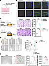 Figure 3
BATexos promote EC migration and tube formation in vitro. (A) Schematic of the internalization of exosomes labeled with PKH26 into ECs. (B) Representative immunofluorescence images showing the uptake of PKH26-labeled exosomes (red) by CD31+ ECs (green). Scale bar: 10 μm. (C) Transwell assay and calculation of the effect of GW4869-mediated inhibition of brown adipocyte exosome release or BATexos on EC migration (n = 3). Scale bar: 100 μm. (D) Scratch assay of ECs treated with BATexos; the migration rate is shown on the right (n = 3). Scale bar: 500 μm. (E) Endothelial tube formation in response to BATexos treatment (n = 3); quantification of the number of junctions and total segment length are shown on the right. Scale bar: 200 μm. The value n represents the number of biologically independent samples, from which all experimental data were obtained. Statistical significance was assessed by 2-tailed Student’s t test. The data are expressed as mean ± SEM. *P < 0.05, **P < 0.01, ****P < 0.0001.
Loss of miR-378 in BAT impairs angiogenesis and delays wound healing. BATexos carry numerous miRNAs that regulate distant organs or tissues (16, 19, 20). To identify the active miRNAs in BATexos involved in promoting angiogenesis and wound healing, we analyzed publicly available microarray data (GSE41306) from NCBI’s Gene Expression Omnibus (GEO), which revealed robust upregulation of several miRNAs in interscapular BAT under cold stimulation (Figure 4A). We subsequently conducted miRNA-Seq of serum from cold-exposed mice and identified a subset of miRNAs that were induced in serum under cold stimulation. Among them, miR-378a-3p, miR-335-5p, miR-446g, and miR-345-5p were significantly upregulated in both serum and interscapular BAT of mice after cold exposure (Figure 4A). Comparative quantification of these 4 candidates in serum showed that miR-378a-3p exhibited the highest expression (Figure 4B). Therefore, we focused primarily on miR-378a-3p in subsequent experiments. To assess the function of miR-378a-3p on vascular repair and wound healing, we created adipose tissue–specific miR-378-KO mice (miR-378Adipo-KO) (Figure 4C). In miR-378Adipo-KO mice, miR-378a-3p expression was significantly reduced in all adipose depots, including BAT and inguinal and epididymal WAT (Figure 4D). In contrast, its expression in the skeletal muscle and liver at either thermoneutral or cold temperatures remained unchanged, demonstrating a selective deletion in adipose tissue (Figure 4D). In the control mice, BAT was the primary source of miR-378a-3p, contributing to 87.62% of its total miR-378a-3p pool in adipose tissue (Figure 4E). In miR-378Adipo-KO mice, miR-378a-3p in BAT showed the greatest reduction (Figure 4, D and E). In addition, circulating levels of miR-378a-3p were markedly reduced in miR-378Adipo-KO mice (Figure 4F), suggesting that BAT serves as a major source of circulating miR-378a-3p.
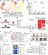 Figure 4
Loss of miR-378 in BAT impairs angiogenesis and delays wound healing. (A) Circled heatmap of miRNA-Seq from BAT (GSE41306, left) and serum (right). Serum was pooled from 5 mice per group after 3-day cold exposure. (B) Expression levels of miRNA candidates in serum of mice kept at room temperature and 4°C (n = 3). (C) Schematic showing creation of miR-378Adipo KO mice. (D) miR-378a-3p expression levels in BAT, inguinal and epididymal WAT, liver, and muscle of control and miR-378Adipo-KO mice (n = 4–6). (E) Bar chart illustrates relative abundance of miR-378 across different adipose depots, including BAT and inguinal and epididymal WAT. (F) qPCR was used to validate miR-378a-3p level in serum of miR-378Adipo–KO mice (n = 3–4). (G) CD31 staining of wounds from the miR-378Adipo-KO and littermate control mice. Quantification of CD31 relative area is shown on the right (n = 3). Scale bar: 500 μm (top) and 100 μm (bottom). (H) Representative images of wounds of control and miR-378Adipo-KO mice. (I) Quantitative analysis of the wound closure rate of control and miR-378Adipo-KO mice (n = 3). (J) Representative images of blood flow at the wounds in miR-378Adipo-KO mice and control mice. The calculated wound microvascular perfusion is shown on the right (n = 3). (K) Representative images of Evans blue staining of injured femoral artery in control mice, miR-378Adipo-KO mice, and AAV-miR-378–treated miR-378Adipo-KO mice (n = 4). Quantification data of percentage of reendothelialization are shown on the right. The value n represents the number of biologically independent samples, from which all experimental data were obtained. Statistical analysis was performed using 2-tailed Student’s t test (B, D, F, G, I, and J) or 1-way ANOVA with Tukey’s multiple-comparison test (K). The data are expressed as mean ± SEM. NS = not significant, *P < 0.05, **P < 0.01, ***P < 0.001, ****P < 0.0001.
We then sought to establish the link between BAT and systemic/wound miR-378a levels. First, we measured circulating miR-378a-3p in the serum of BATectomized mice and sham-operated mice and found it was significantly reduced after BAT removal (Supplemental Figure 4A). Extending this analysis to the wound tissue itself, we observed a concomitant decrease in both miR-378a-3p and miR-378a-5p levels in BATectomized mice compared with sham controls (Supplemental Figure 4B). Notably, the absolute level of miR-378a-5p in wounds was considerably lower than that of miR-378a-3p (Supplemental Figure 4B). To more directly investigate the role of BATexos, we administered the exosome secretion inhibitor GW4869 by local injection into the BAT of mice. This intervention led to a marked drop in serum miR-378a-3p levels (Supplemental Figure 4C). Consistent with these in vivo findings, direct profiling of isolated BAT-derived exosomes revealed a markedly higher content of miR-378a-3p compared with miR-378a-5p (Supplemental Figure 4D). Collectively, these data provide converging lines of evidence that BAT is a primary source of miR-378a-3p, which is efficiently packaged into exosomes for systemic delivery.
To elucidate the pro-angiogenic role of miR-378a-3p loss in BAT on wound healing, we generated full-thickness dorsal skin wounds in miR-378Adipo-KO mice. These mice exhibited reduced CD31 staining at the wound site, indicating impaired angiogenesis (Figure 4G), along with delayed wound closure compared with controls (Figure 4, H and I). Consistently, a reduced microvascular perfusion was observed in miR-378Adipo-KO mice (Figure 4J). Moreover, compared with the control mice, the miR-378Adipo-KO mice displayed decreased reendothelialization by Evans blue staining (Figure 4K). To investigate the specific role of miR-378a-3p in vascular repair, we performed a rescue experiment in a femoral artery injury model using miR-378Adipo-KO mice. Systemic delivery of an adeno-associated virus vector (AAV9-mmu-miR-378a-3p) via tail vein injection significantly restored the impaired reendothelialization observed in miR-378Adipo-KO mice. This result indicates that miR-378a-3p is essential for promoting vascular repair (Figure 4K). Similarly, CD31 staining was more intense in the wound tissue of AAV-miR-378–treated mice compared with that in control mice (Supplemental Figure 5A). Consistently, an accelerated wound healing rate was observed in mice treated with AAV relative to control mice (Supplemental Figure 5, B and C). Of note, the level of AKT phosphorylation and PI3K phosphorylation was significantly higher in the wounds of AAV-miR-378–treated mice than in those of control mice (Supplemental Figure 5D). To further validate the crucial role of miR-378a-3p in wound healing, we treated Rab27a-KO mice with miR-378Adipo-KO BATexos. We found control BATexos enhanced angiogenesis and promoted wound repair of Rab27a-KO mice, whereas miR-378Adipo-KO BATexos had no similar effect (Supplemental Figure 5, E–G). These results demonstrated that adipose-derived miR-378a-3p, predominantly from BAT, is essential for maintaining vascular integrity and promoting efficient wound healing. Loss of miR-378 in BAT impairs angiogenesis and reendothelialization, while exogenous overexpression enhances vascular repair.
miR-378a-3p mediates the pro-angiogenic effect of BATexos on ECs. To further elucidate the mechanism of miR-378a-3p in modulating endothelial angiogenic function, we overexpressed miR-378a-3p in ECs and performed tube formation assays to assess its role in angiogenesis. The results revealed an increased segment length and junctions of ECs after miR-378a-3p overexpression (Figure 5, A and B). In line with these results, the expression of angiogenesis-related genes (such as Tie2, Pecam1, and Fsp1) in the miR-378a-3p–overexpressed ECs was significantly increased compared with controls (Figure 5C). Moreover, to assess its role in cell migration, we performed Transwell assays. miR-378a-3p overexpression in ECs promoted their migration (Figure 5D). In contrast, BATexos isolated from miR-378Adipo-KO mice failed to promote EC migration to the same extent as BATexos from control mice (Figure 5D). Consistently, in scratch wound assays, miR-378a-3p overexpression accelerated EC migration within 24 hours. In contrast, treatment with BATexos from miR-378Adipo-KO mice resulted in impaired EC migration compared with treatment with control BATexos (Figure 5E). Collectively, these data revealed that miR-378a-3p is a critical mediator of the pro-angiogenic effects of BATexos, promoting both EC migration and tube formation in vitro.
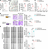 Figure 5
miR-378a-3p mediates the pro-angiogenic effect of BATexos on endothelial cells. (A) qPCR was used to measure the miR-378a-3p level in ECs overexpressing miR-378a-3p (n = 3). (B) Left: representative images of the tube formation assay in ECs overexpressing miR-378a-3p (n = 5). Scale bar: 200 μm. Right: quantification of the number of junctions and total segment length. (C) Relative expression levels of angiogenesis-related genes in ECs after miR-378a-3p overexpression (n = 3). (D) Transwell experiment and quantitative analysis of ECs overexpressing miR-378a-3p or cells cocultured with BATexos from miR-378Adipo-KO mice or littermate control mice (n = 3). Scale bar: 100 μm. (E) Scratch assays were used to assess the migration of ECs overexpressing miR-378a-3p or cells cocultured with BATexos from miR-378Adipo-KO mice (n = 3–4). Scale bar: 500 μm. Quantification of migration rate is shown on the right. The value n represents the number of biologically independent samples, from which all experimental data were obtained. Statistical significance was assessed by 2-tailed Student’s t test. The data are expressed as mean ± SEM. *P < 0.05, **P < 0.01, ***P < 0.001, ****P < 0.0001.
miR-378a-3p enhances endothelial angiogenic function by targeting Pten and activating the PI3K/AKT signaling pathway. To explore potential mechanisms by which miR-378a-3p promoted EC function, we performed mRNA-Seq analysis in ECs transduced with Lenti-miR-378a (miR-378 OE) compared with the control group (Figure 6, A and B). Gene Ontology (GO) analysis revealed that the angiogenesis-related signaling pathway was enriched after miR-378a-3p overexpression (Figure 6C). To identify direct targets of miR-378a-3p, we examined the intersection of angiogenesis-related genes in the miRWalk, DIANA, and TarBase databases, yielding 8 candidate genes (Figure 6D). Among these, Pten and Nufip2 were highly expressed in these cells (Figure 6E). It has been reported that PI3K p110α is a target gene of miR-378a-3p (17); therefore, we also examined its protein level. Additionally, overexpression of miR-378a-3p in ECs led to a reduction in the protein levels of PTEN but not NUFIP2 and PI3K p110α (Figure 6F); thus, we focused on PTEN for further investigations. Consistently, AAV-miR-378 treatment resulted in a marked decrease in PTEN protein abundance within the wound bed compared with the control group (Supplemental Figure 5D). To confirm direct interaction between miR-378a-3p and Pten, we constructed dual-luciferase reporter plasmids containing either WT or mutated Pten 3′UTR (Figure 6, G and H). Cotransfection of miR-378a-3p significantly suppressed luciferase activity in the WT construct but had no effect on the mutant, confirming direct binding (Figure 6H). PTEN is known to inhibit angiogenesis through the PI3K/AKT signaling pathway (21). Subsequently, we examined the phosphorylation of AKT and PI3K in ECs after miR-378a-3p overexpression. As expected, overexpression of miR-378a-3p increased phosphorylation levels of both PI3K and AKT in ECs (Figure 6I). Conversely, an Akt inhibitor MK2206 led to inhibition in AKT phosphorylation and PI3K phosphorylation in ECs, which was partially rescued by miR-378a-3p overexpression (Figure 6I). Functionally, overexpression of PTEN attenuated the enhanced tube formation capacity induced by miR-378a-3p (Supplemental Figure 5H and Figure 6J). To further validate the effects of miR-378a-3p and PI3K/AKT signaling on EC migration, we performed a scratch assay: miR-378a-3p overexpression promoted EC migration and Akt inhibition by MK2206 impaired EC migration, which could be partially reversed by miR-378a-3p overexpression (Figure 6K). Together, our findings demonstrated that miR-378a-3p promoted EC migration and tube formation through the PI3K/AKT signaling pathway via suppressing PTEN.
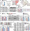 Figure 6
miR-378a-3p enhances endothelial angiogenic function by targeting Pten and activating the PI3K/AKT signaling pathway. (A) qPCR analysis of miR-378a-3p level (n = 3). (B) Schematic of RNA-Seq results for ECs overexpressing miR-378a-3p. (C) GO analysis of genes upregulated in ECs after overexpressing miR-378a-3p. (D) The intersection of the predicted results in the miRWalk database, the DIANA database, the TarBase database, and angiogenesis-related genes. (E) qPCR analysis of candidate target genes in ECs. (F) Western blot analysis of PTEN, NUFIP2, and PI3K p110α in ECs treated with miR-control and miR-378a-3p (n = 3). Right: quantification of the data. (G) Schematic illustration of the predicted binding site and corresponding mutation site between the miR-378a-3p and Pten 3′UTR. (H) Results of dual-luciferase reporter experiments (n = 3). (I) Representative images of tube formation in ECs transfected with miR-control, miR-378 mimic, or miR-378 mimic plus PTEN overexpression plasmid (n = 4). Scale bar: 200 μm. The corresponding quantitative analysis of junction number and tube length is displayed on the bottom. (J) Protein levels of p-AKT, AKT, p-PI3K, and PI3K in ECs under 4 conditions: miR-control, miR-378a-3p overexpression, Akt inhibitor (MK2206) treatment, and combined miR-378a-3p overexpression with MK2206 treatment. Quantification of p-AKT/AKT and p-PI3K/PI3K levels in ECs of 4 groups (n = 3) (right). (K) Scratch assays were used to assess the migration of ECs overexpressing miR-378a-3p or treated with the Akt inhibitor MK2206 (n = 5). Scale bar: 500 μm. The corresponding statistical analysis of migration rate is displayed on the right. The value n represents the number of biologically independent samples, from which all experimental data were obtained. Statistical analysis was performed using 2-tailed Student’s t test (A, F, and H) or 1-way ANOVA with Tukey’s multiple-comparison test (I–K). The data are expressed as mean ± SEM. NS = not significant, *P < 0.05, **P < 0.01, ***P < 0.001, ****P < 0.0001.
Liposome-encapsulated miR-378 mimics promote wound healing in diabetic mice. Diabetic wounds are one of the most challenging types of wounds to heal, and there remains an urgent need for effective therapeutic strategies (22). Given the angiogenic potential of miR-378, we hypothesized that it might enhance wound repair in the diabetic setting. Notably, we detected decreased miR-378 expression in mice with diabetes mellitus (DM) compared with those without DM (Figure 7, A and B). Consistent with previous studies, we also found that wound healing was delayed in mice with DM compared with those without DM (Figure 7, C–E). A substantial decrease of angiogenesis-related genes in wounds was observed in mice with DM (Figure 7F). To test whether exogenous delivery of miR-378 could rescue impaired wound healing in diabetes, we generated liposomes encapsulating synthetic miR-378a-3p mimic (miR-378-loaded liposomes) using lipid nanoparticle (LNP) technology, a clinically safe and customizable delivery platform (23–26). We applied these liposomes to the back skin wounds of a diabetic mouse model to explore the effect of miR-378 on diabetic wound healing, which is one of the refractory ulcers (Figure 7, G and H). An elevated expression level of miR-378 was detected in diabetic wounds after the administration of miR-378-loaded liposomes (Figure 7I). Compared with that in the control group, the wound healing rate was higher after miR-378-loaded liposomes treatment, which was consistent with increased CD31 expression in the wound area after miR-378-loaded liposomes treatment, indicating improved angiogenesis (Figure 7, J–M). Taken together, these findings revealed that liposome-based delivery of an miR-378a-3p mimic promotes vascularization and accelerates diabetic wound healing, highlighting its translational potential as a promising therapeutic approach for chronic diabetic ulcer.
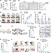 Figure 7
Liposome-encapsulated miR-378 mimics promote wound healing in diabetic mice. (A) Random blood glucose in diabetic (DM) and nondiabetic (NDM) mouse wounds. (B) Relative expression level of miR-378 in DM and NDM mouse wounds. (C) CD31 staining of DM and NDM wounds. Scale bar: 200 μm. Quantification of CD31 relative area is shown on the right (n = 4). (D) Representative images of DM and NDM wounds. (E) Quantitative analysis of wound healing rates in DM and NDM groups. (F) Angiogenesis-related genes detected in wounds of DM and NDM mice. (G) Schematic illustration of the preparation of miR-378a-3p LNPs. (H) Schematic illustration of the DM wound model and treatment with miR-378 LNPs. (I) Relative miR-378 levels in DM wounds. (J) CD31 staining of DM wounds treated with miR-378 LNPs. Scale bar: 200 μm (top) and 50 μm (bottom). (K) Quantification of CD31 relative area in DM wounds treated with control or miR-378 LNPs (n = 4–5). (L) Representative images of DM wounds treated with miR-378 LNPs (n = 3). (M) Quantification of wound closure rate in mice treated with miR-378 LNPs (n = 3). The value n represents the number of biologically independent samples, from which all experimental data were obtained. Statistical significance was assessed by 2-tailed Student’s t test. The data are expressed as mean ± SEM. *P < 0.05, **P < 0.01, ***P < 0.001, ****P < 0.0001.


