
Research ArticleInflammationMetabolism
Open Access |  10.1172/jci.insight.198123
A weight loss fibrotic response in visceral fat is driven by defective lysosome-dependent collagenolysis
Clémentine Rebière,1 Amanda Oliveira,1,2 Sébastien Dussaud,1 Iman Haddad,3 Agatha Assis-Ferreira,1,4 Adma M. Babetto,1,2 Christine Rouault,1 Fatiha Merabtene,1 Cheïma Benbida,1 Jimon Abatan,1 Simone Vargas da Silva,1,4 Laís Bhering Martins,1,2 Licia Torres,1,2 Ana Letícia M. Silveira,1,2 Cindy Rose,1 Laurent Genser,1,5 Fabrizio Andreelli,1,6 Isabelle Dugail,1 Marina Chaves de Oliveira,1,2 Joelle Vinh,3 Simon Lecoutre,1 Adaliene V.M. Ferreira,2 Karine Clément,1,7 Emmanuel L. Gautier,1 and Genevieve Marcelin1
1Sorbonne Université, INSERM, Nutrition and obesities: systemic approach research group, Nutriomics, Paris, France.
2Immunometabolism, Department of Nutrition, Nursing School, Universidade Federal de Minas Gerais, Belo Horizonte, Brazil.
3ESPCI Paris, Paris Sciences et Lettres University, UAR CNRS 2051, Biological Mass Spectrometry and Proteomics, Paris, France.
4Cellular and Molecular Pharmacology, Department of Cell Biology, Institute of Biology, Universidade do Estado do Rio de Janeiro, Rio de Janeiro, Brazil.
5Visceral Surgery Department,
6Diabetology Department, and
7Nutrition Department, Centre de Recherche en Nutrition Humaine d’Ile-de-France, Assistance Publique Hôpitaux de Paris, Pitié-Salpêtrière Hospital, Paris, France.
Address for correspondence. Geneviève Marcelin, Emmanuel L. Gautier, or Karine Clément, Sorbonne Université & INSERM, 91 boulevard de l’Hopital 75013 Paris, France. Email: genevieve.marcelin@inserm.fr (GM); emmanuel-laurent.gautier@inserm.fr (ELG); karine.clement@inserm.fr (KC).
Authorship note: C Rebière and AO contributed equally to this work.
Find articles by Rebière, C. in: PubMed | Google Scholar
1Sorbonne Université, INSERM, Nutrition and obesities: systemic approach research group, Nutriomics, Paris, France.
2Immunometabolism, Department of Nutrition, Nursing School, Universidade Federal de Minas Gerais, Belo Horizonte, Brazil.
3ESPCI Paris, Paris Sciences et Lettres University, UAR CNRS 2051, Biological Mass Spectrometry and Proteomics, Paris, France.
4Cellular and Molecular Pharmacology, Department of Cell Biology, Institute of Biology, Universidade do Estado do Rio de Janeiro, Rio de Janeiro, Brazil.
5Visceral Surgery Department,
6Diabetology Department, and
7Nutrition Department, Centre de Recherche en Nutrition Humaine d’Ile-de-France, Assistance Publique Hôpitaux de Paris, Pitié-Salpêtrière Hospital, Paris, France.
Address for correspondence. Geneviève Marcelin, Emmanuel L. Gautier, or Karine Clément, Sorbonne Université & INSERM, 91 boulevard de l’Hopital 75013 Paris, France. Email: genevieve.marcelin@inserm.fr (GM); emmanuel-laurent.gautier@inserm.fr (ELG); karine.clement@inserm.fr (KC).
Authorship note: C Rebière and AO contributed equally to this work.
Find articles by Oliveira, A. in: PubMed | Google Scholar
1Sorbonne Université, INSERM, Nutrition and obesities: systemic approach research group, Nutriomics, Paris, France.
2Immunometabolism, Department of Nutrition, Nursing School, Universidade Federal de Minas Gerais, Belo Horizonte, Brazil.
3ESPCI Paris, Paris Sciences et Lettres University, UAR CNRS 2051, Biological Mass Spectrometry and Proteomics, Paris, France.
4Cellular and Molecular Pharmacology, Department of Cell Biology, Institute of Biology, Universidade do Estado do Rio de Janeiro, Rio de Janeiro, Brazil.
5Visceral Surgery Department,
6Diabetology Department, and
7Nutrition Department, Centre de Recherche en Nutrition Humaine d’Ile-de-France, Assistance Publique Hôpitaux de Paris, Pitié-Salpêtrière Hospital, Paris, France.
Address for correspondence. Geneviève Marcelin, Emmanuel L. Gautier, or Karine Clément, Sorbonne Université & INSERM, 91 boulevard de l’Hopital 75013 Paris, France. Email: genevieve.marcelin@inserm.fr (GM); emmanuel-laurent.gautier@inserm.fr (ELG); karine.clement@inserm.fr (KC).
Authorship note: C Rebière and AO contributed equally to this work.
Find articles by Dussaud, S. in: PubMed | Google Scholar
1Sorbonne Université, INSERM, Nutrition and obesities: systemic approach research group, Nutriomics, Paris, France.
2Immunometabolism, Department of Nutrition, Nursing School, Universidade Federal de Minas Gerais, Belo Horizonte, Brazil.
3ESPCI Paris, Paris Sciences et Lettres University, UAR CNRS 2051, Biological Mass Spectrometry and Proteomics, Paris, France.
4Cellular and Molecular Pharmacology, Department of Cell Biology, Institute of Biology, Universidade do Estado do Rio de Janeiro, Rio de Janeiro, Brazil.
5Visceral Surgery Department,
6Diabetology Department, and
7Nutrition Department, Centre de Recherche en Nutrition Humaine d’Ile-de-France, Assistance Publique Hôpitaux de Paris, Pitié-Salpêtrière Hospital, Paris, France.
Address for correspondence. Geneviève Marcelin, Emmanuel L. Gautier, or Karine Clément, Sorbonne Université & INSERM, 91 boulevard de l’Hopital 75013 Paris, France. Email: genevieve.marcelin@inserm.fr (GM); emmanuel-laurent.gautier@inserm.fr (ELG); karine.clement@inserm.fr (KC).
Authorship note: C Rebière and AO contributed equally to this work.
Find articles by Haddad, I. in: PubMed | Google Scholar
1Sorbonne Université, INSERM, Nutrition and obesities: systemic approach research group, Nutriomics, Paris, France.
2Immunometabolism, Department of Nutrition, Nursing School, Universidade Federal de Minas Gerais, Belo Horizonte, Brazil.
3ESPCI Paris, Paris Sciences et Lettres University, UAR CNRS 2051, Biological Mass Spectrometry and Proteomics, Paris, France.
4Cellular and Molecular Pharmacology, Department of Cell Biology, Institute of Biology, Universidade do Estado do Rio de Janeiro, Rio de Janeiro, Brazil.
5Visceral Surgery Department,
6Diabetology Department, and
7Nutrition Department, Centre de Recherche en Nutrition Humaine d’Ile-de-France, Assistance Publique Hôpitaux de Paris, Pitié-Salpêtrière Hospital, Paris, France.
Address for correspondence. Geneviève Marcelin, Emmanuel L. Gautier, or Karine Clément, Sorbonne Université & INSERM, 91 boulevard de l’Hopital 75013 Paris, France. Email: genevieve.marcelin@inserm.fr (GM); emmanuel-laurent.gautier@inserm.fr (ELG); karine.clement@inserm.fr (KC).
Authorship note: C Rebière and AO contributed equally to this work.
Find articles by Assis-Ferreira, A. in: PubMed | Google Scholar
1Sorbonne Université, INSERM, Nutrition and obesities: systemic approach research group, Nutriomics, Paris, France.
2Immunometabolism, Department of Nutrition, Nursing School, Universidade Federal de Minas Gerais, Belo Horizonte, Brazil.
3ESPCI Paris, Paris Sciences et Lettres University, UAR CNRS 2051, Biological Mass Spectrometry and Proteomics, Paris, France.
4Cellular and Molecular Pharmacology, Department of Cell Biology, Institute of Biology, Universidade do Estado do Rio de Janeiro, Rio de Janeiro, Brazil.
5Visceral Surgery Department,
6Diabetology Department, and
7Nutrition Department, Centre de Recherche en Nutrition Humaine d’Ile-de-France, Assistance Publique Hôpitaux de Paris, Pitié-Salpêtrière Hospital, Paris, France.
Address for correspondence. Geneviève Marcelin, Emmanuel L. Gautier, or Karine Clément, Sorbonne Université & INSERM, 91 boulevard de l’Hopital 75013 Paris, France. Email: genevieve.marcelin@inserm.fr (GM); emmanuel-laurent.gautier@inserm.fr (ELG); karine.clement@inserm.fr (KC).
Authorship note: C Rebière and AO contributed equally to this work.
Find articles by Babetto, A. in: PubMed | Google Scholar
1Sorbonne Université, INSERM, Nutrition and obesities: systemic approach research group, Nutriomics, Paris, France.
2Immunometabolism, Department of Nutrition, Nursing School, Universidade Federal de Minas Gerais, Belo Horizonte, Brazil.
3ESPCI Paris, Paris Sciences et Lettres University, UAR CNRS 2051, Biological Mass Spectrometry and Proteomics, Paris, France.
4Cellular and Molecular Pharmacology, Department of Cell Biology, Institute of Biology, Universidade do Estado do Rio de Janeiro, Rio de Janeiro, Brazil.
5Visceral Surgery Department,
6Diabetology Department, and
7Nutrition Department, Centre de Recherche en Nutrition Humaine d’Ile-de-France, Assistance Publique Hôpitaux de Paris, Pitié-Salpêtrière Hospital, Paris, France.
Address for correspondence. Geneviève Marcelin, Emmanuel L. Gautier, or Karine Clément, Sorbonne Université & INSERM, 91 boulevard de l’Hopital 75013 Paris, France. Email: genevieve.marcelin@inserm.fr (GM); emmanuel-laurent.gautier@inserm.fr (ELG); karine.clement@inserm.fr (KC).
Authorship note: C Rebière and AO contributed equally to this work.
Find articles by Rouault, C. in: PubMed | Google Scholar
1Sorbonne Université, INSERM, Nutrition and obesities: systemic approach research group, Nutriomics, Paris, France.
2Immunometabolism, Department of Nutrition, Nursing School, Universidade Federal de Minas Gerais, Belo Horizonte, Brazil.
3ESPCI Paris, Paris Sciences et Lettres University, UAR CNRS 2051, Biological Mass Spectrometry and Proteomics, Paris, France.
4Cellular and Molecular Pharmacology, Department of Cell Biology, Institute of Biology, Universidade do Estado do Rio de Janeiro, Rio de Janeiro, Brazil.
5Visceral Surgery Department,
6Diabetology Department, and
7Nutrition Department, Centre de Recherche en Nutrition Humaine d’Ile-de-France, Assistance Publique Hôpitaux de Paris, Pitié-Salpêtrière Hospital, Paris, France.
Address for correspondence. Geneviève Marcelin, Emmanuel L. Gautier, or Karine Clément, Sorbonne Université & INSERM, 91 boulevard de l’Hopital 75013 Paris, France. Email: genevieve.marcelin@inserm.fr (GM); emmanuel-laurent.gautier@inserm.fr (ELG); karine.clement@inserm.fr (KC).
Authorship note: C Rebière and AO contributed equally to this work.
Find articles by Merabtene, F. in: PubMed | Google Scholar
1Sorbonne Université, INSERM, Nutrition and obesities: systemic approach research group, Nutriomics, Paris, France.
2Immunometabolism, Department of Nutrition, Nursing School, Universidade Federal de Minas Gerais, Belo Horizonte, Brazil.
3ESPCI Paris, Paris Sciences et Lettres University, UAR CNRS 2051, Biological Mass Spectrometry and Proteomics, Paris, France.
4Cellular and Molecular Pharmacology, Department of Cell Biology, Institute of Biology, Universidade do Estado do Rio de Janeiro, Rio de Janeiro, Brazil.
5Visceral Surgery Department,
6Diabetology Department, and
7Nutrition Department, Centre de Recherche en Nutrition Humaine d’Ile-de-France, Assistance Publique Hôpitaux de Paris, Pitié-Salpêtrière Hospital, Paris, France.
Address for correspondence. Geneviève Marcelin, Emmanuel L. Gautier, or Karine Clément, Sorbonne Université & INSERM, 91 boulevard de l’Hopital 75013 Paris, France. Email: genevieve.marcelin@inserm.fr (GM); emmanuel-laurent.gautier@inserm.fr (ELG); karine.clement@inserm.fr (KC).
Authorship note: C Rebière and AO contributed equally to this work.
Find articles by Benbida, C. in: PubMed | Google Scholar
1Sorbonne Université, INSERM, Nutrition and obesities: systemic approach research group, Nutriomics, Paris, France.
2Immunometabolism, Department of Nutrition, Nursing School, Universidade Federal de Minas Gerais, Belo Horizonte, Brazil.
3ESPCI Paris, Paris Sciences et Lettres University, UAR CNRS 2051, Biological Mass Spectrometry and Proteomics, Paris, France.
4Cellular and Molecular Pharmacology, Department of Cell Biology, Institute of Biology, Universidade do Estado do Rio de Janeiro, Rio de Janeiro, Brazil.
5Visceral Surgery Department,
6Diabetology Department, and
7Nutrition Department, Centre de Recherche en Nutrition Humaine d’Ile-de-France, Assistance Publique Hôpitaux de Paris, Pitié-Salpêtrière Hospital, Paris, France.
Address for correspondence. Geneviève Marcelin, Emmanuel L. Gautier, or Karine Clément, Sorbonne Université & INSERM, 91 boulevard de l’Hopital 75013 Paris, France. Email: genevieve.marcelin@inserm.fr (GM); emmanuel-laurent.gautier@inserm.fr (ELG); karine.clement@inserm.fr (KC).
Authorship note: C Rebière and AO contributed equally to this work.
Find articles by Abatan, J. in: PubMed | Google Scholar
1Sorbonne Université, INSERM, Nutrition and obesities: systemic approach research group, Nutriomics, Paris, France.
2Immunometabolism, Department of Nutrition, Nursing School, Universidade Federal de Minas Gerais, Belo Horizonte, Brazil.
3ESPCI Paris, Paris Sciences et Lettres University, UAR CNRS 2051, Biological Mass Spectrometry and Proteomics, Paris, France.
4Cellular and Molecular Pharmacology, Department of Cell Biology, Institute of Biology, Universidade do Estado do Rio de Janeiro, Rio de Janeiro, Brazil.
5Visceral Surgery Department,
6Diabetology Department, and
7Nutrition Department, Centre de Recherche en Nutrition Humaine d’Ile-de-France, Assistance Publique Hôpitaux de Paris, Pitié-Salpêtrière Hospital, Paris, France.
Address for correspondence. Geneviève Marcelin, Emmanuel L. Gautier, or Karine Clément, Sorbonne Université & INSERM, 91 boulevard de l’Hopital 75013 Paris, France. Email: genevieve.marcelin@inserm.fr (GM); emmanuel-laurent.gautier@inserm.fr (ELG); karine.clement@inserm.fr (KC).
Authorship note: C Rebière and AO contributed equally to this work.
Find articles by Vargas da Silva, S. in: PubMed | Google Scholar
1Sorbonne Université, INSERM, Nutrition and obesities: systemic approach research group, Nutriomics, Paris, France.
2Immunometabolism, Department of Nutrition, Nursing School, Universidade Federal de Minas Gerais, Belo Horizonte, Brazil.
3ESPCI Paris, Paris Sciences et Lettres University, UAR CNRS 2051, Biological Mass Spectrometry and Proteomics, Paris, France.
4Cellular and Molecular Pharmacology, Department of Cell Biology, Institute of Biology, Universidade do Estado do Rio de Janeiro, Rio de Janeiro, Brazil.
5Visceral Surgery Department,
6Diabetology Department, and
7Nutrition Department, Centre de Recherche en Nutrition Humaine d’Ile-de-France, Assistance Publique Hôpitaux de Paris, Pitié-Salpêtrière Hospital, Paris, France.
Address for correspondence. Geneviève Marcelin, Emmanuel L. Gautier, or Karine Clément, Sorbonne Université & INSERM, 91 boulevard de l’Hopital 75013 Paris, France. Email: genevieve.marcelin@inserm.fr (GM); emmanuel-laurent.gautier@inserm.fr (ELG); karine.clement@inserm.fr (KC).
Authorship note: C Rebière and AO contributed equally to this work.
Find articles by Martins, L. in: PubMed | Google Scholar
1Sorbonne Université, INSERM, Nutrition and obesities: systemic approach research group, Nutriomics, Paris, France.
2Immunometabolism, Department of Nutrition, Nursing School, Universidade Federal de Minas Gerais, Belo Horizonte, Brazil.
3ESPCI Paris, Paris Sciences et Lettres University, UAR CNRS 2051, Biological Mass Spectrometry and Proteomics, Paris, France.
4Cellular and Molecular Pharmacology, Department of Cell Biology, Institute of Biology, Universidade do Estado do Rio de Janeiro, Rio de Janeiro, Brazil.
5Visceral Surgery Department,
6Diabetology Department, and
7Nutrition Department, Centre de Recherche en Nutrition Humaine d’Ile-de-France, Assistance Publique Hôpitaux de Paris, Pitié-Salpêtrière Hospital, Paris, France.
Address for correspondence. Geneviève Marcelin, Emmanuel L. Gautier, or Karine Clément, Sorbonne Université & INSERM, 91 boulevard de l’Hopital 75013 Paris, France. Email: genevieve.marcelin@inserm.fr (GM); emmanuel-laurent.gautier@inserm.fr (ELG); karine.clement@inserm.fr (KC).
Authorship note: C Rebière and AO contributed equally to this work.
Find articles by
Torres, L.
in:
PubMed
|
Google Scholar
|

1Sorbonne Université, INSERM, Nutrition and obesities: systemic approach research group, Nutriomics, Paris, France.
2Immunometabolism, Department of Nutrition, Nursing School, Universidade Federal de Minas Gerais, Belo Horizonte, Brazil.
3ESPCI Paris, Paris Sciences et Lettres University, UAR CNRS 2051, Biological Mass Spectrometry and Proteomics, Paris, France.
4Cellular and Molecular Pharmacology, Department of Cell Biology, Institute of Biology, Universidade do Estado do Rio de Janeiro, Rio de Janeiro, Brazil.
5Visceral Surgery Department,
6Diabetology Department, and
7Nutrition Department, Centre de Recherche en Nutrition Humaine d’Ile-de-France, Assistance Publique Hôpitaux de Paris, Pitié-Salpêtrière Hospital, Paris, France.
Address for correspondence. Geneviève Marcelin, Emmanuel L. Gautier, or Karine Clément, Sorbonne Université & INSERM, 91 boulevard de l’Hopital 75013 Paris, France. Email: genevieve.marcelin@inserm.fr (GM); emmanuel-laurent.gautier@inserm.fr (ELG); karine.clement@inserm.fr (KC).
Authorship note: C Rebière and AO contributed equally to this work.
Find articles by Silveira, A. in: PubMed | Google Scholar
1Sorbonne Université, INSERM, Nutrition and obesities: systemic approach research group, Nutriomics, Paris, France.
2Immunometabolism, Department of Nutrition, Nursing School, Universidade Federal de Minas Gerais, Belo Horizonte, Brazil.
3ESPCI Paris, Paris Sciences et Lettres University, UAR CNRS 2051, Biological Mass Spectrometry and Proteomics, Paris, France.
4Cellular and Molecular Pharmacology, Department of Cell Biology, Institute of Biology, Universidade do Estado do Rio de Janeiro, Rio de Janeiro, Brazil.
5Visceral Surgery Department,
6Diabetology Department, and
7Nutrition Department, Centre de Recherche en Nutrition Humaine d’Ile-de-France, Assistance Publique Hôpitaux de Paris, Pitié-Salpêtrière Hospital, Paris, France.
Address for correspondence. Geneviève Marcelin, Emmanuel L. Gautier, or Karine Clément, Sorbonne Université & INSERM, 91 boulevard de l’Hopital 75013 Paris, France. Email: genevieve.marcelin@inserm.fr (GM); emmanuel-laurent.gautier@inserm.fr (ELG); karine.clement@inserm.fr (KC).
Authorship note: C Rebière and AO contributed equally to this work.
Find articles by Rose, C. in: PubMed | Google Scholar
1Sorbonne Université, INSERM, Nutrition and obesities: systemic approach research group, Nutriomics, Paris, France.
2Immunometabolism, Department of Nutrition, Nursing School, Universidade Federal de Minas Gerais, Belo Horizonte, Brazil.
3ESPCI Paris, Paris Sciences et Lettres University, UAR CNRS 2051, Biological Mass Spectrometry and Proteomics, Paris, France.
4Cellular and Molecular Pharmacology, Department of Cell Biology, Institute of Biology, Universidade do Estado do Rio de Janeiro, Rio de Janeiro, Brazil.
5Visceral Surgery Department,
6Diabetology Department, and
7Nutrition Department, Centre de Recherche en Nutrition Humaine d’Ile-de-France, Assistance Publique Hôpitaux de Paris, Pitié-Salpêtrière Hospital, Paris, France.
Address for correspondence. Geneviève Marcelin, Emmanuel L. Gautier, or Karine Clément, Sorbonne Université & INSERM, 91 boulevard de l’Hopital 75013 Paris, France. Email: genevieve.marcelin@inserm.fr (GM); emmanuel-laurent.gautier@inserm.fr (ELG); karine.clement@inserm.fr (KC).
Authorship note: C Rebière and AO contributed equally to this work.
Find articles by Genser, L. in: PubMed | Google Scholar
1Sorbonne Université, INSERM, Nutrition and obesities: systemic approach research group, Nutriomics, Paris, France.
2Immunometabolism, Department of Nutrition, Nursing School, Universidade Federal de Minas Gerais, Belo Horizonte, Brazil.
3ESPCI Paris, Paris Sciences et Lettres University, UAR CNRS 2051, Biological Mass Spectrometry and Proteomics, Paris, France.
4Cellular and Molecular Pharmacology, Department of Cell Biology, Institute of Biology, Universidade do Estado do Rio de Janeiro, Rio de Janeiro, Brazil.
5Visceral Surgery Department,
6Diabetology Department, and
7Nutrition Department, Centre de Recherche en Nutrition Humaine d’Ile-de-France, Assistance Publique Hôpitaux de Paris, Pitié-Salpêtrière Hospital, Paris, France.
Address for correspondence. Geneviève Marcelin, Emmanuel L. Gautier, or Karine Clément, Sorbonne Université & INSERM, 91 boulevard de l’Hopital 75013 Paris, France. Email: genevieve.marcelin@inserm.fr (GM); emmanuel-laurent.gautier@inserm.fr (ELG); karine.clement@inserm.fr (KC).
Authorship note: C Rebière and AO contributed equally to this work.
Find articles by Andreelli, F. in: PubMed | Google Scholar
1Sorbonne Université, INSERM, Nutrition and obesities: systemic approach research group, Nutriomics, Paris, France.
2Immunometabolism, Department of Nutrition, Nursing School, Universidade Federal de Minas Gerais, Belo Horizonte, Brazil.
3ESPCI Paris, Paris Sciences et Lettres University, UAR CNRS 2051, Biological Mass Spectrometry and Proteomics, Paris, France.
4Cellular and Molecular Pharmacology, Department of Cell Biology, Institute of Biology, Universidade do Estado do Rio de Janeiro, Rio de Janeiro, Brazil.
5Visceral Surgery Department,
6Diabetology Department, and
7Nutrition Department, Centre de Recherche en Nutrition Humaine d’Ile-de-France, Assistance Publique Hôpitaux de Paris, Pitié-Salpêtrière Hospital, Paris, France.
Address for correspondence. Geneviève Marcelin, Emmanuel L. Gautier, or Karine Clément, Sorbonne Université & INSERM, 91 boulevard de l’Hopital 75013 Paris, France. Email: genevieve.marcelin@inserm.fr (GM); emmanuel-laurent.gautier@inserm.fr (ELG); karine.clement@inserm.fr (KC).
Authorship note: C Rebière and AO contributed equally to this work.
Find articles by
Dugail, I.
in:
PubMed
|
Google Scholar
|
1Sorbonne Université, INSERM, Nutrition and obesities: systemic approach research group, Nutriomics, Paris, France.
2Immunometabolism, Department of Nutrition, Nursing School, Universidade Federal de Minas Gerais, Belo Horizonte, Brazil.
3ESPCI Paris, Paris Sciences et Lettres University, UAR CNRS 2051, Biological Mass Spectrometry and Proteomics, Paris, France.
4Cellular and Molecular Pharmacology, Department of Cell Biology, Institute of Biology, Universidade do Estado do Rio de Janeiro, Rio de Janeiro, Brazil.
5Visceral Surgery Department,
6Diabetology Department, and
7Nutrition Department, Centre de Recherche en Nutrition Humaine d’Ile-de-France, Assistance Publique Hôpitaux de Paris, Pitié-Salpêtrière Hospital, Paris, France.
Address for correspondence. Geneviève Marcelin, Emmanuel L. Gautier, or Karine Clément, Sorbonne Université & INSERM, 91 boulevard de l’Hopital 75013 Paris, France. Email: genevieve.marcelin@inserm.fr (GM); emmanuel-laurent.gautier@inserm.fr (ELG); karine.clement@inserm.fr (KC).
Authorship note: C Rebière and AO contributed equally to this work.
Find articles by de Oliveira, M. in: PubMed | Google Scholar
1Sorbonne Université, INSERM, Nutrition and obesities: systemic approach research group, Nutriomics, Paris, France.
2Immunometabolism, Department of Nutrition, Nursing School, Universidade Federal de Minas Gerais, Belo Horizonte, Brazil.
3ESPCI Paris, Paris Sciences et Lettres University, UAR CNRS 2051, Biological Mass Spectrometry and Proteomics, Paris, France.
4Cellular and Molecular Pharmacology, Department of Cell Biology, Institute of Biology, Universidade do Estado do Rio de Janeiro, Rio de Janeiro, Brazil.
5Visceral Surgery Department,
6Diabetology Department, and
7Nutrition Department, Centre de Recherche en Nutrition Humaine d’Ile-de-France, Assistance Publique Hôpitaux de Paris, Pitié-Salpêtrière Hospital, Paris, France.
Address for correspondence. Geneviève Marcelin, Emmanuel L. Gautier, or Karine Clément, Sorbonne Université & INSERM, 91 boulevard de l’Hopital 75013 Paris, France. Email: genevieve.marcelin@inserm.fr (GM); emmanuel-laurent.gautier@inserm.fr (ELG); karine.clement@inserm.fr (KC).
Authorship note: C Rebière and AO contributed equally to this work.
Find articles by Vinh, J. in: PubMed | Google Scholar
1Sorbonne Université, INSERM, Nutrition and obesities: systemic approach research group, Nutriomics, Paris, France.
2Immunometabolism, Department of Nutrition, Nursing School, Universidade Federal de Minas Gerais, Belo Horizonte, Brazil.
3ESPCI Paris, Paris Sciences et Lettres University, UAR CNRS 2051, Biological Mass Spectrometry and Proteomics, Paris, France.
4Cellular and Molecular Pharmacology, Department of Cell Biology, Institute of Biology, Universidade do Estado do Rio de Janeiro, Rio de Janeiro, Brazil.
5Visceral Surgery Department,
6Diabetology Department, and
7Nutrition Department, Centre de Recherche en Nutrition Humaine d’Ile-de-France, Assistance Publique Hôpitaux de Paris, Pitié-Salpêtrière Hospital, Paris, France.
Address for correspondence. Geneviève Marcelin, Emmanuel L. Gautier, or Karine Clément, Sorbonne Université & INSERM, 91 boulevard de l’Hopital 75013 Paris, France. Email: genevieve.marcelin@inserm.fr (GM); emmanuel-laurent.gautier@inserm.fr (ELG); karine.clement@inserm.fr (KC).
Authorship note: C Rebière and AO contributed equally to this work.
Find articles by Lecoutre, S. in: PubMed | Google Scholar
1Sorbonne Université, INSERM, Nutrition and obesities: systemic approach research group, Nutriomics, Paris, France.
2Immunometabolism, Department of Nutrition, Nursing School, Universidade Federal de Minas Gerais, Belo Horizonte, Brazil.
3ESPCI Paris, Paris Sciences et Lettres University, UAR CNRS 2051, Biological Mass Spectrometry and Proteomics, Paris, France.
4Cellular and Molecular Pharmacology, Department of Cell Biology, Institute of Biology, Universidade do Estado do Rio de Janeiro, Rio de Janeiro, Brazil.
5Visceral Surgery Department,
6Diabetology Department, and
7Nutrition Department, Centre de Recherche en Nutrition Humaine d’Ile-de-France, Assistance Publique Hôpitaux de Paris, Pitié-Salpêtrière Hospital, Paris, France.
Address for correspondence. Geneviève Marcelin, Emmanuel L. Gautier, or Karine Clément, Sorbonne Université & INSERM, 91 boulevard de l’Hopital 75013 Paris, France. Email: genevieve.marcelin@inserm.fr (GM); emmanuel-laurent.gautier@inserm.fr (ELG); karine.clement@inserm.fr (KC).
Authorship note: C Rebière and AO contributed equally to this work.
Find articles by Ferreira, A. in: PubMed | Google Scholar
1Sorbonne Université, INSERM, Nutrition and obesities: systemic approach research group, Nutriomics, Paris, France.
2Immunometabolism, Department of Nutrition, Nursing School, Universidade Federal de Minas Gerais, Belo Horizonte, Brazil.
3ESPCI Paris, Paris Sciences et Lettres University, UAR CNRS 2051, Biological Mass Spectrometry and Proteomics, Paris, France.
4Cellular and Molecular Pharmacology, Department of Cell Biology, Institute of Biology, Universidade do Estado do Rio de Janeiro, Rio de Janeiro, Brazil.
5Visceral Surgery Department,
6Diabetology Department, and
7Nutrition Department, Centre de Recherche en Nutrition Humaine d’Ile-de-France, Assistance Publique Hôpitaux de Paris, Pitié-Salpêtrière Hospital, Paris, France.
Address for correspondence. Geneviève Marcelin, Emmanuel L. Gautier, or Karine Clément, Sorbonne Université & INSERM, 91 boulevard de l’Hopital 75013 Paris, France. Email: genevieve.marcelin@inserm.fr (GM); emmanuel-laurent.gautier@inserm.fr (ELG); karine.clement@inserm.fr (KC).
Authorship note: C Rebière and AO contributed equally to this work.
Find articles by Clément, K. in: PubMed | Google Scholar
1Sorbonne Université, INSERM, Nutrition and obesities: systemic approach research group, Nutriomics, Paris, France.
2Immunometabolism, Department of Nutrition, Nursing School, Universidade Federal de Minas Gerais, Belo Horizonte, Brazil.
3ESPCI Paris, Paris Sciences et Lettres University, UAR CNRS 2051, Biological Mass Spectrometry and Proteomics, Paris, France.
4Cellular and Molecular Pharmacology, Department of Cell Biology, Institute of Biology, Universidade do Estado do Rio de Janeiro, Rio de Janeiro, Brazil.
5Visceral Surgery Department,
6Diabetology Department, and
7Nutrition Department, Centre de Recherche en Nutrition Humaine d’Ile-de-France, Assistance Publique Hôpitaux de Paris, Pitié-Salpêtrière Hospital, Paris, France.
Address for correspondence. Geneviève Marcelin, Emmanuel L. Gautier, or Karine Clément, Sorbonne Université & INSERM, 91 boulevard de l’Hopital 75013 Paris, France. Email: genevieve.marcelin@inserm.fr (GM); emmanuel-laurent.gautier@inserm.fr (ELG); karine.clement@inserm.fr (KC).
Authorship note: C Rebière and AO contributed equally to this work.
Find articles by Gautier, E. in: PubMed | Google Scholar
1Sorbonne Université, INSERM, Nutrition and obesities: systemic approach research group, Nutriomics, Paris, France.
2Immunometabolism, Department of Nutrition, Nursing School, Universidade Federal de Minas Gerais, Belo Horizonte, Brazil.
3ESPCI Paris, Paris Sciences et Lettres University, UAR CNRS 2051, Biological Mass Spectrometry and Proteomics, Paris, France.
4Cellular and Molecular Pharmacology, Department of Cell Biology, Institute of Biology, Universidade do Estado do Rio de Janeiro, Rio de Janeiro, Brazil.
5Visceral Surgery Department,
6Diabetology Department, and
7Nutrition Department, Centre de Recherche en Nutrition Humaine d’Ile-de-France, Assistance Publique Hôpitaux de Paris, Pitié-Salpêtrière Hospital, Paris, France.
Address for correspondence. Geneviève Marcelin, Emmanuel L. Gautier, or Karine Clément, Sorbonne Université & INSERM, 91 boulevard de l’Hopital 75013 Paris, France. Email: genevieve.marcelin@inserm.fr (GM); emmanuel-laurent.gautier@inserm.fr (ELG); karine.clement@inserm.fr (KC).
Authorship note: C Rebière and AO contributed equally to this work.
Find articles by Marcelin, G. in: PubMed | Google Scholar
Published November 9, 2026 - More info
JCI Insight. 2026;11(21):e198123. https://doi.org/10.1172/jci.insight.198123.
© 2026 Rebière et al. This work is licensed under the Creative Commons Attribution 4.0 International License. To view a copy of this license, visit http://creativecommons.org/licenses/by/4.0/.
Received: July 18, 2025; Accepted: September 21, 2026


-
Results
vWAT dysfunctions persist after WL after obesity. WL remains the cornerstone of obesity management. However, accumulating evidence suggests that vWAT does not fully recover its functional integrity after obesity. In particular, although sustained impairments in postobese vWAT resilience have been described (22–24), the mechanisms underlying the persistence of vWAT fibrosis following WL remain largely unexplored. To address this question, we used the C3H/HeOuJ (C3H) mouse strain, a well-established model of obesity-associated fibrosis characterized by robust ECM deposition in the vWAT (8, 9). This model provides a unique opportunity to investigate the mechanisms governing fibrosis persistence and ECM remodeling following WL. We therefore performed an in-depth characterization of vWAT remodeling and function during the postobese state.
To this end, we analyzed C3H mice fed a high-fat diet (HFD) for 4 weeks, after which mice were fed chow diet for 4 weeks to induce WL (resulting in formerly obese [FOb] animals) (Figure 1A). FOb animals were compared with age-matched lean animals and to baseline obese mice before WL initiation (Ob4w; Figure 1A). Following the nutritional switch, body weight, body composition, and fat depot masses in FOb mice normalized (Figure 1, B–E, and Supplemental Figure 1A; supplemental material available online with this article; https://doi.org/10.1172/jci.insight.198123DS1). Accordingly, adipocyte size in both depots returned to levels observed in lean animals (Figure 1F and Supplemental Figure 1, B and C) and glucose tolerance improved, reaching values comparable to those of lean mice (Figure 1G), contrasting with the impaired glucose clearance seen in Ob4w animals (Supplemental Figure 1, D and E).
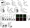 Figure 1
EpiWAT exhibited alterations in formerly obese mice. (A) Experimental design showing dietary interventions (chow or high-fat diet, as indicated) and the timeline for obesity induction and weight loss. Arrows indicate the feeding durations for the lean (Le), obese (Ob4w), and formerly obese (FOb) groups. (B) Body weight, (C) fat mass, (D) lean mass curves and (E) EpiWAT mass in Le, Ob4w, and FOb mice (n = 10). (F) Adipocyte size distribution and mean adipocyte area in EpiWAT of Le, Ob4w, and FOb mice (n = 6). (G) Glucose tolerance test in age-matched Le and FOb mice after 8 weeks of dietary intervention (n = 6). (H) Relative mRNA expression of adipocyte markers in EpiWAT measured by quantitative PCR (n = 7–10). (I) Representative immunofluorescence staining of EpiWAT sections from Le, Ob4w, and FOb mice with anti-Perilipin 1 (red) and DAPI (blue); tissue autofluorescence (green) was used to assess structural integrity (scale bar: 200 μm). The percentage of Perilipin-negative adipocytes was quantified in EpiWAT sections (n = 4–9). (J) Left: Basal and CL-316,243–stimulated (CL-stimulated) glycerol release from EpiWAT explants, normalized to adipocyte number in Le, Ob4w, and FOb mice (n = 6). Right: fold change in CL-induced lipolysis over basal glycerol release in EpiWAT explants from Le, Ob4w, and FOb mice (n = 6). (B–D, and G) Statistical significance was determined by Student’s t test; (E, F, and H–J) 1-way ANOVA followed by Tukey’s multiple comparisons test. *P < 0.05; **P < 0.01; ***P < 0.001.
In addition to this normalization of fat mass and glucose homeostasis following WL, we investigated whether vWAT function was also restored. We first assessed molecular and cellular alterations in vWAT (epididymal fat depot [EpiWAT]) following WL. Gene expression analysis revealed, with the exception of Lep, persistent downregulation of key adipocyte genes (Adipoq, Hsl, Pnpla2, Plin1, and Adrb3) in EpiWAT of FOb mice compared with lean mice (Figure 1H). These defects were specific to EpiWAT, as the subcutaneous WAT (inguinal fat depot [IngWAT]) showed no significant changes for these genes in FOb mice compared with lean mice (Supplemental Figure 1F). Immunofluorescent staining further revealed a reduction in perilipin-1 expression in adipocytes from FOb animals compared with that in lean mice, which was even more pronounced than that in Ob4w mice (Figure 1I). Together with the reduced Plin1 transcript levels (Figure 1H), these findings are consistent with persistent adipocyte dysfunction following WL. To determine whether these defects translated into functional abnormalities, we next examined the lipolytic capacity of adipocytes. EpiWAT from FOb mice exhibited persistently elevated basal lipolysis (Figure 1J), a hallmark feature of adipocyte dysfunction in obesity (25). Moreover, β3-adrenergic receptor–stimulated lipolysis, critical for catecholamine-driven lipid mobilization in mice (26), remained blunted (Figure 1J), consistent with sustained downregulation of Adrb3, which encodes β3-adrenergic receptor (Figure 1H). Again, these defects were specific to EpiWAT, as IngWAT showed no significant alterations in stimulated lipolysis or Adrb3 expression (Supplemental Figure 1, F–H).
In summary, despite systemic metabolic improvements, visceral EpiWAT in FOb mice retains significant molecular and functional defects after WL, highlighting a persistent impairment in adipose tissue recovery.
WL exacerbates fibrosis in visceral adipose tissue. As fibrosis contributes to WAT dysfunction (4, 27), we therefore examined the fibrotic remodeling after WL. Histological analysis revealed increased collagen deposition in the EpiWAT of FOb mice compared with Ob4w animals, with fibrosis worsening over the course of the WL period (Figure 2A). Hydroxyproline content, a biochemical proxy for total collagen (28), was quantified and presented as per gram of vWAT (Figure 2B) or in the entire fat pad (Figure 2C). Both analysis confirmed the increased fibrosis accumulation during WL. In contrast, no such changes were observed in IngWAT, indicating a depot-specific response (Supplemental Figure 1B).
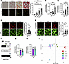 Figure 2
Fibrosis deposition in EpiWAT of FOb mice. (A) Representative Picrosirius red-stained histological sections of EpiWAT from lean (Le), obese (Ob4w), and formerly obese (FOb) mice, imaged under bright-field and polarized light (scale bar: 100 μm). We analyzed 2 FOb groups, both fed a high-fat diet (HFD) for 4 weeks, and then switched to a chow diet for either 4 weeks (named FOb or FOb 4w) or 10 weeks (FOb 10w). Quantification of fibrosis area normalized to adipocyte number (relative values; lean group set to 1), based on Picrosirius red staining (n = 7–12). (B) Hydroxyproline content in EpiWAT normalized to fat pad mass (μg/mg) and (C) total hydroxyproline content per fat pad (n = 5–10). (D) Representative immunofluorescence staining of EpiWAT sections from Le, Ob4w, and FOb mice using an anti-ICTP antibody (red). Tissue autofluorescence (green) highlights structural integrity (scale bar: 50 μm). Quantification (arbitrary units [AU]) is normalized to adipocyte number (n = 5–6). (E) Representative immunofluorescence staining of EpiWAT using an anti-Nε-(γ-L-glutamyl)-L-lysine antibody (red); autofluorescence (green) highlights tissue integrity (scale bar: 50 μm). Quantification (AU) is normalized to adipocyte number (n = 6). (F) Experimental design showing dietary interventions (chow or high-fat diet, as indicated) and the timeline for obesity induction and weight loss. Arrows indicate feeding durations for the groups: lean (Le, orange), formerly obese (FOb, purple) and obese mice fed HFD for 4 weeks (Ob4w, green) or 8 weeks (Ob8w, blue). (G and H) Fibrosis quantification in EpiWAT of FOb and Ob8w mice by measuring: (G) hydroxyproline content normalized to fat pad mass (μg/mg) and (H) total hydroxyproline content per fat pad (n = 5–10). (I) Principal component analysis of extracellular matrix (ECM) proteins from Le, Ob4w, Ob8w and FOb EpiWAT (n = 5). (A–E) Statistical testing was performed using 1-way ANOVA with Tukey’s multiple comparisons test. (G and H) Statistical significance was assessed using Student’s t test. *P < 0.05; **P < 0.01; ***P < 0.001.
While sexual dimorphism is well recognized in obesity, most studies have focused on differences in susceptibility to weight gain and metabolic dysfunction (29), whereas less attention has been paid to WAT fibrosis. In this setting, we analyzed female C3H mice subjected to the same experimental design as male mice (Figure 1A). Body weight, body composition, and fat pad mass increased in HFD-fed female mice and were normalized following WL (Supplemental Figure 2, A–E), as observed in male mice. Female C3H mice displayed body weight and fat mass values comparable to male mice after 4 weeks of HFD feeding (Supplemental Figure 2, A and B). In females, 4 weeks of HFD feeding induced glucose intolerance (Supplemental Figure 2F), which was normalized after 4 weeks of WL (Supplemental Figure 2G). In agreement with our previous observations (9), obesity also increased hydroxyproline content in the vWAT of females (periovarian fat depot [ovaWAT]), indicating the development of fibrosis (Supplemental Figure 2, H and I). However, the magnitude of fibrosis was lower than that observed in males (Supplemental Figure 2J). Importantly, in contrast to our observations in male mice, we did not detect further fibrosis accumulation following WL in female mice. The apparent increased hydroxyproline content (Supplemental Figure 2H), which was normalized to fat mass after WL, most likely reflected the reduction in ovaWAT mass (Supplemental Figure 2E) rather than additional collagen deposition. Indeed, total hydroxyproline content per fat pad was similar between obese and postobese females (Supplemental Figure 2I). These findings suggest that WL does not promote additional fibrosis accumulation in ovaWAT.
Given that posttranslational crosslinking of ECM proteins, such as collagens, critically shapes ECM architecture and contributes to fibrotic stiffness and tissue dysfunction (30), we next assessed two key crosslinking products, the carboxy terminal cross-linked telopeptide of type I collagen (ICTP) and Nε-(γ-L-glutamyl)-L-lysine, by immunostaining (31, 32) in males. Both markers were significantly elevated in obese mice (Ob4w) compared with lean mice and remained high in FOb males (Figure 2, D and E), indicating persistent ECM stiffening despite WL.
Because WAT fibrosis was minimal after only 4 weeks of HFD in males (Figure 2, A–C), the subsequent collagen accumulation observed in FOb mice likely reflects active remodeling rather than residual fibrosis from early obesity. To further delineate the dynamics of ECM remodeling, we compared EpiWAT from lean male mice (8 weeks on chow), Ob4w and Ob8w animals (4 and 8 weeks of HFD, respectively), and FOb mice (4 weeks on HFD followed by 4 weeks on chow, FOb) (Figure 2F). Hydroxyproline content in FOb and Ob8w mice was comparable (Figure 2, G and H), suggesting that WL induces fibrotic changes of similar magnitude to those observed in advanced obesity.
To gain further insight into the ECM composition in these weight gain–WL conditions, we performed proteomic profiling of the EpiWAT matrisome. Mass spectrometry identified 158 matrisome proteins, including 68 core matrisome and 90 matrisome-associated proteins (33). Principal component analysis revealed that samples from FOb and Ob8w mice clustered together, distinct from lean and Ob4w groups (Figure 2I), consistent with a convergent fibrotic signature driven by either sustained obesity or WL. Accordingly, no significant differences in individual protein levels were detected between FOb and Ob8w mice (Supplemental Figure 3). Of the 158 identified matrisome proteins, 104 were differentially expressed in FOb and Ob8w mice relative to lean (Supplemental Figure 3A). Notably, 70% of these changes were shared between FOb and Ob8w mice (Supplemental Figure 3, A and C), while only 5.8% and 22.1% were specific to Ob8w and FOb, respectively (Supplemental Figure 3, A, B, and D). Downregulated proteins included basement membrane components (e.g., LAMB1, COL4A1, NID1) and protease inhibitors, suggesting compromised matrix integrity. In parallel, FOb and Ob8w mice showed increased expression of cysteine proteases (CTSS, CTSB) and collagen-stabilizing enzymes (LOXL1, P4HA1), consistent with active ECM remodeling and fiber crosslinking.
Together, these findings reveal that EpiWAT fibrosis is not only retained but intensified following WL. Importantly, this remodeling is characterized by impaired basement membrane architecture and increased ECM crosslinking, potentially contributing to major persistent tissue dysfunction in the FOb state.
Macrophage persistence and reprogramming in the EpiWAT after WL following obesity. As proinflammatory immune cell recruitment contributes to obesity-induced fibrosis (4, 5, 8), we next assessed whether obesity-associated macrophage accumulation was maintained after WL. Flow cytometry analysis of EpiWAT revealed that total macrophage numbers (Supplemental Figure 4, A and B) were elevated approximately 4-fold in both Ob4w and FOb mice compared with lean controls (Figure 3A). However, owing to the reduced EpiWAT mass in FOb mice, macrophage density was significantly higher than in Ob4w mice (Figure 3B), indicating a proportionally greater macrophage burden after WL. To further define macrophage phenotypes, we sorted EpiWAT macrophages from lean, Ob4w, and FOb mice for RNA-seq. Prior studies have identified a distinct subpopulation of monocyte-derived macrophages, termed lipid-associated macrophages (LAMs), characterized by high CD9 expression and a transcriptional program enriched in lipid metabolism, lysosomal function, and inflammatory pathways (34). Gene set enrichment analysis (GSEA) revealed that LAM-associated transcripts were enriched not only in Ob4w versus lean mice (data not shown), but also in FOb versus Ob4w macrophages (Figure 3C). Flow cytometry confirmed an increased proportion of CD9+ LAMs in FOb mice compared with Ob4w mice (Figure 3, D–F), suggesting that the obesity-associated macrophage program is maintained in adipose tissue even after WL.
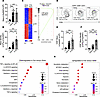 Figure 3
Lipid-associated macrophage accumulation persisted in EpiWAT of FOb mice. (A) Total macrophage number (CD45+CD64+CD31–) in EpiWAT and (B) macrophage number per gram of EpiWAT from lean (Le), obese (Ob4w), and formerly obese (FOb) mice, quantified by flow cytometry (n = 10). (C) Gene set enrichment analysis (GSEA) of the lipid-associated macrophage (LAM) gene signature within the total macrophage population (CD45+CD64+CD31–) from Ob4w and FOb mice (n = 4). (D) Representative FACS plot and mean percentage (± SEM) of CD9+ macrophages (Macs) in EpiWAT from Le, Ob4w, and FOb C3H mice (n = 10). (E) Total CD9+ macrophage number in EpiWAT and (F) CD9+ macrophage number per gram of EpiWAT in Le, Ob4w, and FOb mice, quantified by flow cytometry (n = 10). (G) Pathways enriched in macrophages isolated from Ob4w mice compared with those from FOb mice. (H) Pathways enriched in macrophages isolated from FOb mice compared with those from Ob4w mice. (A, B, and D–F) Statistical significance was assessed using 1-way ANOVA with Tukey’s multiple comparisons test. (D) Statistical significance was defined as P < 0.001 for Lean vs. Ob4w and P < 0.001 Ob4w vs. FOb. (C) LIMMA analysis was used to identify differentially expressed genes, followed by GSEA of the CD9+ macrophage gene signature using Phantasus software. (G and H) Pathway enrichment was analyzed using ClusterProfiler, with FDR correction and adjusted P values calculated using the Benjamini-Hochberg procedure. *P < 0.05; **P < 0.01; ***P < 0.001
TLR4 signaling in bone marrow–derived myeloid cells has previously been implicated in adipose tissue fibrosis in the C3H mouse strain (8). We therefore tested whether pharmacological inhibition of TLR4 might protect adipose tissue from fibrosis during WL. We administered TAK242 — a TLR4 inhibitor with antiinflammatory properties previously shown to reduce adipose tissue fibrosis in obese C3H mice (8), at the time of the diet switch (Supplemental Figure 5A). TAK242 treatment had no effect on body weight or composition (Supplemental Figure 5B), EpiWAT mass (Supplemental Figure 5C), or collagen content (Supplemental Figure 5, D and E). Thus, fibrosis persistence after WL occurs independently from TLR4 signaling.
We next examined the transcriptional landscape of adipose tissue macrophages after WL using RNA-seq. This revealed a marked shift in macrophage gene expression in FOb animals. Compared with Ob4w, FOb macrophages showed reduced inflammatory activation and antigen presentation, as indicated by downregulation of genes in the TNFA/NF-κB signaling pathway (Figure 3G and Supplemental Figure 6), as well as genes encoding MHC class II components, including H2-Ab1 (fold change = 0.33, P = 5.10–7), H2-Eb1 (fold change = 0.30, P = 2.10–6), and Cd74 (fold change = 0.34, P = 1.10–6). In addition, FOb macrophages displayed increased expression of genes associated with oxidative and metabolic stress (Hmox1, Gclc, Gclm, Txn1, Txnrd1, Cat, Srxn1) and glycolysis and one-carbon metabolism (Slc2a1, Hk2, Aldoa, Pkm, Mthfd2) compared with Ob4w macrophages (Figure 3H). Finally, several immunoregulatory molecules (Cd5l, Havcr2, Lilrb4a, Adgre5) were enriched in FOb macrophages (Supplemental Figure 6).
Together, these data indicate that adipose tissue macrophages undergo transcriptional reprogramming after WL, which is characterized by attenuation of proinflammatory and antigen-presenting programs, and the acquisition of stress-adapted and immunomodulatory features. This shift in FOb macrophage transcriptional state further suggests that specific mechanisms may underlie WL-associated adverse adipose tissue remodeling.
WL uncouples ECM production from fibrosis in visceral fat. We next evaluated a dual antifibrotic strategy targeting tyrosine kinase and COX2 signaling, using nintedanib (35, 36) and celecoxib (37), agents with established efficacy in EpiWAT fibrosis in our previous study (7). Coadministration of these compounds similarly failed to reduce collagen accumulation during WL (Supplemental Figure 5, F–J), mirroring the inefficacy of TAK242. To elucidate the mechanisms underlying the differential sensitivity to antifibrotic therapies between obesity-associated and WL–associated fibrosis, we analyzed ECM-related gene expression in EpiWAT. We aimed to uncover molecular differences in fibrotic remodeling that may influence treatment efficacy in these two physiologically distinct contexts. While these ECM-related genes were upregulated in Ob4w mice, they were downregulated in FOb animals, reaching levels comparable to those in lean mice (Figure 4, A and B). Thus, despite the persistent and even increased fibrosis observed in the FOb EpiWAT (Figure 2, A–C), the transcriptional program driving ECM synthesis was suppressed after WL. This likely explains the failure of TAK242 and the nintedanib-celecoxib combination in this context, as WL itself is sufficient to suppress ECM gene expression, making further inhibition ineffective.
 Figure 4
Dissociation of the tissue fibrosis signature from fibroinflammatory progenitor persistence. (A and B) Relative mRNA expression of (A) fibrosis-related ECM components and (B) fibrogenic cytokines measured by quantitative PCR in EpiWAT of lean (Le), obese (Ob4w), and formerly obese (FOb) C3H mice (n = 5–10). (C) Total fibroinflammatory progenitor (FIP) (PDGFRα+PDPN+CD9+Ly-6C+CD45–CD31–) count in EpiWAT (n = 10) and (D) FIP count per gram of EpiWAT (n = 10) quantified by flow cytometry. (E) Left: Representative FACS plots showing Ki-67 expression in FIPs from EpiWAT stromal vascular fraction (SVF) of Le, Ob4w, and FOb C3H mice. Right: Percentage (mean ± SEM) of Ki-67+ FIPs in EpiWAT (n = 5). Statistical testing was performed using 1-way ANOVA with Tukey’s multiple comparisons test. *P < 0.05; **P < 0.01; ***P < 0.001
We next turned our attention to fibroinflammatory progenitors (FIPs), a specific subset of CD9+Ly-6C+ PDGFRα+ adipose stromal progenitor cells (Supplemental Figure 4, A and C) that expand during obesity and acquire profibrotic and proinflammatory features (6, 9). Given their central role in obesity-associated fibrosis through increased ECM production (6, 9), we investigated whether the activation of this population was modulated after WL. Flow cytometry revealed elevated FIP abundance in both Ob4w and FOb mice relative to lean animals (Figure 4C), with FIP density (cell count per gram of fat) further increased in FOb animals owing to adipose mass reduction (Figure 4D). FIP proliferation was enhanced in Ob4w mice but returned to baseline after WL (Figure 4E), probably indicating a halt in expansion although FIPs persisted and became more densely concentrated within EpiWAT of FOb mice. To further characterize FIP states in these different contexts, we performed transcriptomic profiling of sorted FIPs from lean, Ob4w, and FOb mice. Principal component analysis revealed that FIPs from FOb mice clustered separately from both lean and Ob4w mice (Figure 5A). K-means clustering of differentially expressed genes between lean and FOb FIPs identified 5 distinct modules (Figure 5B). Cluster 1 included genes encoding ECM proteins, which were selectively downregulated in FOb FIPs compared with both lean and Ob4w animals. Cluster 2 comprised genes involved in epithelial-mesenchymal transition (EMT) and ECM remodeling, which were upregulated in Ob4w and FOb mice but attenuated in FOb mice relative to Ob4w mice, reflecting the tissue-level suppression of fibrotic markers (Figure 4, A and B). These two clusters indicate that the fibrogenic activation of FIPs is significantly reduced following WL. To quantify this reversal, we focused on 46 EMT-related genes upregulated in Ob4w FIPs versus lean FIPs. Of these, 32 were significantly downregulated in FOb versus Ob4w FIPs and 15 returned to lean levels (Figure 5C), demonstrating broad suppression of the fibrogenic program within FIPs after WL. Cluster 3 included inflammatory genes, which were upregulated in both Ob4w and FOb FIPs compared with lean mice, suggesting persistent inflammatory activation. Cluster 4, linked to BMP signaling and ECM organization, was downregulated in Ob4w mice and remained suppressed in FOb mice. Notably, Cluster 5 enriched for NRF2 signaling, glutathione metabolism, and lysosomal function, showed stronger induction in FOb FIPs than in Ob4w FIPs, pointing to elevated oxidative stress after WL.
 Figure 5
The fibroinflammatory progenitors revert to an inactive phenotype during weight loss. (A) Principal component analysis (PCA) of fibroinflammatory progenitor (FIP) transcriptomes from Le, Ob4w, and FOb mice (n = 6). (B) K-means clustering analysis of FIPs from Le, Ob4w, and FOb mice identified 5 clusters. Representative genes and enriched pathways for each cluster are shown (n = 6). (C) Heatmap showing expression of genes in the epithelial-mesenchymal transition (EMT) pathway that are upregulated in FIPs from Ob4w vs. Le mice (fold change >1.5, adjusted P < 0.05; n = 6). (B and C) A LIMMA analysis was conducted to select differentially expressed genes between (B) Le and FOb and (C) Le and Ob4w (fold change >1.5, adjusted P value for multiple testing).
In summary, these findings reveal a striking uncoupling between ECM gene expression and ECM protein accumulation in the EpiWAT of FOb mice. Despite continued collagen deposition, the transcriptional program within FIPs that drives ECM synthesis is largely silenced after WL. This decoupling likely underlies the inefficacy of therapies targeting ECM production (7, 8) and suggests that postobesity fibrosis is maintained by mechanisms beyond the production of ECM components. We therefore hypothesized that the incremental fibrosis observed during WL may arise from impaired matrix turnover or clearance, rather than ongoing ECM production.
Lysosomal stress links impaired collagen degradation to fibrosis during WL. To investigate whether defective collagen degradation contributes to the worsening of EpiWAT fibrosis during WL, we developed an ex vivo assay using the dye quenched (DQ) collagen I, a fluorogenic substrate that emits FITC fluorescence upon proteolytic cleavage (38, 39). While FIPs are central to fibrosis development through enhanced ECM production, the entire progenitor cell population could participate to collagen degradation and fibrosis resolution after WL. We therefore did not restrict the analysis to the FIP subset, as ECM degradation and turnover are not necessarily confined to this population.
In lean EpiWAT, collagenolytic activity, as indicated by FITC fluorescence, was primarily observed in macrophages (CD45+CD64+) and progenitors (PDGFRα+), while endothelial cells (CD31+CD45–PDGFRα–) showed minimal signal (Figure 6, A and B). When comparing lean, Ob4w, and FOb mice, both macrophages and progenitors from Ob4w and FOb mice exhibited significantly reduced collagen-degrading capacities relative to those of lean mice (Figure 6, C and D), indicating that collagenolysis is impaired during obesity and remains defective after WL. We next assessed collagenolytic activity in macrophages and progenitor cells from IngWAT in males (Supplemental Figure 7, A and B) and periovarian WAT (OvaWAT) in females (Supplemental Figure 7, C and D). Unlike EpiWAT in males, neither depot exhibited alterations in collagen degradation capacity following obesity or WL, with comparable collagenolytic activity observed in lean, Ob4w, and FOb mice (Supplemental Figure 7, A–D).
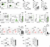 Figure 6
Reduced collagen 1 degradation and decreased lysosome signal in macrophages and adipose progenitors of FOb C3H mice. (A) Representative FACS plots showing DQ-COL1+ macrophages (Macs), adipose progenitors (PDGFRα+; Progs), and endothelial cells (Endo) incubated at 4°C (negative control, black) or 37°C (green). (B) Mean percentage of DQ-COL1+ cells among Macs, Progs, and Endos (n = 3). (C and D) Percentage of DQ-COL1+ cells in (C) Macs and (D) Progs isolated from EpiWAT of lean (Le), obese (Ob4w), and formerly obese (FOb) C3H mice (n = 6–9). (E) Representative FACS plot showing the gating strategy to identify LysoTlo and LysoThi Macs (based on Lysotracker staining) from lean EpiWAT and corresponding DQ-COL1 fluorescence intensity (n = 3). (F) Same analysis as in E for Progs (n = 3). (G) Quantification of LysoT+ Macs isolated from EpiWAT SVF of Le, Ob4w, and FOb mice in the presence (red) or absence (black) of Lysotracker staining (n = 4). (H) Quantification of LysoT+ Progs isolated from EpiWAT SVF of Le, Ob4w, and FOb mice in the presence (red) or absence (black) of Lysotracker staining (n = 4). (I) FACS analysis of LAMP1 expression in Macs from EpiWAT SVF of Le and FOb mice (n = 4). (J) FACS analysis of LAMP1 expression in Progs from EpiWAT SVF of Le and FOb mice (n = 4). (B–D, G, and H) Statistical testing was performed using 1-way ANOVA followed by Tukey’s multiple comparisons test. (E, F, I, and J) Statistical significance was assessed using Student’s t test. *P < 0.05; **P < 0.01; ***P < 0.001.
To gain further insight into the alterations observed in EpiWAT, we analyzed the expression of genes encoding regulators of collagen degradation (40). At the whole-tissue level, the expression of these collagenolytic regulators remained largely unchanged across groups (Supplemental Figure 8A). Yet, a few genes (e.g., Mmp2, Mmp3, Mmp14, Timp1) that were elevated during obesity returned to baseline (i.e., lean) in FOb mice. Also, genes involved in collagen uptake were comparably expressed in FIPs (Supplemental Figure 8B) and macrophages (Supplemental Figure 8C) from lean and FOb mice, suggesting that uptake mechanisms are not responsible for the impaired collagenolysis.
We next examined whether lysosomal dysfunction could underlie this defect, as lysosomal pathways are essential for intracellular collagen degradation (39, 41). Revisiting the RNA-seq data obtained from macrophages and FIPs isolated from the EpiWAT of lean, Ob4w, and FOb mice, GSEA revealed significant enrichment of lysosome-associated genes (KEGG lysosome pathway, 127 genes) in both macrophages (Supplemental Figure 9, A and B) and FIPs (Supplemental Figure 9, C and D) from Ob4w and FOb mice compared with lean animals. Given that lysosomal gene induction can reflect stress-induced compensatory responses (42), these findings point to potential lysosomal dysfunction.
To directly evaluate lysosomal function, we used LysoTracker, a pH-sensitive dye that accumulates in acidic lysosomes (43). Flow cytometry enables quantitative assessment of LysoTracker signal in large numbers of freshly isolated stromal vascular fraction (SVF) cells, while minimizing manipulation and preserving their tissue-derived phenotype. In both macrophages and Progs, LysoTracker intensity strongly correlated with DQ collagen degradation (Figure 6, E and F), confirming the requirement of lysosomal acidification for collagenolysis. Importantly, LysoTracker staining was reduced in Ob4w mice and further diminished in FOb mice in both macrophages (Figure 6G) and Progs (Figure 6H), indicating lysosomal alteration with obesity and WL. However, the lysosomal membrane protein LAMP1 (43), showed unchanged expression in macrophages and Progs from FOb animals compared with lean mice (Figure 6, I and J), suggesting that lysosome abundance is preserved and that the defect lies specifically in acidification capacity. We also assessed LysoTracker staining in macrophages and progenitor cells from IngWAT in males (Supplemental Figure 7, E and F) and ovaWAT in females (Supplemental Figure 7, G and H). In agreement with the preserved collagen degradation capacity in these depots, LysoTracker fluorescence intensity remained unchanged across lean, Ob4w, and FOb mice (Supplemental Figure 7, E–H).
Together these findings identify impaired lysosomal acidification as a key mechanism driving defective collagenolysis after WL in EpiWAT. This deficiency likely contributes to the accumulation of fibrotic ECM components in the postobese state, despite suppression of collagen gene expression.


