
Research ArticleCardiologyVascular biology
Open Access |  10.1172/jci.insight.195761
USP10 mitigates Ang II–induced atrial remodeling and atrial fibrillation susceptibility by deubiquitinating NDUFS1
Wanrong Fu,1 Xiao-Xu Tian,1 Jianghua Zhou,2 Yu-Xu Huang,3 Huan Li,1 Zhenya Wang,1 Tong-You Wade Wei,4 Li Li,5 and Guo-Jun Zhao1
1Department of Cardiology, The First Affiliated Hospital of Zhengzhou University, Zhengzhou, China.
2Department of Cardiology, The First Affiliated Hospital of Wenzhou Medical University, Wenzhou, China.
3Department of Medicine, JingGangShan University, Ji’an, China.
4Division of Cardiology, Department of Medicine, UCSD, La Jolla, California, USA.
5Department of Cardiovascular Surgery, The First Affiliated Hospital of Zhengzhou University, Zhengzhou, China.
Address correspondence to: Guo-Jun Zhao, Department of Cardiology, The First Affiliated Hospital of Zhengzhou University, Zhengzhou, China. Email: zhaoguojun@zzu.edu.cn.
Authorship note: WF, XXT, and JZ contributed equally to this work.
Find articles by Fu, W. in: PubMed | Google Scholar
1Department of Cardiology, The First Affiliated Hospital of Zhengzhou University, Zhengzhou, China.
2Department of Cardiology, The First Affiliated Hospital of Wenzhou Medical University, Wenzhou, China.
3Department of Medicine, JingGangShan University, Ji’an, China.
4Division of Cardiology, Department of Medicine, UCSD, La Jolla, California, USA.
5Department of Cardiovascular Surgery, The First Affiliated Hospital of Zhengzhou University, Zhengzhou, China.
Address correspondence to: Guo-Jun Zhao, Department of Cardiology, The First Affiliated Hospital of Zhengzhou University, Zhengzhou, China. Email: zhaoguojun@zzu.edu.cn.
Authorship note: WF, XXT, and JZ contributed equally to this work.
Find articles by Tian, X. in: PubMed | Google Scholar
1Department of Cardiology, The First Affiliated Hospital of Zhengzhou University, Zhengzhou, China.
2Department of Cardiology, The First Affiliated Hospital of Wenzhou Medical University, Wenzhou, China.
3Department of Medicine, JingGangShan University, Ji’an, China.
4Division of Cardiology, Department of Medicine, UCSD, La Jolla, California, USA.
5Department of Cardiovascular Surgery, The First Affiliated Hospital of Zhengzhou University, Zhengzhou, China.
Address correspondence to: Guo-Jun Zhao, Department of Cardiology, The First Affiliated Hospital of Zhengzhou University, Zhengzhou, China. Email: zhaoguojun@zzu.edu.cn.
Authorship note: WF, XXT, and JZ contributed equally to this work.
Find articles by Zhou, J. in: PubMed | Google Scholar
1Department of Cardiology, The First Affiliated Hospital of Zhengzhou University, Zhengzhou, China.
2Department of Cardiology, The First Affiliated Hospital of Wenzhou Medical University, Wenzhou, China.
3Department of Medicine, JingGangShan University, Ji’an, China.
4Division of Cardiology, Department of Medicine, UCSD, La Jolla, California, USA.
5Department of Cardiovascular Surgery, The First Affiliated Hospital of Zhengzhou University, Zhengzhou, China.
Address correspondence to: Guo-Jun Zhao, Department of Cardiology, The First Affiliated Hospital of Zhengzhou University, Zhengzhou, China. Email: zhaoguojun@zzu.edu.cn.
Authorship note: WF, XXT, and JZ contributed equally to this work.
Find articles by Huang, Y. in: PubMed | Google Scholar
1Department of Cardiology, The First Affiliated Hospital of Zhengzhou University, Zhengzhou, China.
2Department of Cardiology, The First Affiliated Hospital of Wenzhou Medical University, Wenzhou, China.
3Department of Medicine, JingGangShan University, Ji’an, China.
4Division of Cardiology, Department of Medicine, UCSD, La Jolla, California, USA.
5Department of Cardiovascular Surgery, The First Affiliated Hospital of Zhengzhou University, Zhengzhou, China.
Address correspondence to: Guo-Jun Zhao, Department of Cardiology, The First Affiliated Hospital of Zhengzhou University, Zhengzhou, China. Email: zhaoguojun@zzu.edu.cn.
Authorship note: WF, XXT, and JZ contributed equally to this work.
Find articles by Li, H. in: PubMed | Google Scholar
1Department of Cardiology, The First Affiliated Hospital of Zhengzhou University, Zhengzhou, China.
2Department of Cardiology, The First Affiliated Hospital of Wenzhou Medical University, Wenzhou, China.
3Department of Medicine, JingGangShan University, Ji’an, China.
4Division of Cardiology, Department of Medicine, UCSD, La Jolla, California, USA.
5Department of Cardiovascular Surgery, The First Affiliated Hospital of Zhengzhou University, Zhengzhou, China.
Address correspondence to: Guo-Jun Zhao, Department of Cardiology, The First Affiliated Hospital of Zhengzhou University, Zhengzhou, China. Email: zhaoguojun@zzu.edu.cn.
Authorship note: WF, XXT, and JZ contributed equally to this work.
Find articles by Wang, Z. in: PubMed | Google Scholar
1Department of Cardiology, The First Affiliated Hospital of Zhengzhou University, Zhengzhou, China.
2Department of Cardiology, The First Affiliated Hospital of Wenzhou Medical University, Wenzhou, China.
3Department of Medicine, JingGangShan University, Ji’an, China.
4Division of Cardiology, Department of Medicine, UCSD, La Jolla, California, USA.
5Department of Cardiovascular Surgery, The First Affiliated Hospital of Zhengzhou University, Zhengzhou, China.
Address correspondence to: Guo-Jun Zhao, Department of Cardiology, The First Affiliated Hospital of Zhengzhou University, Zhengzhou, China. Email: zhaoguojun@zzu.edu.cn.
Authorship note: WF, XXT, and JZ contributed equally to this work.
Find articles by Wei, T. in: PubMed | Google Scholar
1Department of Cardiology, The First Affiliated Hospital of Zhengzhou University, Zhengzhou, China.
2Department of Cardiology, The First Affiliated Hospital of Wenzhou Medical University, Wenzhou, China.
3Department of Medicine, JingGangShan University, Ji’an, China.
4Division of Cardiology, Department of Medicine, UCSD, La Jolla, California, USA.
5Department of Cardiovascular Surgery, The First Affiliated Hospital of Zhengzhou University, Zhengzhou, China.
Address correspondence to: Guo-Jun Zhao, Department of Cardiology, The First Affiliated Hospital of Zhengzhou University, Zhengzhou, China. Email: zhaoguojun@zzu.edu.cn.
Authorship note: WF, XXT, and JZ contributed equally to this work.
Find articles by Li, L. in: PubMed | Google Scholar
1Department of Cardiology, The First Affiliated Hospital of Zhengzhou University, Zhengzhou, China.
2Department of Cardiology, The First Affiliated Hospital of Wenzhou Medical University, Wenzhou, China.
3Department of Medicine, JingGangShan University, Ji’an, China.
4Division of Cardiology, Department of Medicine, UCSD, La Jolla, California, USA.
5Department of Cardiovascular Surgery, The First Affiliated Hospital of Zhengzhou University, Zhengzhou, China.
Address correspondence to: Guo-Jun Zhao, Department of Cardiology, The First Affiliated Hospital of Zhengzhou University, Zhengzhou, China. Email: zhaoguojun@zzu.edu.cn.
Authorship note: WF, XXT, and JZ contributed equally to this work.
Find articles by Zhao, G. in: PubMed | Google Scholar
Published August 24, 2026 - More info
JCI Insight. 2026;11(16):e195761. https://doi.org/10.1172/jci.insight.195761.
© 2026 Fu et al. This work is licensed under the Creative Commons Attribution 4.0 International License. To view a copy of this license, visit http://creativecommons.org/licenses/by/4.0/.
Received: May 21, 2025; Accepted: June 24, 2026


-
Results
USP10 is decreased in AF. To investigate the involvement of USP10 in AF, we analyzed an NCBI Gene Expression Omnibus (GEO) RNA-seq dataset (GSE2240) derived from atrial tissues of patients with AF and sinus rhythm (SR) controls (21). Our analysis revealed that USP10 levels were lower in atrial tissues from patients with AF compared with SR controls (Figure 1A). To validate this finding, we collected atrial tissues from patients with AF and SR, assessing USP10 mRNA and protein levels. Consistently, both mRNA and protein levels of USP10 were significantly reduced in atrial tissues from AF patients compared with SR controls (Figure 1, B and C). To determine whether this observation is recapitulated in experimental models, we examined whether this reduction is reproduced in an Ang II–induced mouse model of AF. Consistent with findings in human samples, USP10 mRNA and protein levels were markedly reduced in the atrial tissues of Ang II–treated mice compared with controls (Figure 1, D and E). Immunostaining further confirmed that the reduction in USP10 was specific to cardiomyocytes (Figure 1F). Moreover, cultured HL-1 atrial myocytes demonstrated a significant downregulation of USP10 following Ang II treatment (Figure 1, G and H). Collectively, these findings indicate that USP10 is consistently downregulated in atrial tissues from patients with AF and Ang II–treated mouse models, suggesting a potential contributory role of USP10 deficiency in the pathogenesis of AF.
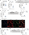 Figure 1
USP10 is decreased in patients with AF and Ang II–treated atrial cardiomyocytes. (A)The expression levels of USP10 in atrial tissues from patients with AF (n = 10) and SR controls (n = 20). (B) qPCR analysis of USP10 mRNA levels in atrial tissues from patients with AF and SR controls (n = 5/group). (C) Western blot and quantification of USP10 protein levels in atrial tissues from patients with AF and SR controls (n = 5/group). (D) qPCR analysis of Usp10 mRNA levels in atrial tissues from mice treated with Ang II or Veh (n = 4/group). (E) Representative Western blots and quantification of USP10 in atrial tissues from Ang II– or Veh-treated mice (n = 4/group). (F) Representative immunostaining images of atrial tissues from Ang II– or Veh-treated mice. Scale bar: 50 μm. (G) qPCR analysis of Usp10 mRNA levels in HL-1 atrial myocytes treated with Ang II or Veh (n = 3/group). (H) Representative Western blots and quantification of USP10 protein levels in Ang II– or Veh-treated HL-1 atrial myocytes (n = 3/group). Data represent mean ± SEM. All statistical comparisons were performed using a 2-tailed Student’s t test. **P < 0.01. USP10, ubiquitin-specific peptidase 10; FPKM, fragments per kilobase of exon per million mapped reads; qPCR, quantitative PCR; AF, atrial fibrillation; SR, sinus rhythm; Veh, vehicle; Ang II, angiotensin II.
USP10 ameliorates Ang II–induced atrial remodeling and reduces AF susceptibility. Given the decreased levels of USP10 in atrial cardiomyocytes, we utilized cardiomyocyte-specific Usp10-transgenic (Usp10-CTg) mice to investigate the role of USP10 in atrial remodeling and AF susceptibility. As illustrated in Figure 2, A–C, Ang II treatment resulted in a significant increase in left atrial size, while overexpression of Usp10 notably attenuated this Ang II–induced atrial enlargement. Additionally, we assessed atrial fibrosis, a key marker of atrial remodeling, using Masson’s trichrome staining. This analysis revealed that Usp10 overexpression effectively suppressed Ang II–induced atrial fibrosis (Figure 2, D and E). To further evaluate the impact of USP10 on AF susceptibility, we conducted transesophageal electrode catheter burst stimulation experiments. Ang II treatment increased both the incidence and duration of AF (Figure 2, F–H). Crucially, overexpression of Usp10 significantly reduced the incidence rate and duration of Ang II–induced AF (Figure 2, F–H). These results demonstrate that USP10 overexpression mitigates Ang II–induced atrial remodeling and decreases AF susceptibility.
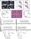 Figure 2
USP10 overexpression ameliorates Ang II–induced atrial remodeling and AF susceptibility. (A and B) Representative echocardiography images and quantification of left atrial size in WT and Usp10-CTg mice subjected to Veh or Ang II treatment (n = 7–9/group). (C–E) Representative images and quantification of Masson’s trichrome staining in left atrial sections from Usp10-CTg and WT control mice treated with Veh or Ang II (n = 4/group). Scale bars: 1 mm (C) and 100 μm (D). (F) Representative limb lead ECG traces from Usp10-CTg and WT control mice in response to Veh or Ang II treatment. (G) Fraction of mice with successful AF induction relative to the total number. (H) Quantification of AF duration in Usp10-CTg and WT control mice treated with Ang II (n = 23/group). Data are presented as mean ± SEM and were analyzed using 2-way ANOVA followed by Tukey’s multiple-comparison test (B and E), χ2 test (G), and 2-tailed Student’s t test (H). *P < 0.05, **P < 0.01. USP10, ubiquitin-specific peptidase 10; AF, atrial fibrillation; Ang II, angiotensin II; ECG, electrocardiogram; CTg, cardiomyocyte-specific transgenic.
USP10 overexpression reduces delayed afterdepolarizations and improves calcium handling. To assess the functional impact of USP10 overexpression on atrial myocytes, we examined calcium handling and proarrhythmic states using whole-cell patch-clamp techniques in atrial cells isolated from WT and Usp10-CTg mice treated with either vehicle or Ang II (22). USP10 overexpression significantly reduced the frequency of delayed afterdepolarizations (DADs) induced by Ang II (Figure 3, A and B), highlighting its protective role in maintaining atrial electrophysiological stability.
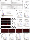 Figure 3
USP10 overexpression mitigates abnormal calcium handling and DADs. (A) Representative DAD images recorded from atrial cardiomyocytes obtained from WT and Usp10-CTg mice in response to vehicle or Ang II. (B) Statistical results of DADs in the indicated groups (n = 19–23/group). (C) Representative Ca2+ transients (CaTs) evoked by 10 mM caffeine in Ca2+-free conditions in atrial cells isolated from WT and Usp10-CTg mice treated with vehicle or Ang II. (D) Statistical results of CaT amplitudes in the indicated groups (n = 15–22/group). (E) Representative Ca²+ wave images in atrial cardiomyocytes from WT and Usp10-CTg mice, recorded following 1-Hz pacing after treatment with either vehicle or Ang II. (F) Statistical results of amplitude and 50% decay time of Ca2+ waves in the indicated groups (n = 22–37/group). (G) Representative Ca2+ spark images in atrial cardiomyocytes obtained from WT and Usp10-CTg mice in response to vehicle or Ang II. Scale bar: 0.1 seconds. (H) Statistical results of Ca2+ spark–induced Ca2+ leak, Ca2+ spark frequency, Ca2+ spark amplitude, and Ca2+ spark mass in the indicated groups (n = 26–31/group). Data are presented as mean ± SEM and were analyzed using the χ2 test (B) and 2-way ANOVA followed by Tukey’s multiple-comparisons test (D, F, and H). *P < 0.05; **P < 0.01. USP10, ubiquitin-specific peptidase 10; AF, atrial fibrillation; Ang II, angiotensin II; CaTs, Ca2+ transients; DAD, delayed afterdepolarization.
DADs are often associated with Ca2+-handling abnormalities that promote cellular triggered activity in AF. To evaluate the effect of USP10 on Ca2+ handling, we assessed caffeine-induced calcium transients (CaTs), an indicator of sarcoplasmic reticulum Ca²+ content. Sarcoplasmic reticulum Ca²+ homeostasis has a critical role in maintaining myocardial electrical stability (23). Ang II treatment increased the amplitude of caffeine-induced CaTs; however, USP10 overexpression reversed this increase compared with Ang II–treated cardiomyocytes (Figure 3, C and D). Following 1-Hz pacing, Ang II–treated atrial myocytes exhibited a higher susceptibility to spontaneous Ca2+ waves compared with vehicle controls (Figure 3E). Furthermore, the frequency of spontaneous Ca2+ waves was lower in USP10-overexpressing cardiomyocytes compared with Ang II–treated cells (Figure 3E). USP10 overexpression also reduced both the amplitude and 50% decay time of spontaneous Ca2+ waves (Figure 3F). Since spontaneous Ca2+ waves are often linked to enhanced sarcoplasmic reticulum Ca2+ release via RyR2 channels, we measured Ca2+ sparks as an indication of RyR2 function. USP10 overexpression significantly reduced Ca2+ spark–mediated leak, Ca2+ spark frequency, and Ca2+ spark mass (Figure 3, G and H). These findings suggest that USP10 overexpression ameliorates Ang II–induced abnormal calcium handling, thereby reducing AF susceptibility.
USP10 contributes to the restoration of mitochondrial function. To elucidate the mechanisms by which USP10 regulates AF susceptibility, we performed RNA-seq on HL-1 cardiomyocytes infected with adenovirus carrying either Usp10 (Ad-USP10) or vector DNA (Ad-Vector) and treated with Ang II. Sample-to-sample clustering analysis revealed significant transcriptional differences between USP10-overexpressing and control cells (Figure 4A). Gene Ontology (GO) analysis identified several AF-associated cellular processes affected by USP10 expression, including mitochondrial function, calcium ion transport, cardiac remodeling, and cardiac rhythm (Figure 4B). Given the critical role of mitochondrial function in AF initiation and progression, we further analyzed genes related to mitochondrial function affected by USP10 overexpression using pathway analysis. GO enrichment analysis showed significant enrichment of genes associated with fatty acid β-oxidation, oxidative phosphorylation, and the tricarboxylic acid (TCA) cycle following USP10 overexpression, suggesting broad effects on mitochondrial energy metabolism (Figure 4C).
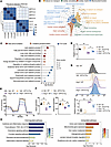 Figure 4
USP10 overexpression contributes to the restoration of mitochondrial function. (A) Sample-to-sample distance plot of HL-1 atrial myocytes expressing Ad-USP10 or Ad-Vector analyzed by RNA-seq (n = 4/group). (B) GO enrichment analysis showing enriched pathways related to mitochondrial function, calcium ion transport, and cardiac rhythm based on RNA-seq data (n = 4/group). (C) GO enrichment analysis showing enriched pathways related to fatty acid β-oxidation, oxidative phosphorylation, and the TCA cycle (n = 4/group). (D) ATP content in atrial cardiomyocytes obtained from WT and Usp10-CTg mice in response to vehicle or Ang II (n = 6/group). (E) OCR profiles indicating mitochondrial respiratory function in atrial cells isolated from WT and Usp10-CTg mice treated with vehicle or Ang II. (F) Quantification of basal respiration and maximal respiration in atrial cells isolated from WT and Usp10-CTg mice treated with vehicle or Ang II (n = 6/group). (G) MitoSOX-based flow cytometry of ROS in atrial cells isolated from WT and Usp10-CTg mice treated with vehicle or Ang II, combining a schematic illustration of the method with quantitative data (bottom; n = 6). (H) WikiPathways enrichment analysis of differentially expressed genes (DEGs) identified from RNA-seq by comparing Ang II–treated HL-1 cells expressing Ad-USP10 with those expressing Ad-Vector. Upregulated and downregulated DEGs were analyzed separately. Data are presented as mean ± SEM and were analyzed using 2-way ANOVA followed by Tukey’s multiple-comparison test (D, F, and G). *P < 0.05; **P < 0.01. USP10, ubiquitin-specific peptidase 10; Ang II, angiotensin II; GO, Gene Ontology; OCR, oxygen consumption rate; TCA cycle, tricarboxylic acid cycle; ROS, reactive oxygen species.
To validate these findings, we measured ATP content in atrial cells isolated from WT and Usp10-CTg mice treated with vehicle or Ang II. As anticipated, ATP levels decreased in response to Ang II stimulation, but USP10 overexpression significantly restored ATP levels (Figure 4D). Furthermore, while Ang II stimulation reduced mitochondrial basal and maximal respiratory capacity, USP10 overexpression significantly enhanced mitochondrial respiratory function, as evidenced by an increased mitochondrial oxygen consumption rate (OCR) (Figure 4, E and F). Using MitoSOX-based flow cytometry, we assessed mitochondrial ROS production in atrial cells isolated from WT and Usp10-CTg mice treated with either vehicle or Ang II, and found that USP10 overexpression significantly suppressed mitochondrial ROS levels (Figure 4G).
We also investigated the global impact of USP10 on pathways related to cardiac remodeling and AF progression. Pathway enrichment analysis of differentially expressed genes (DEGs) identified by comparing Ang II–treated HL-1 cells expressing Ad-USP10 with those expressing Ad-Vector revealed that genes downregulated by USP10 overexpression were significantly enriched in pathways associated with cytokine and inflammatory responses, focal adhesion/PI3K/Akt/mTOR signaling, chemokine signaling, G protein signaling, and glycolysis/gluconeogenesis (Figure 4H). In contrast, genes upregulated in USP10-overexpressing cells were enriched in pathways involved in amino acid metabolism, mitochondrial gene expression, 1-carbon metabolism, adipogenesis-related genes, exercise-induced circadian regulation, and glutathione/1-carbon metabolism (Figure 4H). Collectively, these results suggest that USP10 contributes to reducing AF susceptibility, potentially by enhancing mitochondrial function.
USP10 interacts with and deubiquitinates NDUFS1. To uncover the underlying mechanisms by which USP10 influences mitochondrial function, we performed immunoprecipitation followed by mass spectrometry (IP-MS) to identify USP10-interacting proteins (Figure 5A). GO analysis of these interacting proteins revealed that many are associated with mitochondrial function, suggesting a close link between USP10 and mitochondrial regulation (Figure 5B). An UpSet plot analysis highlighted NDUFS1 as a key protein appearing in multiple intersections across various biological processes, indicating its involvement in USP10’s regulation of cellular functions (Figure 5C). NDUFS1, a crucial component of complex I in the mitochondrial electron transport chain, is essential for efficient energy production and mitochondrial function (24). Co-IP experiments confirmed the interaction between USP10 and NDUFS1 (Figure 5, D and E). Given that USP10 is a deubiquitinase known for regulating protein degradation by removing lysine 48–linked (K48-linked) ubiquitin from target proteins, we investigated the impact of USP10 on NDUFS1 protein levels. Both loss- and gain-of-function studies demonstrated that USP10 increases NDUFS1 protein expression (Figure 5, F and G). Furthermore, USP10 overexpression reversed the Ang II–induced reduction in NDUFS1 levels in mouse atrial tissues (Figure 5H). To further validate this mechanism, we used USP10-C424A, an enzymatically inactive mutant that lacks deubiquitinating activity (25). As shown in Figure 5I, while WT USP10 overexpression reduced K48-linked ubiquitination of NDUFS1, the USP10-C424A mutant had no effect on NDUFS1 ubiquitination. Consistently, Western blot analysis showed that the increase in NDUFS1 protein levels induced by USP10 overexpression was not observed in cells expressing USP10-C424A (Figure 5J). These findings identify NDUFS1 as a direct target of USP10 and suggest that NDUFS1 plays a critical role in mediating USP10’s regulatory effects on mitochondrial function.
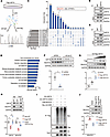 Figure 5
USP10 interacts with and deubiquitinates NDUFS1. (A) Diagram illustrating the experimental design used to explore USP10-interacting proteins by IP-MS in HL-1 cells. (B) GO analysis showing the top 10 enriched terms of USP10-binding proteins. (C) UpSet plot illustrating the intersections across different biological events of USP10-binding proteins. (D and E) Co-IP and Western blot analyses showing the interaction between USP10 and NDUFS1 in HL-1 cells. (F) Western blot analysis of USP10 and NDUFS1 protein levels in HL-1 atrial myocytes expressing Ad-shUsp10 or Ad-shRNA (n = 4/group). (G) Western blot analysis of USP10 and NDUFS1 protein levels in HL-1 atrial myocytes expressing different amounts of Ad-Flag-USP10 adenovirus (n = 4/group). (H) Western blot analysis of USP10 and NDUFS1 protein levels in mouse atrial tissues in the indicated groups (n = 4/group). (I) Representative Western blot images of K48-type ubiquitination of NDUFS1 in HL-1 cells in the indicated groups. (J) Western blot analysis of NDUFS1 protein levels in HEK293T cells expressing different plasmids (n = 3/group). Data are presented as mean ± SEM and were analyzed using an unpaired, 2-tailed Student’s t test (F) and 1-way ANOVA with Tukey’s post hoc test (G, H, and J). *P < 0.05; **P < 0.01. USP10, ubiquitin-specific peptidase 10; NDUFS1, NADH:ubiquinone oxidoreductase subunit S1; Ang II, angiotensin II; GO, Gene Ontology.
USP10 deubiquitinates NDUFS1 at K621. Following the identification of NDUFS1 as a direct target of USP10, we further investigated the specific ubiquitination sites on NDUFS1. Using GPS-Uber, a widely used ubiquitination prediction tool (26), we identified multiple potential ubiquitination sites on NDUFS1 (Figure 6A). Previous studies have demonstrated that MDM2, an E3 ubiquitin ligase, interacts with NDUFS1 (27). We analyzed the spatial relationship between MDM2 and the predicted ubiquitination sites on NDUFS1, focusing specifically on those sites in close proximity to MDM2. The predicted docking models identified 2 key interaction sites — Lys311 (K311) and Lys621 (K621) — between NDUFS1 and MDM2, while other predicted ubiquitination sites were located farther away (Figure 6B). To experimentally validate these ubiquitination sites, we generated NDUFS1-K311R and NDUFS1-K621R mutants by substituting lysine (K) with arginine (R) at positions 311 and 621, respectively. As shown in Figure 6C, the ubiquitination level of NDUFS1-K311R was similar to that of WT NDUFS1. In contrast, the ubiquitination level of NDUFS1-K621R was significantly reduced, demonstrating that MDM2 specifically ubiquitinates NDUFS1 at K621. Consistently, NDUFS1 expression was reduced in MDM2-transfected cells, but MDM2 did not further decrease the protein level of NDUFS1-K621R mutant (Figure 6D).
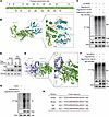 Figure 6
USP10 deubiquitinates NDUFS1 at K621. (A) Predicted ubiquitination sites of NDUFS1 using GPS-Uber. (B) Molecular docking simulation and calculation of the interaction sites between MDM2 (blue) and NDUFS1 (green). (C) Western blot analysis of K48-type ubiquitination of NDUFS1 in the indicated groups. (D) Representative Western blots showing Flag-NDUFS1 protein levels in the indicated groups. (E) Molecular docking simulation and calculation of the interaction sites between USP10 (purple) and NDUFS1 (green). (F) Western blot analysis of K48-type ubiquitination of NDUFS1 in the indicated groups. (G) Western blot analysis of the ubiquitination of WT and K621R mutant immunoprecipitated with NDUFS1. (H) Alignment of K621 and adjacent amino acids of NDUFS1 across different species. USP10, ubiquitin-specific peptidase 10; NDUFS1, NADH:ubiquinone oxidoreductase subunit S1.
To confirm that USP10 deubiquitinates NDUFS1 specifically at K621, we assessed the spatial interaction between USP10 and NDUFS1. Predicted docking models revealed a direct interaction between USP10 and NDUFS1 at K621 (Figure 6E). Supporting this, a ubiquitination assay demonstrated that USP10 was unable to further reduce the ubiquitination level of NDUFS1-K621R compared with WT NDUFS1 (Figure 6F). Ubiquitination analysis of immunoprecipitated NDUFS1 (WT and K621R mutant) confirmed that K621 is the primary ubiquitination site (Figure 6G). Notably, the K621 ubiquitination site and the surrounding protein sequence are conserved among various vertebrates, including humans, mice, rats, and cattle (Figure 6H).
NDUFS1-K621R attenuates abnormal calcium handling, DADs, and AF susceptibility. To directly assess the functional role of NDUFS1 in AF, we generated adeno-associated virus–mediated (AAV-mediated) NDUFS1 and NDUFS1-K621R overexpression mouse models and evaluated Ca2+ handling, proarrhythmic activity, and AF susceptibility.
NDUFS1 overexpression significantly reduced Ang II–induced Ca²+ sparks, calcium leak levels, and spark area, whereas the K621R variant produced a further reduction in these parameters (Supplemental Figure 1, A and B; supplemental material available online with this article; https://doi.org/10.1172/jci.insight.195761DS1). Consistently, NDUFS1 overexpression decreased the frequency of DADs under Ang II stimulation, with a more pronounced effect observed in the K621R group (Supplemental Figure 1, C and D). In vivo, both NDUFS1 and its K621R variant attenuated Ang II–induced atrial fibrosis (Supplemental Figure 1, E and F) and reduced AF incidence and duration (Supplemental Figure 1, G and H), with the K621R variant consistently exhibiting a stronger effect across all assays.
Collectively, these findings demonstrate that NDUFS1 exerts a protective effect against Ang II–induced atrial remodeling and arrhythmogenesis, and that the K621R mutation enhances its functional activity.
USP10 knockdown exacerbates abnormal calcium handling, DADs, and AF susceptibility. Given that USP10 overexpression mitigates abnormal calcium handling, DADs, atrial remodeling, and AF susceptibility in mice, we hypothesized that USP10 knockdown would have the inverse effects. To test this hypothesis, we performed a series of loss-of-function experiments. Consistent with our expectations, USP10 knockdown promoted DADs, as evidenced by an increased frequency (Figure 7, A and B). Additionally, USP10 knockdown significantly increased the amplitude of Ang II–induced CaTs (Figure 7C) and the incidence of Ca2+ sparks (Figure 7, D and E).
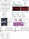 Figure 7
USP10 knockdown exacerbates abnormal calcium handling, DADs, and AF susceptibility. (A) Representative DAD images recorded from HL-1 cells expressing Ad-shRNA or Ad-shUsp10 under Ang II treatment. (B) Statistical results of DAD incidence. (C) Representative Ca2+ transient (CaT) images and statistical results of CaT amplitudes in the indicated groups (n = 15–22/group). (D) Representative Ca2+ spark images obtained from HL-1 cells expressing Ad-shRNA or Ad-shUsp10 in response to Ang II stimulation. Scale bar: 0.1 seconds. (E) Statistical results of Ca2+ spark–induced Ca2+ leak, Ca2+ spark frequency, and Ca2+ spark area in the indicated groups (n = 20–31/group). (F) Representative echocardiography images and quantification of left atrial size from WT and Usp10-CKO mice under Ang II treatment (n = 7–8/group). (G) Representative images and quantitative results of Masson’s trichrome staining on left atrial sections of Usp10-CKO and WT control mice in response to Ang II treatment (n = 4/group). (H) Representative limb lead ECG traces recorded from Usp10-CKO and WT control mice in response to Ang II treatment. (I) Fraction of mice with successful AF induction relative to the total number and AF duration in Usp10-CKO and WT control mice in response to Ang II treatment (n = 19/group). Data are presented as mean ± SEM and were analyzed using the χ2 test (B and I [left]) and the 2-tailed Student’s t test (C, E–G, and I [right]). *P < 0.05; **P < 0.01. NS, not significantly different. USP10, ubiquitin-specific peptidase 10; AF, atrial fibrillation; Ang II, angiotensin II; CaTs, Ca2+ transients; DAD, delayed afterdepolarization; ECG, electrocardiogram; CKO, cardiomyocyte-specific knockout.
To further assess the impact of Usp10 loss, we utilized cardiomyocyte-specific Usp10-knockout (Usp10-CKO) mice to evaluate whether Usp10 KO would exacerbate Ang II–induced atrial remodeling and AF susceptibility. As anticipated, Usp10 KO significantly amplified Ang II–induced left atrial enlargement, atrial fibrosis, and the incidence and duration of AF (Figure 7, F–I). Collectively, these findings demonstrate that the loss of Usp10 exacerbates Ang II–induced abnormal calcium handling, DADs, and AF susceptibility, highlighting the critical role of USP10 in maintaining normal electrophysiological and structural integrity.


