
Research ArticleAgingCardiology
Open Access |  10.1172/jci.insight.195508
PCPE-1 promotes cardiac fibrosis with aging and obesity
Yung-Ting Hsiao,1 Yohko Yoshida,2,3 Hirotsugu Tsuchimochi,4 Jingyuan Tang,1 Tin May Aung,1 Chun-Han Chang,1 Agian Jeffilano Barinda,1 Zhihong Li,1,5 Nur Syakirah Binti Othman,1 Tom Yoshizaki,1 Yiwei Ling,6 Shujiro Okuda,6 Manabu Abe,7,8 Seiya Mizuno,9 Satoru Takahashi,9 Takayuki Inomata,10 Hidetaka Kioka,11 Yasushi Sakata,11 Daichi Maeda,2 Yuya Matsue,2 Takaaki Furihata,2 Hiroshi Iwata,2 James T. Pearson,4 Kinya Otsu,5,12 Kenneth Walsh,13 Akihito Ishigami,14 Tohru Minamino,2 and Ippei Shimizu1,15
1Department of Cardiovascular Aging, National Cerebral and Cardiovascular Center Research Institute, Osaka, Japan.
2Department of Cardiovascular Biology and Medicine, and
3Department of Advanced Senotherapeutics, Juntendo University Graduate School of Medicine, Tokyo, Japan.
4Department of Cardiac Physiology, National Cerebral and Cardiovascular Center Research Institute, Osaka, Japan.
5Department of Cardiovascular Pathophysiology and Therapeutics, The University of Osaka Graduate School of Medicine, Osaka, Japan.
6Division of Bioinformatics, Niigata University Graduate School of Medical and Dental Sciences, Niigata, Japan.
7Department of Cellular Neurobiology, Brain Research Institute, and
8Department of Animal Model Development, Brain Research Institute, Niigata University, Niigata, Japan.
9Laboratory Animal Resource Center in Transborder Medical Research Center, Institute of Medicine, University of Tsukuba, Ibaraki, Japan.
10Department of Cardiovascular Medicine, Niigata University Graduate School of Medical and Dental Sciences, Niigata, Japan.
11Department of Cardiovascular Medicine, The University of Osaka Graduate School of Medicine, Osaka, Japan.
12National Cerebral and Cardiovascular Center Research Institute, Osaka, Japan.
13Division of Cardiovascular Medicine, Robert M. Berne Cardiovascular Research Center, University of Virginia School of Medicine, Charlottesville, Virginia, USA.
14Molecular Regulation of Aging, Tokyo Metropolitan Institute for Geriatrics and Gerontology, Tokyo, Japan.
15Department of Cardiovascular Medicine, National Cerebral and Cardiovascular Center, Osaka, Japan.
Address correspondence to: Ippei Shimizu, National Cerebral and Cardiovascular Center, 6-1 Kishibe-Shimmachi, Suita, Osaka, 564-8565, Japan. Phone: 81.6.6170.1069 ext. 21004; Email: shimizu.ippei@ncvc.go.jp.
Find articles by Hsiao, Y. in: PubMed | Google Scholar
1Department of Cardiovascular Aging, National Cerebral and Cardiovascular Center Research Institute, Osaka, Japan.
2Department of Cardiovascular Biology and Medicine, and
3Department of Advanced Senotherapeutics, Juntendo University Graduate School of Medicine, Tokyo, Japan.
4Department of Cardiac Physiology, National Cerebral and Cardiovascular Center Research Institute, Osaka, Japan.
5Department of Cardiovascular Pathophysiology and Therapeutics, The University of Osaka Graduate School of Medicine, Osaka, Japan.
6Division of Bioinformatics, Niigata University Graduate School of Medical and Dental Sciences, Niigata, Japan.
7Department of Cellular Neurobiology, Brain Research Institute, and
8Department of Animal Model Development, Brain Research Institute, Niigata University, Niigata, Japan.
9Laboratory Animal Resource Center in Transborder Medical Research Center, Institute of Medicine, University of Tsukuba, Ibaraki, Japan.
10Department of Cardiovascular Medicine, Niigata University Graduate School of Medical and Dental Sciences, Niigata, Japan.
11Department of Cardiovascular Medicine, The University of Osaka Graduate School of Medicine, Osaka, Japan.
12National Cerebral and Cardiovascular Center Research Institute, Osaka, Japan.
13Division of Cardiovascular Medicine, Robert M. Berne Cardiovascular Research Center, University of Virginia School of Medicine, Charlottesville, Virginia, USA.
14Molecular Regulation of Aging, Tokyo Metropolitan Institute for Geriatrics and Gerontology, Tokyo, Japan.
15Department of Cardiovascular Medicine, National Cerebral and Cardiovascular Center, Osaka, Japan.
Address correspondence to: Ippei Shimizu, National Cerebral and Cardiovascular Center, 6-1 Kishibe-Shimmachi, Suita, Osaka, 564-8565, Japan. Phone: 81.6.6170.1069 ext. 21004; Email: shimizu.ippei@ncvc.go.jp.
Find articles by Yoshida, Y. in: PubMed | Google Scholar
1Department of Cardiovascular Aging, National Cerebral and Cardiovascular Center Research Institute, Osaka, Japan.
2Department of Cardiovascular Biology and Medicine, and
3Department of Advanced Senotherapeutics, Juntendo University Graduate School of Medicine, Tokyo, Japan.
4Department of Cardiac Physiology, National Cerebral and Cardiovascular Center Research Institute, Osaka, Japan.
5Department of Cardiovascular Pathophysiology and Therapeutics, The University of Osaka Graduate School of Medicine, Osaka, Japan.
6Division of Bioinformatics, Niigata University Graduate School of Medical and Dental Sciences, Niigata, Japan.
7Department of Cellular Neurobiology, Brain Research Institute, and
8Department of Animal Model Development, Brain Research Institute, Niigata University, Niigata, Japan.
9Laboratory Animal Resource Center in Transborder Medical Research Center, Institute of Medicine, University of Tsukuba, Ibaraki, Japan.
10Department of Cardiovascular Medicine, Niigata University Graduate School of Medical and Dental Sciences, Niigata, Japan.
11Department of Cardiovascular Medicine, The University of Osaka Graduate School of Medicine, Osaka, Japan.
12National Cerebral and Cardiovascular Center Research Institute, Osaka, Japan.
13Division of Cardiovascular Medicine, Robert M. Berne Cardiovascular Research Center, University of Virginia School of Medicine, Charlottesville, Virginia, USA.
14Molecular Regulation of Aging, Tokyo Metropolitan Institute for Geriatrics and Gerontology, Tokyo, Japan.
15Department of Cardiovascular Medicine, National Cerebral and Cardiovascular Center, Osaka, Japan.
Address correspondence to: Ippei Shimizu, National Cerebral and Cardiovascular Center, 6-1 Kishibe-Shimmachi, Suita, Osaka, 564-8565, Japan. Phone: 81.6.6170.1069 ext. 21004; Email: shimizu.ippei@ncvc.go.jp.
Find articles by
Tsuchimochi, H.
in:
PubMed
|
Google Scholar
|

1Department of Cardiovascular Aging, National Cerebral and Cardiovascular Center Research Institute, Osaka, Japan.
2Department of Cardiovascular Biology and Medicine, and
3Department of Advanced Senotherapeutics, Juntendo University Graduate School of Medicine, Tokyo, Japan.
4Department of Cardiac Physiology, National Cerebral and Cardiovascular Center Research Institute, Osaka, Japan.
5Department of Cardiovascular Pathophysiology and Therapeutics, The University of Osaka Graduate School of Medicine, Osaka, Japan.
6Division of Bioinformatics, Niigata University Graduate School of Medical and Dental Sciences, Niigata, Japan.
7Department of Cellular Neurobiology, Brain Research Institute, and
8Department of Animal Model Development, Brain Research Institute, Niigata University, Niigata, Japan.
9Laboratory Animal Resource Center in Transborder Medical Research Center, Institute of Medicine, University of Tsukuba, Ibaraki, Japan.
10Department of Cardiovascular Medicine, Niigata University Graduate School of Medical and Dental Sciences, Niigata, Japan.
11Department of Cardiovascular Medicine, The University of Osaka Graduate School of Medicine, Osaka, Japan.
12National Cerebral and Cardiovascular Center Research Institute, Osaka, Japan.
13Division of Cardiovascular Medicine, Robert M. Berne Cardiovascular Research Center, University of Virginia School of Medicine, Charlottesville, Virginia, USA.
14Molecular Regulation of Aging, Tokyo Metropolitan Institute for Geriatrics and Gerontology, Tokyo, Japan.
15Department of Cardiovascular Medicine, National Cerebral and Cardiovascular Center, Osaka, Japan.
Address correspondence to: Ippei Shimizu, National Cerebral and Cardiovascular Center, 6-1 Kishibe-Shimmachi, Suita, Osaka, 564-8565, Japan. Phone: 81.6.6170.1069 ext. 21004; Email: shimizu.ippei@ncvc.go.jp.
Find articles by Tang, J. in: PubMed | Google Scholar
1Department of Cardiovascular Aging, National Cerebral and Cardiovascular Center Research Institute, Osaka, Japan.
2Department of Cardiovascular Biology and Medicine, and
3Department of Advanced Senotherapeutics, Juntendo University Graduate School of Medicine, Tokyo, Japan.
4Department of Cardiac Physiology, National Cerebral and Cardiovascular Center Research Institute, Osaka, Japan.
5Department of Cardiovascular Pathophysiology and Therapeutics, The University of Osaka Graduate School of Medicine, Osaka, Japan.
6Division of Bioinformatics, Niigata University Graduate School of Medical and Dental Sciences, Niigata, Japan.
7Department of Cellular Neurobiology, Brain Research Institute, and
8Department of Animal Model Development, Brain Research Institute, Niigata University, Niigata, Japan.
9Laboratory Animal Resource Center in Transborder Medical Research Center, Institute of Medicine, University of Tsukuba, Ibaraki, Japan.
10Department of Cardiovascular Medicine, Niigata University Graduate School of Medical and Dental Sciences, Niigata, Japan.
11Department of Cardiovascular Medicine, The University of Osaka Graduate School of Medicine, Osaka, Japan.
12National Cerebral and Cardiovascular Center Research Institute, Osaka, Japan.
13Division of Cardiovascular Medicine, Robert M. Berne Cardiovascular Research Center, University of Virginia School of Medicine, Charlottesville, Virginia, USA.
14Molecular Regulation of Aging, Tokyo Metropolitan Institute for Geriatrics and Gerontology, Tokyo, Japan.
15Department of Cardiovascular Medicine, National Cerebral and Cardiovascular Center, Osaka, Japan.
Address correspondence to: Ippei Shimizu, National Cerebral and Cardiovascular Center, 6-1 Kishibe-Shimmachi, Suita, Osaka, 564-8565, Japan. Phone: 81.6.6170.1069 ext. 21004; Email: shimizu.ippei@ncvc.go.jp.
Find articles by Aung, T. in: PubMed | Google Scholar
1Department of Cardiovascular Aging, National Cerebral and Cardiovascular Center Research Institute, Osaka, Japan.
2Department of Cardiovascular Biology and Medicine, and
3Department of Advanced Senotherapeutics, Juntendo University Graduate School of Medicine, Tokyo, Japan.
4Department of Cardiac Physiology, National Cerebral and Cardiovascular Center Research Institute, Osaka, Japan.
5Department of Cardiovascular Pathophysiology and Therapeutics, The University of Osaka Graduate School of Medicine, Osaka, Japan.
6Division of Bioinformatics, Niigata University Graduate School of Medical and Dental Sciences, Niigata, Japan.
7Department of Cellular Neurobiology, Brain Research Institute, and
8Department of Animal Model Development, Brain Research Institute, Niigata University, Niigata, Japan.
9Laboratory Animal Resource Center in Transborder Medical Research Center, Institute of Medicine, University of Tsukuba, Ibaraki, Japan.
10Department of Cardiovascular Medicine, Niigata University Graduate School of Medical and Dental Sciences, Niigata, Japan.
11Department of Cardiovascular Medicine, The University of Osaka Graduate School of Medicine, Osaka, Japan.
12National Cerebral and Cardiovascular Center Research Institute, Osaka, Japan.
13Division of Cardiovascular Medicine, Robert M. Berne Cardiovascular Research Center, University of Virginia School of Medicine, Charlottesville, Virginia, USA.
14Molecular Regulation of Aging, Tokyo Metropolitan Institute for Geriatrics and Gerontology, Tokyo, Japan.
15Department of Cardiovascular Medicine, National Cerebral and Cardiovascular Center, Osaka, Japan.
Address correspondence to: Ippei Shimizu, National Cerebral and Cardiovascular Center, 6-1 Kishibe-Shimmachi, Suita, Osaka, 564-8565, Japan. Phone: 81.6.6170.1069 ext. 21004; Email: shimizu.ippei@ncvc.go.jp.
Find articles by Chang, C. in: PubMed | Google Scholar
1Department of Cardiovascular Aging, National Cerebral and Cardiovascular Center Research Institute, Osaka, Japan.
2Department of Cardiovascular Biology and Medicine, and
3Department of Advanced Senotherapeutics, Juntendo University Graduate School of Medicine, Tokyo, Japan.
4Department of Cardiac Physiology, National Cerebral and Cardiovascular Center Research Institute, Osaka, Japan.
5Department of Cardiovascular Pathophysiology and Therapeutics, The University of Osaka Graduate School of Medicine, Osaka, Japan.
6Division of Bioinformatics, Niigata University Graduate School of Medical and Dental Sciences, Niigata, Japan.
7Department of Cellular Neurobiology, Brain Research Institute, and
8Department of Animal Model Development, Brain Research Institute, Niigata University, Niigata, Japan.
9Laboratory Animal Resource Center in Transborder Medical Research Center, Institute of Medicine, University of Tsukuba, Ibaraki, Japan.
10Department of Cardiovascular Medicine, Niigata University Graduate School of Medical and Dental Sciences, Niigata, Japan.
11Department of Cardiovascular Medicine, The University of Osaka Graduate School of Medicine, Osaka, Japan.
12National Cerebral and Cardiovascular Center Research Institute, Osaka, Japan.
13Division of Cardiovascular Medicine, Robert M. Berne Cardiovascular Research Center, University of Virginia School of Medicine, Charlottesville, Virginia, USA.
14Molecular Regulation of Aging, Tokyo Metropolitan Institute for Geriatrics and Gerontology, Tokyo, Japan.
15Department of Cardiovascular Medicine, National Cerebral and Cardiovascular Center, Osaka, Japan.
Address correspondence to: Ippei Shimizu, National Cerebral and Cardiovascular Center, 6-1 Kishibe-Shimmachi, Suita, Osaka, 564-8565, Japan. Phone: 81.6.6170.1069 ext. 21004; Email: shimizu.ippei@ncvc.go.jp.
Find articles by Barinda, A. in: PubMed | Google Scholar
1Department of Cardiovascular Aging, National Cerebral and Cardiovascular Center Research Institute, Osaka, Japan.
2Department of Cardiovascular Biology and Medicine, and
3Department of Advanced Senotherapeutics, Juntendo University Graduate School of Medicine, Tokyo, Japan.
4Department of Cardiac Physiology, National Cerebral and Cardiovascular Center Research Institute, Osaka, Japan.
5Department of Cardiovascular Pathophysiology and Therapeutics, The University of Osaka Graduate School of Medicine, Osaka, Japan.
6Division of Bioinformatics, Niigata University Graduate School of Medical and Dental Sciences, Niigata, Japan.
7Department of Cellular Neurobiology, Brain Research Institute, and
8Department of Animal Model Development, Brain Research Institute, Niigata University, Niigata, Japan.
9Laboratory Animal Resource Center in Transborder Medical Research Center, Institute of Medicine, University of Tsukuba, Ibaraki, Japan.
10Department of Cardiovascular Medicine, Niigata University Graduate School of Medical and Dental Sciences, Niigata, Japan.
11Department of Cardiovascular Medicine, The University of Osaka Graduate School of Medicine, Osaka, Japan.
12National Cerebral and Cardiovascular Center Research Institute, Osaka, Japan.
13Division of Cardiovascular Medicine, Robert M. Berne Cardiovascular Research Center, University of Virginia School of Medicine, Charlottesville, Virginia, USA.
14Molecular Regulation of Aging, Tokyo Metropolitan Institute for Geriatrics and Gerontology, Tokyo, Japan.
15Department of Cardiovascular Medicine, National Cerebral and Cardiovascular Center, Osaka, Japan.
Address correspondence to: Ippei Shimizu, National Cerebral and Cardiovascular Center, 6-1 Kishibe-Shimmachi, Suita, Osaka, 564-8565, Japan. Phone: 81.6.6170.1069 ext. 21004; Email: shimizu.ippei@ncvc.go.jp.
Find articles by Li, Z. in: PubMed | Google Scholar
1Department of Cardiovascular Aging, National Cerebral and Cardiovascular Center Research Institute, Osaka, Japan.
2Department of Cardiovascular Biology and Medicine, and
3Department of Advanced Senotherapeutics, Juntendo University Graduate School of Medicine, Tokyo, Japan.
4Department of Cardiac Physiology, National Cerebral and Cardiovascular Center Research Institute, Osaka, Japan.
5Department of Cardiovascular Pathophysiology and Therapeutics, The University of Osaka Graduate School of Medicine, Osaka, Japan.
6Division of Bioinformatics, Niigata University Graduate School of Medical and Dental Sciences, Niigata, Japan.
7Department of Cellular Neurobiology, Brain Research Institute, and
8Department of Animal Model Development, Brain Research Institute, Niigata University, Niigata, Japan.
9Laboratory Animal Resource Center in Transborder Medical Research Center, Institute of Medicine, University of Tsukuba, Ibaraki, Japan.
10Department of Cardiovascular Medicine, Niigata University Graduate School of Medical and Dental Sciences, Niigata, Japan.
11Department of Cardiovascular Medicine, The University of Osaka Graduate School of Medicine, Osaka, Japan.
12National Cerebral and Cardiovascular Center Research Institute, Osaka, Japan.
13Division of Cardiovascular Medicine, Robert M. Berne Cardiovascular Research Center, University of Virginia School of Medicine, Charlottesville, Virginia, USA.
14Molecular Regulation of Aging, Tokyo Metropolitan Institute for Geriatrics and Gerontology, Tokyo, Japan.
15Department of Cardiovascular Medicine, National Cerebral and Cardiovascular Center, Osaka, Japan.
Address correspondence to: Ippei Shimizu, National Cerebral and Cardiovascular Center, 6-1 Kishibe-Shimmachi, Suita, Osaka, 564-8565, Japan. Phone: 81.6.6170.1069 ext. 21004; Email: shimizu.ippei@ncvc.go.jp.
Find articles by Othman, N. in: PubMed | Google Scholar
1Department of Cardiovascular Aging, National Cerebral and Cardiovascular Center Research Institute, Osaka, Japan.
2Department of Cardiovascular Biology and Medicine, and
3Department of Advanced Senotherapeutics, Juntendo University Graduate School of Medicine, Tokyo, Japan.
4Department of Cardiac Physiology, National Cerebral and Cardiovascular Center Research Institute, Osaka, Japan.
5Department of Cardiovascular Pathophysiology and Therapeutics, The University of Osaka Graduate School of Medicine, Osaka, Japan.
6Division of Bioinformatics, Niigata University Graduate School of Medical and Dental Sciences, Niigata, Japan.
7Department of Cellular Neurobiology, Brain Research Institute, and
8Department of Animal Model Development, Brain Research Institute, Niigata University, Niigata, Japan.
9Laboratory Animal Resource Center in Transborder Medical Research Center, Institute of Medicine, University of Tsukuba, Ibaraki, Japan.
10Department of Cardiovascular Medicine, Niigata University Graduate School of Medical and Dental Sciences, Niigata, Japan.
11Department of Cardiovascular Medicine, The University of Osaka Graduate School of Medicine, Osaka, Japan.
12National Cerebral and Cardiovascular Center Research Institute, Osaka, Japan.
13Division of Cardiovascular Medicine, Robert M. Berne Cardiovascular Research Center, University of Virginia School of Medicine, Charlottesville, Virginia, USA.
14Molecular Regulation of Aging, Tokyo Metropolitan Institute for Geriatrics and Gerontology, Tokyo, Japan.
15Department of Cardiovascular Medicine, National Cerebral and Cardiovascular Center, Osaka, Japan.
Address correspondence to: Ippei Shimizu, National Cerebral and Cardiovascular Center, 6-1 Kishibe-Shimmachi, Suita, Osaka, 564-8565, Japan. Phone: 81.6.6170.1069 ext. 21004; Email: shimizu.ippei@ncvc.go.jp.
Find articles by Yoshizaki, T. in: PubMed | Google Scholar
1Department of Cardiovascular Aging, National Cerebral and Cardiovascular Center Research Institute, Osaka, Japan.
2Department of Cardiovascular Biology and Medicine, and
3Department of Advanced Senotherapeutics, Juntendo University Graduate School of Medicine, Tokyo, Japan.
4Department of Cardiac Physiology, National Cerebral and Cardiovascular Center Research Institute, Osaka, Japan.
5Department of Cardiovascular Pathophysiology and Therapeutics, The University of Osaka Graduate School of Medicine, Osaka, Japan.
6Division of Bioinformatics, Niigata University Graduate School of Medical and Dental Sciences, Niigata, Japan.
7Department of Cellular Neurobiology, Brain Research Institute, and
8Department of Animal Model Development, Brain Research Institute, Niigata University, Niigata, Japan.
9Laboratory Animal Resource Center in Transborder Medical Research Center, Institute of Medicine, University of Tsukuba, Ibaraki, Japan.
10Department of Cardiovascular Medicine, Niigata University Graduate School of Medical and Dental Sciences, Niigata, Japan.
11Department of Cardiovascular Medicine, The University of Osaka Graduate School of Medicine, Osaka, Japan.
12National Cerebral and Cardiovascular Center Research Institute, Osaka, Japan.
13Division of Cardiovascular Medicine, Robert M. Berne Cardiovascular Research Center, University of Virginia School of Medicine, Charlottesville, Virginia, USA.
14Molecular Regulation of Aging, Tokyo Metropolitan Institute for Geriatrics and Gerontology, Tokyo, Japan.
15Department of Cardiovascular Medicine, National Cerebral and Cardiovascular Center, Osaka, Japan.
Address correspondence to: Ippei Shimizu, National Cerebral and Cardiovascular Center, 6-1 Kishibe-Shimmachi, Suita, Osaka, 564-8565, Japan. Phone: 81.6.6170.1069 ext. 21004; Email: shimizu.ippei@ncvc.go.jp.
Find articles by Ling, Y. in: PubMed | Google Scholar
1Department of Cardiovascular Aging, National Cerebral and Cardiovascular Center Research Institute, Osaka, Japan.
2Department of Cardiovascular Biology and Medicine, and
3Department of Advanced Senotherapeutics, Juntendo University Graduate School of Medicine, Tokyo, Japan.
4Department of Cardiac Physiology, National Cerebral and Cardiovascular Center Research Institute, Osaka, Japan.
5Department of Cardiovascular Pathophysiology and Therapeutics, The University of Osaka Graduate School of Medicine, Osaka, Japan.
6Division of Bioinformatics, Niigata University Graduate School of Medical and Dental Sciences, Niigata, Japan.
7Department of Cellular Neurobiology, Brain Research Institute, and
8Department of Animal Model Development, Brain Research Institute, Niigata University, Niigata, Japan.
9Laboratory Animal Resource Center in Transborder Medical Research Center, Institute of Medicine, University of Tsukuba, Ibaraki, Japan.
10Department of Cardiovascular Medicine, Niigata University Graduate School of Medical and Dental Sciences, Niigata, Japan.
11Department of Cardiovascular Medicine, The University of Osaka Graduate School of Medicine, Osaka, Japan.
12National Cerebral and Cardiovascular Center Research Institute, Osaka, Japan.
13Division of Cardiovascular Medicine, Robert M. Berne Cardiovascular Research Center, University of Virginia School of Medicine, Charlottesville, Virginia, USA.
14Molecular Regulation of Aging, Tokyo Metropolitan Institute for Geriatrics and Gerontology, Tokyo, Japan.
15Department of Cardiovascular Medicine, National Cerebral and Cardiovascular Center, Osaka, Japan.
Address correspondence to: Ippei Shimizu, National Cerebral and Cardiovascular Center, 6-1 Kishibe-Shimmachi, Suita, Osaka, 564-8565, Japan. Phone: 81.6.6170.1069 ext. 21004; Email: shimizu.ippei@ncvc.go.jp.
Find articles by
Okuda, S.
in:
PubMed
|
Google Scholar
|
1Department of Cardiovascular Aging, National Cerebral and Cardiovascular Center Research Institute, Osaka, Japan.
2Department of Cardiovascular Biology and Medicine, and
3Department of Advanced Senotherapeutics, Juntendo University Graduate School of Medicine, Tokyo, Japan.
4Department of Cardiac Physiology, National Cerebral and Cardiovascular Center Research Institute, Osaka, Japan.
5Department of Cardiovascular Pathophysiology and Therapeutics, The University of Osaka Graduate School of Medicine, Osaka, Japan.
6Division of Bioinformatics, Niigata University Graduate School of Medical and Dental Sciences, Niigata, Japan.
7Department of Cellular Neurobiology, Brain Research Institute, and
8Department of Animal Model Development, Brain Research Institute, Niigata University, Niigata, Japan.
9Laboratory Animal Resource Center in Transborder Medical Research Center, Institute of Medicine, University of Tsukuba, Ibaraki, Japan.
10Department of Cardiovascular Medicine, Niigata University Graduate School of Medical and Dental Sciences, Niigata, Japan.
11Department of Cardiovascular Medicine, The University of Osaka Graduate School of Medicine, Osaka, Japan.
12National Cerebral and Cardiovascular Center Research Institute, Osaka, Japan.
13Division of Cardiovascular Medicine, Robert M. Berne Cardiovascular Research Center, University of Virginia School of Medicine, Charlottesville, Virginia, USA.
14Molecular Regulation of Aging, Tokyo Metropolitan Institute for Geriatrics and Gerontology, Tokyo, Japan.
15Department of Cardiovascular Medicine, National Cerebral and Cardiovascular Center, Osaka, Japan.
Address correspondence to: Ippei Shimizu, National Cerebral and Cardiovascular Center, 6-1 Kishibe-Shimmachi, Suita, Osaka, 564-8565, Japan. Phone: 81.6.6170.1069 ext. 21004; Email: shimizu.ippei@ncvc.go.jp.
Find articles by
Abe, M.
in:
PubMed
|
Google Scholar
|
1Department of Cardiovascular Aging, National Cerebral and Cardiovascular Center Research Institute, Osaka, Japan.
2Department of Cardiovascular Biology and Medicine, and
3Department of Advanced Senotherapeutics, Juntendo University Graduate School of Medicine, Tokyo, Japan.
4Department of Cardiac Physiology, National Cerebral and Cardiovascular Center Research Institute, Osaka, Japan.
5Department of Cardiovascular Pathophysiology and Therapeutics, The University of Osaka Graduate School of Medicine, Osaka, Japan.
6Division of Bioinformatics, Niigata University Graduate School of Medical and Dental Sciences, Niigata, Japan.
7Department of Cellular Neurobiology, Brain Research Institute, and
8Department of Animal Model Development, Brain Research Institute, Niigata University, Niigata, Japan.
9Laboratory Animal Resource Center in Transborder Medical Research Center, Institute of Medicine, University of Tsukuba, Ibaraki, Japan.
10Department of Cardiovascular Medicine, Niigata University Graduate School of Medical and Dental Sciences, Niigata, Japan.
11Department of Cardiovascular Medicine, The University of Osaka Graduate School of Medicine, Osaka, Japan.
12National Cerebral and Cardiovascular Center Research Institute, Osaka, Japan.
13Division of Cardiovascular Medicine, Robert M. Berne Cardiovascular Research Center, University of Virginia School of Medicine, Charlottesville, Virginia, USA.
14Molecular Regulation of Aging, Tokyo Metropolitan Institute for Geriatrics and Gerontology, Tokyo, Japan.
15Department of Cardiovascular Medicine, National Cerebral and Cardiovascular Center, Osaka, Japan.
Address correspondence to: Ippei Shimizu, National Cerebral and Cardiovascular Center, 6-1 Kishibe-Shimmachi, Suita, Osaka, 564-8565, Japan. Phone: 81.6.6170.1069 ext. 21004; Email: shimizu.ippei@ncvc.go.jp.
Find articles by Mizuno, S. in: PubMed | Google Scholar
1Department of Cardiovascular Aging, National Cerebral and Cardiovascular Center Research Institute, Osaka, Japan.
2Department of Cardiovascular Biology and Medicine, and
3Department of Advanced Senotherapeutics, Juntendo University Graduate School of Medicine, Tokyo, Japan.
4Department of Cardiac Physiology, National Cerebral and Cardiovascular Center Research Institute, Osaka, Japan.
5Department of Cardiovascular Pathophysiology and Therapeutics, The University of Osaka Graduate School of Medicine, Osaka, Japan.
6Division of Bioinformatics, Niigata University Graduate School of Medical and Dental Sciences, Niigata, Japan.
7Department of Cellular Neurobiology, Brain Research Institute, and
8Department of Animal Model Development, Brain Research Institute, Niigata University, Niigata, Japan.
9Laboratory Animal Resource Center in Transborder Medical Research Center, Institute of Medicine, University of Tsukuba, Ibaraki, Japan.
10Department of Cardiovascular Medicine, Niigata University Graduate School of Medical and Dental Sciences, Niigata, Japan.
11Department of Cardiovascular Medicine, The University of Osaka Graduate School of Medicine, Osaka, Japan.
12National Cerebral and Cardiovascular Center Research Institute, Osaka, Japan.
13Division of Cardiovascular Medicine, Robert M. Berne Cardiovascular Research Center, University of Virginia School of Medicine, Charlottesville, Virginia, USA.
14Molecular Regulation of Aging, Tokyo Metropolitan Institute for Geriatrics and Gerontology, Tokyo, Japan.
15Department of Cardiovascular Medicine, National Cerebral and Cardiovascular Center, Osaka, Japan.
Address correspondence to: Ippei Shimizu, National Cerebral and Cardiovascular Center, 6-1 Kishibe-Shimmachi, Suita, Osaka, 564-8565, Japan. Phone: 81.6.6170.1069 ext. 21004; Email: shimizu.ippei@ncvc.go.jp.
Find articles by
Takahashi, S.
in:
PubMed
|
Google Scholar
|
1Department of Cardiovascular Aging, National Cerebral and Cardiovascular Center Research Institute, Osaka, Japan.
2Department of Cardiovascular Biology and Medicine, and
3Department of Advanced Senotherapeutics, Juntendo University Graduate School of Medicine, Tokyo, Japan.
4Department of Cardiac Physiology, National Cerebral and Cardiovascular Center Research Institute, Osaka, Japan.
5Department of Cardiovascular Pathophysiology and Therapeutics, The University of Osaka Graduate School of Medicine, Osaka, Japan.
6Division of Bioinformatics, Niigata University Graduate School of Medical and Dental Sciences, Niigata, Japan.
7Department of Cellular Neurobiology, Brain Research Institute, and
8Department of Animal Model Development, Brain Research Institute, Niigata University, Niigata, Japan.
9Laboratory Animal Resource Center in Transborder Medical Research Center, Institute of Medicine, University of Tsukuba, Ibaraki, Japan.
10Department of Cardiovascular Medicine, Niigata University Graduate School of Medical and Dental Sciences, Niigata, Japan.
11Department of Cardiovascular Medicine, The University of Osaka Graduate School of Medicine, Osaka, Japan.
12National Cerebral and Cardiovascular Center Research Institute, Osaka, Japan.
13Division of Cardiovascular Medicine, Robert M. Berne Cardiovascular Research Center, University of Virginia School of Medicine, Charlottesville, Virginia, USA.
14Molecular Regulation of Aging, Tokyo Metropolitan Institute for Geriatrics and Gerontology, Tokyo, Japan.
15Department of Cardiovascular Medicine, National Cerebral and Cardiovascular Center, Osaka, Japan.
Address correspondence to: Ippei Shimizu, National Cerebral and Cardiovascular Center, 6-1 Kishibe-Shimmachi, Suita, Osaka, 564-8565, Japan. Phone: 81.6.6170.1069 ext. 21004; Email: shimizu.ippei@ncvc.go.jp.
Find articles by Inomata, T. in: PubMed | Google Scholar
1Department of Cardiovascular Aging, National Cerebral and Cardiovascular Center Research Institute, Osaka, Japan.
2Department of Cardiovascular Biology and Medicine, and
3Department of Advanced Senotherapeutics, Juntendo University Graduate School of Medicine, Tokyo, Japan.
4Department of Cardiac Physiology, National Cerebral and Cardiovascular Center Research Institute, Osaka, Japan.
5Department of Cardiovascular Pathophysiology and Therapeutics, The University of Osaka Graduate School of Medicine, Osaka, Japan.
6Division of Bioinformatics, Niigata University Graduate School of Medical and Dental Sciences, Niigata, Japan.
7Department of Cellular Neurobiology, Brain Research Institute, and
8Department of Animal Model Development, Brain Research Institute, Niigata University, Niigata, Japan.
9Laboratory Animal Resource Center in Transborder Medical Research Center, Institute of Medicine, University of Tsukuba, Ibaraki, Japan.
10Department of Cardiovascular Medicine, Niigata University Graduate School of Medical and Dental Sciences, Niigata, Japan.
11Department of Cardiovascular Medicine, The University of Osaka Graduate School of Medicine, Osaka, Japan.
12National Cerebral and Cardiovascular Center Research Institute, Osaka, Japan.
13Division of Cardiovascular Medicine, Robert M. Berne Cardiovascular Research Center, University of Virginia School of Medicine, Charlottesville, Virginia, USA.
14Molecular Regulation of Aging, Tokyo Metropolitan Institute for Geriatrics and Gerontology, Tokyo, Japan.
15Department of Cardiovascular Medicine, National Cerebral and Cardiovascular Center, Osaka, Japan.
Address correspondence to: Ippei Shimizu, National Cerebral and Cardiovascular Center, 6-1 Kishibe-Shimmachi, Suita, Osaka, 564-8565, Japan. Phone: 81.6.6170.1069 ext. 21004; Email: shimizu.ippei@ncvc.go.jp.
Find articles by Kioka, H. in: PubMed | Google Scholar
1Department of Cardiovascular Aging, National Cerebral and Cardiovascular Center Research Institute, Osaka, Japan.
2Department of Cardiovascular Biology and Medicine, and
3Department of Advanced Senotherapeutics, Juntendo University Graduate School of Medicine, Tokyo, Japan.
4Department of Cardiac Physiology, National Cerebral and Cardiovascular Center Research Institute, Osaka, Japan.
5Department of Cardiovascular Pathophysiology and Therapeutics, The University of Osaka Graduate School of Medicine, Osaka, Japan.
6Division of Bioinformatics, Niigata University Graduate School of Medical and Dental Sciences, Niigata, Japan.
7Department of Cellular Neurobiology, Brain Research Institute, and
8Department of Animal Model Development, Brain Research Institute, Niigata University, Niigata, Japan.
9Laboratory Animal Resource Center in Transborder Medical Research Center, Institute of Medicine, University of Tsukuba, Ibaraki, Japan.
10Department of Cardiovascular Medicine, Niigata University Graduate School of Medical and Dental Sciences, Niigata, Japan.
11Department of Cardiovascular Medicine, The University of Osaka Graduate School of Medicine, Osaka, Japan.
12National Cerebral and Cardiovascular Center Research Institute, Osaka, Japan.
13Division of Cardiovascular Medicine, Robert M. Berne Cardiovascular Research Center, University of Virginia School of Medicine, Charlottesville, Virginia, USA.
14Molecular Regulation of Aging, Tokyo Metropolitan Institute for Geriatrics and Gerontology, Tokyo, Japan.
15Department of Cardiovascular Medicine, National Cerebral and Cardiovascular Center, Osaka, Japan.
Address correspondence to: Ippei Shimizu, National Cerebral and Cardiovascular Center, 6-1 Kishibe-Shimmachi, Suita, Osaka, 564-8565, Japan. Phone: 81.6.6170.1069 ext. 21004; Email: shimizu.ippei@ncvc.go.jp.
Find articles by Sakata, Y. in: PubMed | Google Scholar
1Department of Cardiovascular Aging, National Cerebral and Cardiovascular Center Research Institute, Osaka, Japan.
2Department of Cardiovascular Biology and Medicine, and
3Department of Advanced Senotherapeutics, Juntendo University Graduate School of Medicine, Tokyo, Japan.
4Department of Cardiac Physiology, National Cerebral and Cardiovascular Center Research Institute, Osaka, Japan.
5Department of Cardiovascular Pathophysiology and Therapeutics, The University of Osaka Graduate School of Medicine, Osaka, Japan.
6Division of Bioinformatics, Niigata University Graduate School of Medical and Dental Sciences, Niigata, Japan.
7Department of Cellular Neurobiology, Brain Research Institute, and
8Department of Animal Model Development, Brain Research Institute, Niigata University, Niigata, Japan.
9Laboratory Animal Resource Center in Transborder Medical Research Center, Institute of Medicine, University of Tsukuba, Ibaraki, Japan.
10Department of Cardiovascular Medicine, Niigata University Graduate School of Medical and Dental Sciences, Niigata, Japan.
11Department of Cardiovascular Medicine, The University of Osaka Graduate School of Medicine, Osaka, Japan.
12National Cerebral and Cardiovascular Center Research Institute, Osaka, Japan.
13Division of Cardiovascular Medicine, Robert M. Berne Cardiovascular Research Center, University of Virginia School of Medicine, Charlottesville, Virginia, USA.
14Molecular Regulation of Aging, Tokyo Metropolitan Institute for Geriatrics and Gerontology, Tokyo, Japan.
15Department of Cardiovascular Medicine, National Cerebral and Cardiovascular Center, Osaka, Japan.
Address correspondence to: Ippei Shimizu, National Cerebral and Cardiovascular Center, 6-1 Kishibe-Shimmachi, Suita, Osaka, 564-8565, Japan. Phone: 81.6.6170.1069 ext. 21004; Email: shimizu.ippei@ncvc.go.jp.
Find articles by Maeda, D. in: PubMed | Google Scholar
1Department of Cardiovascular Aging, National Cerebral and Cardiovascular Center Research Institute, Osaka, Japan.
2Department of Cardiovascular Biology and Medicine, and
3Department of Advanced Senotherapeutics, Juntendo University Graduate School of Medicine, Tokyo, Japan.
4Department of Cardiac Physiology, National Cerebral and Cardiovascular Center Research Institute, Osaka, Japan.
5Department of Cardiovascular Pathophysiology and Therapeutics, The University of Osaka Graduate School of Medicine, Osaka, Japan.
6Division of Bioinformatics, Niigata University Graduate School of Medical and Dental Sciences, Niigata, Japan.
7Department of Cellular Neurobiology, Brain Research Institute, and
8Department of Animal Model Development, Brain Research Institute, Niigata University, Niigata, Japan.
9Laboratory Animal Resource Center in Transborder Medical Research Center, Institute of Medicine, University of Tsukuba, Ibaraki, Japan.
10Department of Cardiovascular Medicine, Niigata University Graduate School of Medical and Dental Sciences, Niigata, Japan.
11Department of Cardiovascular Medicine, The University of Osaka Graduate School of Medicine, Osaka, Japan.
12National Cerebral and Cardiovascular Center Research Institute, Osaka, Japan.
13Division of Cardiovascular Medicine, Robert M. Berne Cardiovascular Research Center, University of Virginia School of Medicine, Charlottesville, Virginia, USA.
14Molecular Regulation of Aging, Tokyo Metropolitan Institute for Geriatrics and Gerontology, Tokyo, Japan.
15Department of Cardiovascular Medicine, National Cerebral and Cardiovascular Center, Osaka, Japan.
Address correspondence to: Ippei Shimizu, National Cerebral and Cardiovascular Center, 6-1 Kishibe-Shimmachi, Suita, Osaka, 564-8565, Japan. Phone: 81.6.6170.1069 ext. 21004; Email: shimizu.ippei@ncvc.go.jp.
Find articles by Matsue, Y. in: PubMed | Google Scholar
1Department of Cardiovascular Aging, National Cerebral and Cardiovascular Center Research Institute, Osaka, Japan.
2Department of Cardiovascular Biology and Medicine, and
3Department of Advanced Senotherapeutics, Juntendo University Graduate School of Medicine, Tokyo, Japan.
4Department of Cardiac Physiology, National Cerebral and Cardiovascular Center Research Institute, Osaka, Japan.
5Department of Cardiovascular Pathophysiology and Therapeutics, The University of Osaka Graduate School of Medicine, Osaka, Japan.
6Division of Bioinformatics, Niigata University Graduate School of Medical and Dental Sciences, Niigata, Japan.
7Department of Cellular Neurobiology, Brain Research Institute, and
8Department of Animal Model Development, Brain Research Institute, Niigata University, Niigata, Japan.
9Laboratory Animal Resource Center in Transborder Medical Research Center, Institute of Medicine, University of Tsukuba, Ibaraki, Japan.
10Department of Cardiovascular Medicine, Niigata University Graduate School of Medical and Dental Sciences, Niigata, Japan.
11Department of Cardiovascular Medicine, The University of Osaka Graduate School of Medicine, Osaka, Japan.
12National Cerebral and Cardiovascular Center Research Institute, Osaka, Japan.
13Division of Cardiovascular Medicine, Robert M. Berne Cardiovascular Research Center, University of Virginia School of Medicine, Charlottesville, Virginia, USA.
14Molecular Regulation of Aging, Tokyo Metropolitan Institute for Geriatrics and Gerontology, Tokyo, Japan.
15Department of Cardiovascular Medicine, National Cerebral and Cardiovascular Center, Osaka, Japan.
Address correspondence to: Ippei Shimizu, National Cerebral and Cardiovascular Center, 6-1 Kishibe-Shimmachi, Suita, Osaka, 564-8565, Japan. Phone: 81.6.6170.1069 ext. 21004; Email: shimizu.ippei@ncvc.go.jp.
Find articles by Furihata, T. in: PubMed | Google Scholar
1Department of Cardiovascular Aging, National Cerebral and Cardiovascular Center Research Institute, Osaka, Japan.
2Department of Cardiovascular Biology and Medicine, and
3Department of Advanced Senotherapeutics, Juntendo University Graduate School of Medicine, Tokyo, Japan.
4Department of Cardiac Physiology, National Cerebral and Cardiovascular Center Research Institute, Osaka, Japan.
5Department of Cardiovascular Pathophysiology and Therapeutics, The University of Osaka Graduate School of Medicine, Osaka, Japan.
6Division of Bioinformatics, Niigata University Graduate School of Medical and Dental Sciences, Niigata, Japan.
7Department of Cellular Neurobiology, Brain Research Institute, and
8Department of Animal Model Development, Brain Research Institute, Niigata University, Niigata, Japan.
9Laboratory Animal Resource Center in Transborder Medical Research Center, Institute of Medicine, University of Tsukuba, Ibaraki, Japan.
10Department of Cardiovascular Medicine, Niigata University Graduate School of Medical and Dental Sciences, Niigata, Japan.
11Department of Cardiovascular Medicine, The University of Osaka Graduate School of Medicine, Osaka, Japan.
12National Cerebral and Cardiovascular Center Research Institute, Osaka, Japan.
13Division of Cardiovascular Medicine, Robert M. Berne Cardiovascular Research Center, University of Virginia School of Medicine, Charlottesville, Virginia, USA.
14Molecular Regulation of Aging, Tokyo Metropolitan Institute for Geriatrics and Gerontology, Tokyo, Japan.
15Department of Cardiovascular Medicine, National Cerebral and Cardiovascular Center, Osaka, Japan.
Address correspondence to: Ippei Shimizu, National Cerebral and Cardiovascular Center, 6-1 Kishibe-Shimmachi, Suita, Osaka, 564-8565, Japan. Phone: 81.6.6170.1069 ext. 21004; Email: shimizu.ippei@ncvc.go.jp.
Find articles by Iwata, H. in: PubMed | Google Scholar
1Department of Cardiovascular Aging, National Cerebral and Cardiovascular Center Research Institute, Osaka, Japan.
2Department of Cardiovascular Biology and Medicine, and
3Department of Advanced Senotherapeutics, Juntendo University Graduate School of Medicine, Tokyo, Japan.
4Department of Cardiac Physiology, National Cerebral and Cardiovascular Center Research Institute, Osaka, Japan.
5Department of Cardiovascular Pathophysiology and Therapeutics, The University of Osaka Graduate School of Medicine, Osaka, Japan.
6Division of Bioinformatics, Niigata University Graduate School of Medical and Dental Sciences, Niigata, Japan.
7Department of Cellular Neurobiology, Brain Research Institute, and
8Department of Animal Model Development, Brain Research Institute, Niigata University, Niigata, Japan.
9Laboratory Animal Resource Center in Transborder Medical Research Center, Institute of Medicine, University of Tsukuba, Ibaraki, Japan.
10Department of Cardiovascular Medicine, Niigata University Graduate School of Medical and Dental Sciences, Niigata, Japan.
11Department of Cardiovascular Medicine, The University of Osaka Graduate School of Medicine, Osaka, Japan.
12National Cerebral and Cardiovascular Center Research Institute, Osaka, Japan.
13Division of Cardiovascular Medicine, Robert M. Berne Cardiovascular Research Center, University of Virginia School of Medicine, Charlottesville, Virginia, USA.
14Molecular Regulation of Aging, Tokyo Metropolitan Institute for Geriatrics and Gerontology, Tokyo, Japan.
15Department of Cardiovascular Medicine, National Cerebral and Cardiovascular Center, Osaka, Japan.
Address correspondence to: Ippei Shimizu, National Cerebral and Cardiovascular Center, 6-1 Kishibe-Shimmachi, Suita, Osaka, 564-8565, Japan. Phone: 81.6.6170.1069 ext. 21004; Email: shimizu.ippei@ncvc.go.jp.
Find articles by
Pearson, J.
in:
PubMed
|
Google Scholar
|
1Department of Cardiovascular Aging, National Cerebral and Cardiovascular Center Research Institute, Osaka, Japan.
2Department of Cardiovascular Biology and Medicine, and
3Department of Advanced Senotherapeutics, Juntendo University Graduate School of Medicine, Tokyo, Japan.
4Department of Cardiac Physiology, National Cerebral and Cardiovascular Center Research Institute, Osaka, Japan.
5Department of Cardiovascular Pathophysiology and Therapeutics, The University of Osaka Graduate School of Medicine, Osaka, Japan.
6Division of Bioinformatics, Niigata University Graduate School of Medical and Dental Sciences, Niigata, Japan.
7Department of Cellular Neurobiology, Brain Research Institute, and
8Department of Animal Model Development, Brain Research Institute, Niigata University, Niigata, Japan.
9Laboratory Animal Resource Center in Transborder Medical Research Center, Institute of Medicine, University of Tsukuba, Ibaraki, Japan.
10Department of Cardiovascular Medicine, Niigata University Graduate School of Medical and Dental Sciences, Niigata, Japan.
11Department of Cardiovascular Medicine, The University of Osaka Graduate School of Medicine, Osaka, Japan.
12National Cerebral and Cardiovascular Center Research Institute, Osaka, Japan.
13Division of Cardiovascular Medicine, Robert M. Berne Cardiovascular Research Center, University of Virginia School of Medicine, Charlottesville, Virginia, USA.
14Molecular Regulation of Aging, Tokyo Metropolitan Institute for Geriatrics and Gerontology, Tokyo, Japan.
15Department of Cardiovascular Medicine, National Cerebral and Cardiovascular Center, Osaka, Japan.
Address correspondence to: Ippei Shimizu, National Cerebral and Cardiovascular Center, 6-1 Kishibe-Shimmachi, Suita, Osaka, 564-8565, Japan. Phone: 81.6.6170.1069 ext. 21004; Email: shimizu.ippei@ncvc.go.jp.
Find articles by Otsu, K. in: PubMed | Google Scholar
1Department of Cardiovascular Aging, National Cerebral and Cardiovascular Center Research Institute, Osaka, Japan.
2Department of Cardiovascular Biology and Medicine, and
3Department of Advanced Senotherapeutics, Juntendo University Graduate School of Medicine, Tokyo, Japan.
4Department of Cardiac Physiology, National Cerebral and Cardiovascular Center Research Institute, Osaka, Japan.
5Department of Cardiovascular Pathophysiology and Therapeutics, The University of Osaka Graduate School of Medicine, Osaka, Japan.
6Division of Bioinformatics, Niigata University Graduate School of Medical and Dental Sciences, Niigata, Japan.
7Department of Cellular Neurobiology, Brain Research Institute, and
8Department of Animal Model Development, Brain Research Institute, Niigata University, Niigata, Japan.
9Laboratory Animal Resource Center in Transborder Medical Research Center, Institute of Medicine, University of Tsukuba, Ibaraki, Japan.
10Department of Cardiovascular Medicine, Niigata University Graduate School of Medical and Dental Sciences, Niigata, Japan.
11Department of Cardiovascular Medicine, The University of Osaka Graduate School of Medicine, Osaka, Japan.
12National Cerebral and Cardiovascular Center Research Institute, Osaka, Japan.
13Division of Cardiovascular Medicine, Robert M. Berne Cardiovascular Research Center, University of Virginia School of Medicine, Charlottesville, Virginia, USA.
14Molecular Regulation of Aging, Tokyo Metropolitan Institute for Geriatrics and Gerontology, Tokyo, Japan.
15Department of Cardiovascular Medicine, National Cerebral and Cardiovascular Center, Osaka, Japan.
Address correspondence to: Ippei Shimizu, National Cerebral and Cardiovascular Center, 6-1 Kishibe-Shimmachi, Suita, Osaka, 564-8565, Japan. Phone: 81.6.6170.1069 ext. 21004; Email: shimizu.ippei@ncvc.go.jp.
Find articles by
Walsh, K.
in:
PubMed
|
Google Scholar
|
1Department of Cardiovascular Aging, National Cerebral and Cardiovascular Center Research Institute, Osaka, Japan.
2Department of Cardiovascular Biology and Medicine, and
3Department of Advanced Senotherapeutics, Juntendo University Graduate School of Medicine, Tokyo, Japan.
4Department of Cardiac Physiology, National Cerebral and Cardiovascular Center Research Institute, Osaka, Japan.
5Department of Cardiovascular Pathophysiology and Therapeutics, The University of Osaka Graduate School of Medicine, Osaka, Japan.
6Division of Bioinformatics, Niigata University Graduate School of Medical and Dental Sciences, Niigata, Japan.
7Department of Cellular Neurobiology, Brain Research Institute, and
8Department of Animal Model Development, Brain Research Institute, Niigata University, Niigata, Japan.
9Laboratory Animal Resource Center in Transborder Medical Research Center, Institute of Medicine, University of Tsukuba, Ibaraki, Japan.
10Department of Cardiovascular Medicine, Niigata University Graduate School of Medical and Dental Sciences, Niigata, Japan.
11Department of Cardiovascular Medicine, The University of Osaka Graduate School of Medicine, Osaka, Japan.
12National Cerebral and Cardiovascular Center Research Institute, Osaka, Japan.
13Division of Cardiovascular Medicine, Robert M. Berne Cardiovascular Research Center, University of Virginia School of Medicine, Charlottesville, Virginia, USA.
14Molecular Regulation of Aging, Tokyo Metropolitan Institute for Geriatrics and Gerontology, Tokyo, Japan.
15Department of Cardiovascular Medicine, National Cerebral and Cardiovascular Center, Osaka, Japan.
Address correspondence to: Ippei Shimizu, National Cerebral and Cardiovascular Center, 6-1 Kishibe-Shimmachi, Suita, Osaka, 564-8565, Japan. Phone: 81.6.6170.1069 ext. 21004; Email: shimizu.ippei@ncvc.go.jp.
Find articles by Ishigami, A. in: PubMed | Google Scholar
1Department of Cardiovascular Aging, National Cerebral and Cardiovascular Center Research Institute, Osaka, Japan.
2Department of Cardiovascular Biology and Medicine, and
3Department of Advanced Senotherapeutics, Juntendo University Graduate School of Medicine, Tokyo, Japan.
4Department of Cardiac Physiology, National Cerebral and Cardiovascular Center Research Institute, Osaka, Japan.
5Department of Cardiovascular Pathophysiology and Therapeutics, The University of Osaka Graduate School of Medicine, Osaka, Japan.
6Division of Bioinformatics, Niigata University Graduate School of Medical and Dental Sciences, Niigata, Japan.
7Department of Cellular Neurobiology, Brain Research Institute, and
8Department of Animal Model Development, Brain Research Institute, Niigata University, Niigata, Japan.
9Laboratory Animal Resource Center in Transborder Medical Research Center, Institute of Medicine, University of Tsukuba, Ibaraki, Japan.
10Department of Cardiovascular Medicine, Niigata University Graduate School of Medical and Dental Sciences, Niigata, Japan.
11Department of Cardiovascular Medicine, The University of Osaka Graduate School of Medicine, Osaka, Japan.
12National Cerebral and Cardiovascular Center Research Institute, Osaka, Japan.
13Division of Cardiovascular Medicine, Robert M. Berne Cardiovascular Research Center, University of Virginia School of Medicine, Charlottesville, Virginia, USA.
14Molecular Regulation of Aging, Tokyo Metropolitan Institute for Geriatrics and Gerontology, Tokyo, Japan.
15Department of Cardiovascular Medicine, National Cerebral and Cardiovascular Center, Osaka, Japan.
Address correspondence to: Ippei Shimizu, National Cerebral and Cardiovascular Center, 6-1 Kishibe-Shimmachi, Suita, Osaka, 564-8565, Japan. Phone: 81.6.6170.1069 ext. 21004; Email: shimizu.ippei@ncvc.go.jp.
Find articles by Minamino, T. in: PubMed | Google Scholar
1Department of Cardiovascular Aging, National Cerebral and Cardiovascular Center Research Institute, Osaka, Japan.
2Department of Cardiovascular Biology and Medicine, and
3Department of Advanced Senotherapeutics, Juntendo University Graduate School of Medicine, Tokyo, Japan.
4Department of Cardiac Physiology, National Cerebral and Cardiovascular Center Research Institute, Osaka, Japan.
5Department of Cardiovascular Pathophysiology and Therapeutics, The University of Osaka Graduate School of Medicine, Osaka, Japan.
6Division of Bioinformatics, Niigata University Graduate School of Medical and Dental Sciences, Niigata, Japan.
7Department of Cellular Neurobiology, Brain Research Institute, and
8Department of Animal Model Development, Brain Research Institute, Niigata University, Niigata, Japan.
9Laboratory Animal Resource Center in Transborder Medical Research Center, Institute of Medicine, University of Tsukuba, Ibaraki, Japan.
10Department of Cardiovascular Medicine, Niigata University Graduate School of Medical and Dental Sciences, Niigata, Japan.
11Department of Cardiovascular Medicine, The University of Osaka Graduate School of Medicine, Osaka, Japan.
12National Cerebral and Cardiovascular Center Research Institute, Osaka, Japan.
13Division of Cardiovascular Medicine, Robert M. Berne Cardiovascular Research Center, University of Virginia School of Medicine, Charlottesville, Virginia, USA.
14Molecular Regulation of Aging, Tokyo Metropolitan Institute for Geriatrics and Gerontology, Tokyo, Japan.
15Department of Cardiovascular Medicine, National Cerebral and Cardiovascular Center, Osaka, Japan.
Address correspondence to: Ippei Shimizu, National Cerebral and Cardiovascular Center, 6-1 Kishibe-Shimmachi, Suita, Osaka, 564-8565, Japan. Phone: 81.6.6170.1069 ext. 21004; Email: shimizu.ippei@ncvc.go.jp.
Find articles by Shimizu, I. in: PubMed | Google Scholar
Published April 9, 2026 - More info
JCI Insight. 2026;11(13):e195508. https://doi.org/10.1172/jci.insight.195508.
© 2026 Hsiao et al. This work is licensed under the Creative Commons Attribution 4.0 International License. To view a copy of this license, visit http://creativecommons.org/licenses/by/4.0/.
Received: May 9, 2025; Accepted: December 18, 2025

-
Results
Characterization of secreted factors from BAT. In an exploratory analysis, we performed bulk RNA-seq on BAT collected from 3-month-old (young) and 19-month-old (aged) C57BL6/NcrSlc male mice fed with a normal chow diet (NCD). We found that transcript Pcolce, the gene encoding the secreted-type profibrotic protein PCPE-1, ranked among the top 4 molecules shown to be preferentially upregulated by aging in BAT (Table 1). This led us to examine transcripts for other well-known adipokines for their expression in aged BAT, which demonstrated that transcript Tnf was mildly increased with aging; however, the expression level change of this molecule was lower than that of Pcolce (Figure 1A). We also found that transcripts for other traditional adipokines, including Il1a, Il1b, Ccl2, Ccl5, Cxcl1, Cxcl2, and Tgfb1, were comparable in expression level between young and aged mice, while that for Cxcl12 was reduced in expression with aging (Figure 1A). In addition to the Pcolce transcript, we found that aged BAT displayed high-level expression of the protein PCPE-1 (Figure 1B). Analysis of single-cell RNA-seq data deposited in the Tabula Muris Senis (https://tabula-muris-senis.sf.czbiohub.org/) showed that transcript Pcolce was predominantly expressed in specific organs, such as adipose tissue, aorta, and heart (Supplemental Figure 1A; supplemental material available online with this article; https://doi.org/10.1172/jci.insight.195508DS1). In BAT, this analysis revealed that mesenchymal stem cells from adipose tissue had an enriched profile for Pcolce expression (Supplemental Figure 1B), and this was higher compared with fibroblasts in the heart (Supplemental Figure 1C). We collected preadipocytes from BAT of neonatal rats and found that their differentiation led to increased Pcolce transcript (Supplemental Figure 1D), and this level was higher compared with cardiac fibroblasts collected from rat neonates (Supplemental Figure 1E). PCPE-1 is an extracellular matrix protein that enhances the cleavage of the C-pro-peptides of procollagen, and this molecule is not considered to mediate biological effects targeting cells. In line with this concept, administration of recombinant PCPE-1 protein did not affect transcripts involved in fibrosis in cultured neonatal rat ventricular myocytes and fibroblasts (Supplemental Figure 1F). Supplementing cultures with this protein also did not change the level of an activation marker in cardiac fibroblasts (Supplemental Figure 1G). Quantitative PCR (qPCR) studies indicated that the transcript Pcolce was reduced in the heart, aorta, and skeletal muscle with age (Supplemental Figure 1H). In contrast, Pcolce expression increased with age in BAT and liver (Supplemental Figure 1H), and the analysis of transcript levels suggested that BAT was the main source of its production with aging (Supplemental Figure 1I). In aging mice, H&E staining showed mild enlargement of intracellular lipids in brown adipocytes (Figure 1C and Supplemental Figure 1J), and an electron microscope study revealed a ballooning phenotype of mitochondria together with a diminished cristae formation within this organelle (Figure 1D and Supplemental Figure 2A). Dihydroethidium staining showed a significant increase in ROS in aged BAT (Figure 1E), together with an increase in oxidative stress–related DNA damage marker, 8-hydroxy-2’-deoxyguanosine (8-OHdG) (Supplemental Figure 2, B and C). Of the anti-ROS molecules evaluated, transcriptome studies showed the highest expression of Gpx3 (coding gene for glutathione peroxidase 3). However, this transcript declined in BAT with aging (Supplemental Figure 2D). Other anti- or pro-ROS markers showed varying results depending on the molecule studied (Supplemental Figure 2D). In this setting, phospho-γH2AX and the expression of DNA damage–related genes Parp3 and Gadd45a increased in the fat pad (Figure 1F and Supplemental Figure 2E), as did its downstream molecules, including c-Fos and c-Jun (Figure 1, G and H). Analysis of the Tabula Muris single-cell RNA-seq data revealed that the cells annotated as adipose tissue mesenchymal stem cells exhibited high Pcolce, Fos, and Jun expression (Supplemental Figure 2F). To further test the role of DNA damage/c-Fos/c-Jun pathway, doxorubicin (DOX) was administered to differentiated brown adipocytes. We found that DOX induced DNA damage (Figure 1, I and J, and Supplemental Figure 2, G–I) and resulted in an increase in phosphorylated c-Fos and c-Jun levels in these cells (Figure 1, K and L, and Supplemental Figure 2, J and K). We also found that DOX increased the expression of the Pcolce transcript, which was abolished by the administration of a c-Fos/c-Jun complex (AP-1) inhibitor (Figure 1, M and N). To further test the role of ROS in the initiation of DNA damage, H2O2 was administered to differentiated brown adipocytes. We found that H2O2 administration led to an increase in the level of the DNA damage marker, as well as the phosphorylation of c-Fos and c-Jun levels (Figure 1, O–R, and Supplemental Figure 3, A–E). H2O2 also increased the Pcolce transcript, which was suppressed by treatment with the AP-1 inhibitor (Figure 1, S and T). Although we previously found that the ER stress–mediated IRE1/JNK/c-Fos/c-Jun pathway increased the PCPE-1 level in obesity (20), the level of phospho-IRE1 remained low in the aged BAT compared with BAT collected from obese mice (Supplemental Figure 3F). Collectively, these results indicated that ROS/DNA damage/c-Fos/c-Jun signaling increased the production of PCPE-1 in brown adipocytes with aging.
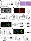 Figure 1
Characterization of secreted factors from BAT. BAT from C57BL/6NCrSlc mice fed an NCD were analyzed. (A) Panel studied young (12–13 weeks) or aged (79 weeks) mice, and other panels tested young (11 weeks) or aged (111–116 weeks) mice. (A) RNA-seq analyzing BAT (GSE274901) (n = 7, 3). (B) BAT PCPE-1 Western blot and quantification (n = 4, 4). (C) H&E staining of BAT. Scale bar: 100 μm. (D and E) Electron microscopic findings (D: Scale bar: 2 μm) and dihydroethidium staining of BAT (E: Scale bar: 50 μm) with quantification (n = 5, 5). (F–H) BAT Western blot analysis of phospho-γH2AX (F), phospho-cFos and c-Fos (G), phospho-c-Jun and c-Jun (H), and their quantification relative to GAPDH (n = 4, 4). (I and O) Immunofluorescent staining for phospho-γH2AX, WGA lectin, and Hoechst in differentiated brown adipocytes administered with DOX (I) or H2O2 (O). Scale bar: 10 μm. (J–L and P–R) For differentiated brown adipocytes, Western blot of phospho-γH2AX (J and P), phospho-cFos and c-Fos (K, L, Q, and R). (M, N, S, and T) qPCR showing transcript Pcolce in differentiated brown adipocytes, administered DOX (M [n = 4], N [n = 6]) or H2O2 (S and T) (n = 6), together with an AP1 inhibitor (AP1-i) (N and T). Housekeeping genes: Actb (M) and 18s (N, S, and T). All data, except for N and T (2-way ANOVA followed by Dunnett’s T3 for N; Tukey’s multiple-comparison test for T), were analyzed by an independent-samples t test. Data information: Representative H&E-stained images or electron microscope images from 1 series of observations (C and D), and other data were obtained from 1 representative analytical experiment out of at least 2 independent experiments showing similar results. *P < 0.05, **P < 0.01. Values are presented as mean ± SEM. NS = not significant.
HFpEF models exhibit high PCPE-1 levels in hearts. Next, we characterized the protein levels of PCPE-1 in humans and found that circulating PCPE-1 increased with age (Figure 2A), and among them, patients hospitalized with heart failure and diagnosed as HFpEF had high levels of this secreted-type profibrotic protein compared with all other cases exhibiting ejection fraction of 50% or greater (Figure 2B). The level of PCPE-1 did not show a positive correlation with BMI (Supplemental Figure 4A). Patients with hypertension had higher circulating PCPE-1 concentrations (Supplemental Figure 4B); however, levels were similar in patients with type 2 diabetes or dyslipidemia (Supplemental Figure 4, C and D). In multiple regression analyses, we found that age, but not hypertension, independently correlated with circulating PCPE-1 (Supplemental Table 1). PCPE-1 also increased in the plasma and heart of aged mice (Figure 2, C and D, and Supplemental Figure 4E) and was associated with LVDD and LV hypertrophy (Figure 2E). Although we found that body and heart weight increased with age (Supplemental Figure 4, F–H) and the aged hearts became fibrotic (Figure 2, F and G, and Supplemental Figure 4I), the transcript levels of Pcolce in the heart were comparable between young and aged mice (Figure 2H). We next characterized middle-aged mice fed with a high-fat diet (HFD) versus an NCD. The HFD increased both body and heart weight (Supplemental Figure 4, J–L), and this condition increased the expression of the DNA damage marker in BAT (Supplemental Figure 4M). In this setting, it was found that PCPE-1 protein levels were increased in the heart (Figure 2I and Supplemental Figure 4N). Echocardiography indicated that LVDD and LV hypertrophy also developed in the HFD condition (Figure 2J). Although histological analysis, biochemical quantification of hydroxyproline, and ELISA findings showed that the heart became fibrotic (Figure 2, K and L, and Supplemental Figure 4O), the cardiac Pcolce transcript was comparable between the HFD and NCD groups (Figure 2M).
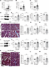 Figure 2
HFpEF models exhibit high PCPE-1 level in hearts. (A and B) Plasma PCPE-1 ELISA in the total cohort (n = 405) (A), and in patients (EF ≥ 50%) hospitalized with heart failure (HHF) (n = 80) or other diagnoses (Con) (n = 219) (B). (C) Plasma PCPE-1 ELISA in young (10–12 weeks) and aged (103–135 weeks) mice (n = 21, 20). (D) Cardiac PCPE-1 Western blot in young (10–12 weeks) and aged (103–135 weeks) mice. (E) Echocardiography in young (8 weeks) and aged (94 weeks) mice: E/e’ (marker for diastolic dysfunction), IVSTd (interventricular septum thickness), LVDs (left ventricular systolic dimension), FS (fractional shortening) (n = 6, 8). (F) Masson’s trichrome stain of the heart (Scale bar: 50 μm) and its quantification (% area/view) (n = 4, 4). (G and H) ELISA for collagen type I (shown as Collagen 1) (G) (n = 8, 7) or transcript Pcolce (H) (n = 6, 6) in the heart of young (10–12 weeks) and aged mice (103–135 weeks). Housekeeping gene: Actb. (I–M) These panels studied C57BL/6NCrSlc mice fed with a normal chow diet (NCD) or a high-fat diet (HFD) aged 44–45 weeks (middle-aged). (I) Cardiac PCPE-1 Western blot. (J) Echocardiography in mice (n = 12, 12). (K) Cardiac Masson’s trichrome stain and quantification (n = 5, 6). Scale bar: 50 μm. (L and M) ELISA for Collagen 1 (L) (n = 8, 8) or transcript Pcolce (M) (n = 6, 8) in the hearts. Housekeeping gene: Rplp0. Logistic curve estimation in A; other panels: independent-samples t test. Data information: Representative Masson’s trichrome–stained images from 1 series of observations (F and K); other data were obtained from 1 representative analytical experiment out of at least 2 independent experiments showing similar results. *P < 0.05, **P < 0.01. Values are presented as mean ± SEM. NS = not significant.
Depletion of PCPE-1 ameliorates HFpEF in obesity and aged mouse models. Next, we characterized systemic middle-aged Pcolce-KO (Pcolce–/–) mice maintained on an NCD or an HFD (Figure 3A and Supplemental Figure 5A). This analysis revealed that both body and heart weight were comparable between genotypes on the different diet regimens (Supplemental Figure 5, B–D). In this setting, the levels of systemic glucose and insulin intolerance were similar between the genotypes in the HFD-fed groups (Supplemental Figure 5, E and F), but we found that PCPE-1 deficiency mitigated LVDD under obesity (Figure 3B and Supplemental Figure 5G) and diminished the degree of cardiac fibrosis (Figure 3, C and D, and Supplemental Figure 5H). Physiological studies also showed an increase in physical activity among the HFD-fed middle-aged Pcolce-KO mice (Figure 3E).
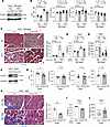 Figure 3
Depletion of PCPE-1 ameliorates HFpEF in obesity and aged mouse models. (A–E) Littermate control (Con) or systemic Pcolce-KO mice fed with a high-fat diet (HFD) from 4 weeks of age or a normal chow diet (NCD) and studied at 41–46 weeks of age (middle-aged). (F–J) Systemic Pcolce-KO mice (74–80 wk) fed with an NCD. (A and F) Western blot analysis of PCPE-1 expression in the heart. (B and G) Echocardiographic findings in the indicated mice: E/e’ (marker for diastolic dysfunction), IVSTd (interventricular septum thickness), LVDs (left ventricular systolic dimension), FS (fractional shortening) (n = 6, 8, 8, 9 for E/e’ and n = 6, 8, 13, 12 for other parameters in B; n = 9, 12 for G). (C and H) Masson’s trichrome stain of the heart. Scale bar: 50 μm. Right panels indicate quantification of relative fibrotic area (C) (n = 7, 5) (H) (n = 4, 5). (D and I) ELISA for cardiac collagen type I (shown as Collagen 1) (n = 10, 9 for D) and (n = 4, 5 for I). (E and J) Treadmill examination test (TMET) showing the running distance (n = 13, 17 for E) and (n = 9, 12 for J). Two-way ANOVA followed by Tukey’s multiple-comparison (B and D) or Dunnett T3 multiple comparison test (C); others: independent-samples t test. Data information: Representative Masson’s trichrome–stained images from 1 series of observations (C and H), and other data were obtained from 1 representative analytical experiment out of at least 2 independent experiments showing similar results. *P < 0.05, **P < 0.01. Values are presented as mean ± SEM. NS = not significant.
In the aging model, the genetic depletion of PCPE-1 resulted in the amelioration of age-associated LVDD (Figure 3, F and G, and Supplemental Figure 6A). Similar to the model of obesity, there were no differences in body weight and heart weight between the genotypes (Supplemental Figure 6, B–D); however, the level of LV fibrosis was diminished in the aged KO mice compared with that in the littermate control group (Figure 3, H and I, and Supplemental Figure 6E). The running distance during the treadmill test also lengthened in the KO group (Figure 3J), and the degrees of systemic insulin and glucose tolerance were found to be comparable between the 2 genotypes (Supplemental Figure 6, F and G). We also found that LV-diastolic dysfunction was significantly ameliorated in the KO group of female mice, suggesting that PCPE-1 mediates the pathogenesis of age-related HFpEF in a sex-independent manner (Supplemental Figure 6H). Transcripts for fibrogenic mediators, including Tgfb1, Tnf, Lgals3, Edn1, and Agt, were comparable in the kidney of C57BL/6NCrSlc-WT mice fed an NCD or HFD (Supplemental Figure 6I), but the Ccn2 transcript increased under this setting (Supplemental Figure 6J). In the HFD-fed Pcolce-KO male mice, transcripts for Ccn2 were reduced compared with HFD-fed littermate control male mice (Supplemental Figure 6, K and L), but there was no detectable effect on the transcripts of other profibrogenic mediators.
The BATokine PCPE-1 contributes to the pathogenesis of HFpEF. To evaluate the contribution of BAT-derived PCPE-1 to the pathogenesis of HFpEF, we specifically depleted PCPE-1 in BAT by crossing UCP-1Cre+/– with Pcolcefl/fl mice (i.e., BAT Pcolce-KO). In these mice, the levels of PCPE-1 in the plasma and heart were reduced compared with levels in the Pcolcefl/fl littermate control mice fed an HFD (Figure 4A and Supplemental Figure 7, A and B). Body, heart weight, levels of systemic glucose/insulin tolerance, and volumes of visceral and subcutaneous fat analyzed by CT were comparable between the genotypes (Supplemental Figure 7, C–H). Similar to the systemic Pcolce-KO mice, the Ccn2 transcript was reduced in the kidney of the BAT Pcolce-KO mice under diet-induced obesity (Supplemental Figure 7I). It was also found that LVDD was ameliorated in BAT-specific Pcolce-KO of the middle-aged mice with obesity (Figure 4B), as was cardiac fibrosis (Figure 4, C and D, and Supplemental Figure 7J). We next generated a BAT-specific PCPE-1 overexpression model by the injection of adeno-associated virus (AAV) encoding Pcolce (AAV-Pcolce) into the BAT fat pad of adult mice (Figure 4E). This led to the induction of Pcolce expression in BAT and an increase in PCPE-1 levels in the heart under conditions of diet-induced obesity (Figure 4F and Supplemental Figure 8A). Body and heart weight were comparable between the groups (Supplemental Figure 8, B–D). Administration of AAV-Pcolce to BAT led to a deterioration of LVDD (Figure 4G) and an aggravation of cardiac fibrosis (Figure 4, H and I, and Supplemental Figure 8E).
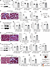 Figure 4
The BATokine PCPE-1 contributes to the pathogenesis of HFpEF. Littermate control (Con) or BAT Pcolce-KO mice were fed with a high-fat diet (HFD) and studied during 41–47 weeks of age (middle-aged). BAT adeno-associated virus (AAV) encoding Pcolce was injected in the BAT of C57BL/6NCrSlc mice fed with an HFD and studied at 15 weeks of age (mature adult). BAT-PCOLCE KI mice were also fed with an HFD and studied at 23–25 weeks of age (mature adult). (A, F, and J) Western blot analysis of mouse (A and F) or human (J) PCPE-1 expression in the heart of the indicated mice. (B, G, and K) Echocardiographic findings in mice: E/e’ (marker for diastolic dysfunction), IVSTd (interventricular septum thickness), LVDs (left ventricular systolic dimension), FS (fractional shortening) (n = 14, 10 for B), (n =7, 7 for G), and (n = 8, 7 for K). (C, H, and L) Masson’s trichrome stain of the heart and its quantification (n =5, 5 for C), (n = 5, 6 for H), and (n = 4, 6 for L) in mice. Scale bar: 50 μm. (D, I, and M) ELISA for collagen type I (shown as Collagen 1) testing hearts in mice (n = 9, 10 for D), (n = 5, 6 for I), and (n = 8, 7 for M). (E) Experimental design of the AAV-Pcolce mouse. (N) Schematic demonstrating that ROS/DNA damage/c-Fos/c-Jun signaling upregulates PCPE-1 expression in BAT with aging. PCPE-1 produced from BAT promotes fibrosis in the heart. All data were analyzed by an independent-samples t test. Data information: Representative Masson’s trichrome–stained images from 1 series of observations (C, H, and L), and other data were obtained from 1 representative analytical experiment out of at least 2 independent experiments showing similar results. *P < 0.05, **P < 0.01. Values are presented as mean ± SEM. NS = not significant.
To further test the role of BAT-derived PCPE-1, we generated BAT-specific human PCOLCE knock-in (KI) mice by crossing UCP1-Cre+/– with PCOLCEKI/KI mice (BAT-PCOLCE-KI) (Supplemental Figure 8F). Analyses of the adult mice revealed that human PCPE-1 protein expression could be detected in hearts of the BAT-PCOLCE-KI mice fed with an HFD (Figure 4J and Supplemental Figure 8G). Body and heart weight were comparable between the genotypes (Supplemental Figure 8, H–J), but echocardiographic analysis revealed that LVDD was enhanced in the KI mice (Figure 4K), as was the progression of LV fibrosis (Figure 4, L and M, and Supplemental Figure 8K).


